|
Diagnostic Microbiology
Diagnostic microbiology is the study of microbial identification. Since the discovery of the germ theory of disease, scientists have been finding ways to harvest specific organisms. Using methods such as differential media or Whole genome sequencing, genome sequencing, physicians and scientists can observe novel functions in organisms for more effective and accurate diagnosis of organisms. Methods used in diagnostic microbiology are often used to take advantage of a particular difference in organisms and attain information about what species it can be identified as, which is often through a reference of previous studies. New studies provide information that others can reference so that scientists can attain a basic understanding of the organism they are examining. Aerobic vs anaerobic Anaerobic organisms require an oxygen-free environment. When culturing anaerobic microbes, broths are often flushed with nitrogen gas to extinguish oxygen present, and growth can also occur on medi ... [...More Info...] [...Related Items...] OR: [Wikipedia] [Google] [Baidu] [Amazon] |
Germ Theory Of Disease
The germ theory of disease is the currently accepted scientific theory for many diseases. It states that microorganisms known as pathogens or "germs" can cause disease. These small organisms, which are too small to be seen without magnification, invade animals, plants, and even bacteria. Their growth and reproduction within their hosts can cause disease. "Germ" refers not just to bacteria but to any type of microorganism, such as protists or fungi, or other pathogens, including parasites, viruses, prions, or viroids. Diseases caused by pathogens are called infectious diseases. Even when a pathogen is the principal cause of a disease, environmental and hereditary factors often influence the severity of the disease, and whether a potential host individual becomes infected when exposed to the pathogen. Pathogens are disease-causing agents that can pass from one individual to another, across multiple domains of life. Basic forms of germ theory were proposed by Girolamo Fracastoro ... [...More Info...] [...Related Items...] OR: [Wikipedia] [Google] [Baidu] [Amazon] |
Helicobacter Pylori
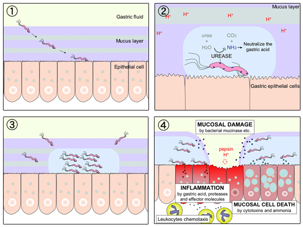
''Helicobacter pylori'', previously known as ''Campylobacter pylori'', is a gram-negative, Flagellum#bacterial, flagellated, Bacterial cellular morphologies#Helical, helical bacterium. Mutants can have a rod or curved rod shape that exhibits less virulence. Its Helix, helical body (from which the genus name ''Helicobacter'' derives) is thought to have evolved to penetrate the gastric mucosa, mucous lining of the stomach, helped by its flagella, and thereby establish infection. While many earlier reports of an association between bacteria and the ulcers had existed, such as the works of John Lykoudis, it was only in 1983 when the bacterium was formally described for the first time in the English-language Western literature as the causal agent of peptic ulcer, gastric ulcers by Australian physician-scientists Barry Marshall and Robin Warren. In 2005, the pair was awarded the Nobel Prize in Physiology or Medicine for their discovery. Infection of the stomach with ''H. pylori'' doe ... [...More Info...] [...Related Items...] OR: [Wikipedia] [Google] [Baidu] [Amazon] |
Ziehl–Neelsen Stain
The Ziehl-Neelsen stain, also known as the acid-fast stain, is a bacteriological staining technique used in cytopathology and microbiology to identify acid-fast bacteria under microscopy, particularly members of the ''Mycobacterium'' genus. This staining method was initially introduced by Paul Ehrlich (1854–1915) and subsequently modified by the German bacteriologists Franz Ziehl (1859–1926) and Friedrich Neelsen (1854–1898) during the late 19th century. The acid-fast staining method, in conjunction with auramine phenol staining, serves as the standard diagnostic tool and is widely accessible for rapidly diagnosing tuberculosis (caused by ''Mycobacterium tuberculosis'') and other diseases caused by atypical mycobacteria, such as leprosy (caused by ''Mycobacterium leprae'') and ''Mycobacterium avium-intracellulare'' infection (caused by ''Mycobacterium avium'' complex) in samples like sputum, gastric washing fluid, and bronchoalveolar lavage fluid. These acid-fast bact ... [...More Info...] [...Related Items...] OR: [Wikipedia] [Google] [Baidu] [Amazon] |
India Ink Stain
India ink (British English: Indian ink; also Chinese ink) is a simple black or coloured ink once widely used for writing and printing and now more commonly used for drawing and outlining, especially when inking comic books and comic strips. India ink is also used in medical applications. Compared to other inks, such as the iron gall ink previously common in Europe, India ink is noted for its deep, rich black color. It is commonly applied with a paintbrush (such as an ink brush) or a dip pen. In East Asian traditions such as ink wash painting and Chinese calligraphy, India ink is commonly used in a solid form called an inkstick. Composition Basic India ink is composed of a variety of fine soot, known as ''lampblack'', combined with water to form a liquid. No binder material is necessary: the carbon molecules are in colloidal suspension and form a waterproof layer after drying. A binding agent such as gelatin or, more commonly, shellac may be added to make the ink more durable ... [...More Info...] [...Related Items...] OR: [Wikipedia] [Google] [Baidu] [Amazon] |
Giemsa Stain
Giemsa stain (), named after German chemist and bacteriologist Gustav Giemsa, is a nucleic acid stain used in cytogenetics and for the histopathological diagnosis of malaria and other parasites. Uses It is specific for the phosphate groups of DNA and attaches itself to regions of DNA where there are high amounts of adenine-thymine bonding. Giemsa stain is used in Giemsa banding, commonly called G-banding, to stain chromosomes and often used to create a karyogram (chromosome map). It can identify chromosomal aberrations such as translocations and rearrangements. It stains the trophozoite ''Trichomonas vaginalis'', which presents with greenish discharge and motile cells on wet prep. Giemsa stain is also a differential stain, such as when it is combined with Wright stain to form Wright–Giemsa stain. It can be used to study the adherence of pathogenic bacteria to human cells. It differentially stains human and bacterial cells purple and pink respectively. It can be used fo ... [...More Info...] [...Related Items...] OR: [Wikipedia] [Google] [Baidu] [Amazon] |
Acid-fast Stain
Acid-fastness is a physical property of certain bacterial and eukaryotic cells, as well as some sub-cellular structures, specifically their resistance to decolorization by acids during laboratory staining procedures. Once stained as part of a sample, these organisms can resist the acid and/or ethanol-based decolorization procedures common in many staining protocols, hence the name ''acid-fast''. The mechanisms of acid-fastness vary by species although the most well-known example is in the genus ''Mycobacterium'', which includes the species responsible for tuberculosis and leprosy. The acid-fastness of ''Mycobacteria'' is due to the high mycolic acid content of their cell walls, which is responsible for the staining pattern of poor absorption followed by high retention. Some bacteria may also be partially acid-fast, such as ''Nocardia''. Acid-fast organisms are difficult to characterize using standard microbiological techniques, though they can be stained using concentrated dyes, ... [...More Info...] [...Related Items...] OR: [Wikipedia] [Google] [Baidu] [Amazon] |
Gram Stain
Gram stain (Gram staining or Gram's method), is a method of staining used to classify bacterial species into two large groups: gram-positive bacteria and gram-negative bacteria. It may also be used to diagnose a fungal infection. The name comes from the Danish bacteriologist Hans Christian Gram, who developed the technique in 1884. Gram staining differentiates bacteria by the chemical and physical properties of their Cell wall#Bacterial cell walls, cell walls. Gram-positive cells have a thick layer of peptidoglycan in the cell wall that retains the primary stain, crystal violet. Gram-negative cells have a thinner peptidoglycan layer that allows the crystal violet to wash out on addition of ethanol. They are stained pink or red by the counterstain, commonly safranin or fuchsine. Lugol's iodine solution is always added after addition of crystal violet to form a stable complex with crystal violet that strengthens the bonds of the stain with the cell wall. Gram staining is almost al ... [...More Info...] [...Related Items...] OR: [Wikipedia] [Google] [Baidu] [Amazon] |
Staining
Staining is a technique used to enhance contrast in samples, generally at the Microscope, microscopic level. Stains and dyes are frequently used in histology (microscopic study of biological tissue (biology), tissues), in cytology (microscopic study of cell (biology), cells), and in the medical fields of histopathology, hematology, and cytopathology that focus on the study and diagnoses of diseases at the microscopic level. Stains may be used to define biological tissues (highlighting, for example, muscle fibers or connective tissue), cell (biology), cell populations (classifying different blood cells), or organelles within individual cells. In biochemistry, it involves adding a class-specific (DNA, proteins, lipids, carbohydrates) dye to a substrate to qualify or quantify the presence of a specific compound. Staining and fluorescent tagging can serve similar purposes. Biological staining is also used to mark cells in flow cytometry, and to flag proteins or nucleic acids in gel ... [...More Info...] [...Related Items...] OR: [Wikipedia] [Google] [Baidu] [Amazon] |
Biopsy
A biopsy is a medical test commonly performed by a surgeon, interventional radiologist, an interventional radiologist, or an interventional cardiology, interventional cardiologist. The process involves the extraction of sampling (medicine), sample Cell (biology), cells or Biological tissue, tissues for examination to determine the presence or extent of a disease. The tissue is then Histopathology, fixed, dehydrated, embedded, sectioned, stained and mounted before it is generally examined under a microscope by a pathologist; it may also be analyzed chemically. When an entire lump or suspicious area is removed, the procedure is called an excisional biopsy. An incisional biopsy or core biopsy samples a portion of the abnormal tissue without attempting to remove the entire lesion or tumor. When a sample of tissue or fluid is removed with a needle in such a way that cells are removed without preserving the histological architecture of the tissue cells, the procedure is called a needle as ... [...More Info...] [...Related Items...] OR: [Wikipedia] [Google] [Baidu] [Amazon] |
Histology
Histology, also known as microscopic anatomy or microanatomy, is the branch of biology that studies the microscopic anatomy of biological tissue (biology), tissues. Histology is the microscopic counterpart to gross anatomy, which looks at larger structures visible without a microscope. Although one may divide microscopic anatomy into ''organology'', the study of organs, ''histology'', the study of tissues, and ''cytology'', the study of cell (biology), cells, modern usage places all of these topics under the field of histology. In medicine, histopathology is the branch of histology that includes the microscopic identification and study of diseased tissue. In the field of paleontology, the term paleohistology refers to the histology of fossil organisms. Biological tissues Animal tissue classification There are four basic types of animal tissues: muscle tissue, nervous tissue, connective tissue, and epithelial tissue. All animal tissues are considered to be subtypes of these ... [...More Info...] [...Related Items...] OR: [Wikipedia] [Google] [Baidu] [Amazon] |
Epitope
An epitope, also known as antigenic determinant, is the part of an antigen that is recognized by the immune system, specifically by antibodies, B cells, or T cells. The part of an antibody that binds to the epitope is called a paratope. Although epitopes are usually non-self proteins, sequences derived from the host that can be recognized (as in the case of autoimmune diseases) are also epitopes. The epitopes of protein antigens are divided into two categories, conformational epitopes and linear epitopes, based on their structure and interaction with the paratope. Conformational and linear epitopes interact with the paratope based on the 3-D conformation adopted by the epitope, which is determined by the surface features of the involved epitope residues and the shape or tertiary structure of other segments of the antigen. A conformational epitope is formed by the 3-D conformation adopted by the interaction of discontiguous amino acid residues. In contrast, a linear epitope i ... [...More Info...] [...Related Items...] OR: [Wikipedia] [Google] [Baidu] [Amazon] |


