|
Descemet Membrane Endothelial Keratoplasty
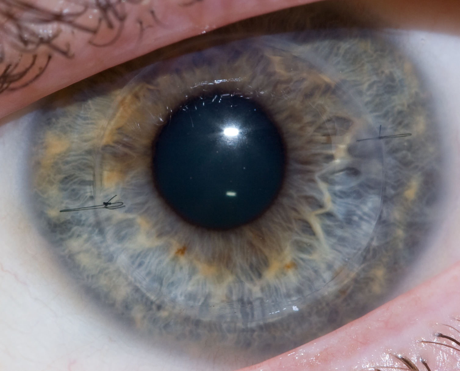
Descemet membrane endothelial keratoplasty (DMEK) is a method of corneal transplantation that involves the removal of a thin sheet of tissue from the posterior (innermost) side of a person's cornea to replace it with the two posterior (innermost) layers of corneal tissue from a donor's eyeball. The two exchanged corneal layers are the Descemet's membrane and the corneal endothelium. The person's corneal tissue is gently excised, peeled off, and replaced with the donor tissue via small 'clear corneal incisions' (small corneal incisions just anterior to the corneal limbus. The donor tissue is tamponaded against the person's exposed posterior corneal stroma by injecting a small air bubble into the anterior chamber. To ensure the air tamponade is effective, people must maintain such a posture that they look up at the ceiling during recovery until the air bubble has fully resorbed. Medical uses Indications for DMEK include: * Corneal dystrophy involving the corneal endothelial layer ... [...More Info...] [...Related Items...] OR: [Wikipedia] [Google] [Baidu] |
Corneal Transplantation
Corneal transplantation, also known as corneal grafting, is a surgical procedure where a damaged or diseased cornea is replaced by donated corneal tissue (the graft). When the entire cornea is replaced it is known as penetrating keratoplasty and when only part of the cornea is replaced it is known as lamellar keratoplasty. Keratoplasty simply means surgery to the cornea. The graft is taken from a recently deceased individual with no known diseases or other factors that may affect the chance of survival of the donated tissue or the health of the recipient. The cornea is the transparent front part of the eye that covers the iris, pupil and anterior chamber. The surgical procedure is performed by ophthalmologists, physicians who specialize in eyes, and is often done on an outpatient basis. Donors can be of any age, as is shown in the case of Janis Babson, who donated her eyes after dying at the age of 10. Corneal transplantation is performed when medicines, keratoconus conser ... [...More Info...] [...Related Items...] OR: [Wikipedia] [Google] [Baidu] |
Posterior Polymorphous Corneal Dystrophy
Posterior polymorphous corneal dystrophy (PPCD; sometimes also ''Schlichting dystrophy'') is a type of corneal dystrophy, characterised by changes in Descemet's membrane and endothelial layer. Symptoms mainly consist of decreased vision due to corneal edema. In some cases they are present from birth, other patients are asymptomatic. Histopathological analysis shows that the cells of endothelium have some characteristics of epithelial cells and have become multilayered. The disease was first described in 1916 by Koeppe as ''keratitis bullosa interna''. Genetics PPCD type 2 is linked to the mutations in COL8A2, and PPCD type 3 mutations in ZEB1 gene, but the underlying genetic disturbance in PPCD type 1 is unknown. Pathophysiology Vacuoles are demonstrated in the posterior parts of the cornea. The vesicles are located on the endothelial surface. The corneal endothelium is normally a single layer of cells that lose their mitotic potential after development is complete. In posterior ... [...More Info...] [...Related Items...] OR: [Wikipedia] [Google] [Baidu] |
Medical Procedures
A medical procedure is a course of action intended to achieve a result in the delivery of healthcare. A medical procedure with the intention of determining, measuring, or diagnosing a patient condition or parameter is also called a medical test. Other common kinds of procedures are therapeutic (i.e., intended to treat, cure, or restore function or structure), such as surgical and physical rehabilitation procedures. Definition *"An activity directed at or performed on an individual with the object of improving health, treating disease or injury, or making a diagnosis."''International Dictionary of Medicine and Biology'', Page 2297. - ''International Dictionary of Medicine and Biology'' *"The act or conduct of diagnosis, treatment, or operation."''Stedman's Medical Dictionary'', 27th ed. Page 1446. - ''Stedman's Medical Dictionary'' by Thomas Lathrop Stedman *"A series of steps by which a desired result is accomplished."''Dorland's Illustrated Medical Dictionary'', 28th ed. Pag ... [...More Info...] [...Related Items...] OR: [Wikipedia] [Google] [Baidu] |
Eye Procedures
An eye is a sensory organ that allows an organism to perceive visual information. It detects light and converts it into electro-chemical impulses in neurons (neurones). It is part of an organism's visual system. In higher organisms, the eye is a complex optical system that collects light from the surrounding environment, regulates its intensity through a diaphragm, focuses it through an adjustable assembly of lenses to form an image, converts this image into a set of electrical signals, and transmits these signals to the brain through neural pathways that connect the eye via the optic nerve to the visual cortex and other areas of the brain. Eyes with resolving power have come in ten fundamentally different forms, classified into compound eyes and non-compound eyes. Compound eyes are made up of multiple small visual units, and are common on insects and crustaceans. Non-compound eyes have a single lens and focus light onto the retina to form a single image. This type of ey ... [...More Info...] [...Related Items...] OR: [Wikipedia] [Google] [Baidu] |
Corneal Transplantation
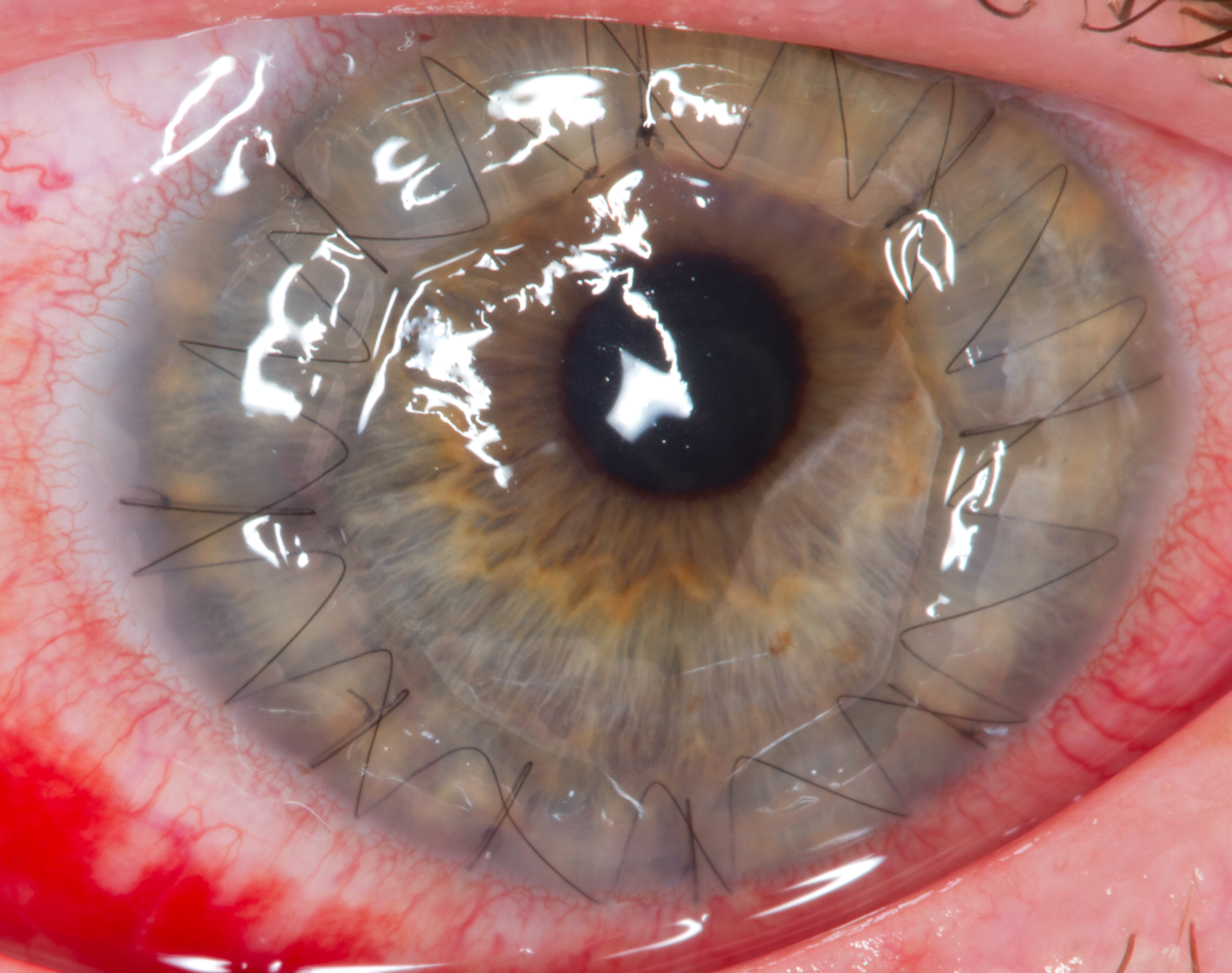
Corneal transplantation, also known as corneal grafting, is a surgical procedure where a damaged or diseased cornea is replaced by donated corneal tissue (the graft). When the entire cornea is replaced it is known as penetrating keratoplasty and when only part of the cornea is replaced it is known as lamellar keratoplasty. Keratoplasty simply means surgery to the cornea. The graft is taken from a recently deceased individual with no known diseases or other factors that may affect the chance of survival of the donated tissue or the health of the recipient. The cornea is the transparent front part of the eye that covers the iris, pupil and anterior chamber. The surgical procedure is performed by ophthalmologists, physicians who specialize in eyes, and is often done on an outpatient basis. Donors can be of any age, as is shown in the case of Janis Babson, who donated her eyes after dying at the age of 10. Corneal transplantation is performed when medicines, keratoconus conser ... [...More Info...] [...Related Items...] OR: [Wikipedia] [Google] [Baidu] |
Femtosecond Laser
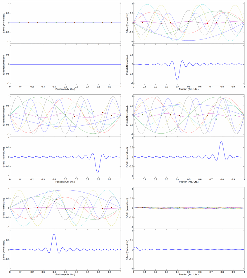
Mode locking is a technique in optics by which a laser can be made to produce pulses of light of extremely short duration, on the order of picoseconds (10−12 s) or femtoseconds (10−15 s). A laser operated in this way is sometimes referred to as a femtosecond laser, for example, in modern refractive surgery. The basis of the technique is to induce a fixed phase relationship between the longitudinal modes of the laser's resonant cavity. Constructive interference between these modes can cause the laser light to be produced as a train of pulses. The laser is then said to be "phase-locked" or "mode-locked". Laser cavity modes Although laser light is perhaps the purest form of light, it is not of a single, pure frequency or wavelength. All lasers produce light over some natural bandwidth or range of frequencies. A laser's bandwidth of operation is determined primarily by the gain medium from which the laser is constructed, and the range of frequencies over which a lase ... [...More Info...] [...Related Items...] OR: [Wikipedia] [Google] [Baidu] |
Iridocorneal Endothelial Syndrome
Iridocorneal endothelial (ICE) syndromes are a spectrum of diseases characterized by slowly progressive abnormalities of the corneal endothelium and features including corneal edema, iris distortion, and secondary angle-closure glaucoma.Weisenthal RW. ''2012-2013 Basic and Clinical Science Course, Section 8, Chapter 12: External Disease and Cornea'' (pp 344–345). San Francisco CA: American Academy of Ophthalmology The Eye M.D. Association ICE syndromes are predominantly unilateral and nonhereditary. The condition occurs in predominantly middle-aged women.Iridocorneal Endothelial (ICE) syndrome presents a unique set of challenges for both patients and ophthalmologists, and effective treatment of this group of rare ocular diseases requires a combination of diagnostic and therapeutic complexity. It's important to understand Signs and symptoms Many cases are asymptomatic, however patients many have decreased vision, glare, monocular diplopia or polyopia, and noticeable iris cha ... [...More Info...] [...Related Items...] OR: [Wikipedia] [Google] [Baidu] |
Bullous Keratopathy
Bullous keratopathy, also known as pseudophakic bullous keratopathy (PBK), is a pathological condition in which small vesicles, or '' bullae'', are formed in the cornea due to endothelial dysfunction. In a healthy cornea, endothelial cells keeps the tissue from excess fluid absorption, pumping it back into the aqueous humor. When affected by some reason, such as Fuchs' dystrophy Fuchs dystrophy, also referred to as Fuchs endothelial corneal dystrophy (FECD) and Fuchs endothelial dystrophy (FED), is a slowly progressing corneal dystrophy that usually affects both eyes and is slightly more common in women than in men. Althou ... or a trauma during cataract removal, endothelial cells suffer mortality or damage. The corneal endothelial cells normally do not undergo mitotic cell division, and cell loss results in permanent loss of function. When endothelial cell counts drop too low, the pump starts failing to function and fluid moves anterior into the stroma and epithelium. The excess ... [...More Info...] [...Related Items...] OR: [Wikipedia] [Google] [Baidu] |
Fuchs' Endothelial Dystrophy
Fuchs dystrophy, also referred to as Fuchs endothelial corneal dystrophy (FECD) and Fuchs endothelial dystrophy (FED), is a slowly progressing corneal dystrophy that usually affects both eyes and is slightly more common in women than in men. Although early signs of Fuchs dystrophy are sometimes seen in people in their 30s and 40s, the disease rarely affects vision until people reach their 50s and 60s. Signs and symptoms As a progressive, chronic condition, signs and symptoms of Fuchs dystrophy gradually progress over decades of life, starting in middle age. Early symptoms include blurry vision upon wakening which improves during the morning, as fluid retained in the cornea is unable to evaporate through the surface of the eye when the lids are closed overnight. As the disease worsens, the interval of blurry morning vision extends from minutes to hours. In moderate stages of the disease, an increase in guttae and swelling in the cornea can contribute to changes in vision and decreas ... [...More Info...] [...Related Items...] OR: [Wikipedia] [Google] [Baidu] |
Descemet's Membrane
Descemet's membrane ( or the Descemet membrane) is the basement membrane that lies between the corneal proper substance, also called stroma, and the endothelial layer of the cornea. It is composed of different kinds of collagen (Type IV and VIII) than the stroma. The endothelial layer is located at the posterior of the cornea. Descemet's membrane, as the basement membrane for the endothelial layer, is secreted by the single layer of squamous epithelial cells that compose the endothelial layer of the cornea. Structure Its thickness ranges from 3 μm at birth to 8–10 μm in adults.Johnson DH, Bourne WM, Campbell RJ: The ultrastructure of Descemet's membrane. I. Changes with age in normal cornea. Arch Ophthalmol 100:1942, 1982 The corneal endothelium is a single layer of squamous cells covering the surface of the cornea that faces the anterior chamber. Clinical significance Significant damage to the membrane may require a corneal transplant. Damage caused by the hereditary c ... [...More Info...] [...Related Items...] OR: [Wikipedia] [Google] [Baidu] |
Corneal Dystrophy
Corneal dystrophy is a group of rare hereditary disorders characterised by bilateral abnormal deposition of substances in the transparent front part of the eye called the cornea. Signs and symptoms Corneal dystrophy may not significantly affect vision in the early stages. However, it does require proper evaluation and treatment for restoration of optimal vision. Corneal dystrophies usually manifest themselves during the first or second decade but sometimes later. It appears as grayish white lines, circles, or clouding of the cornea. Corneal dystrophy can also have a crystalline appearance. There are over 20 corneal dystrophies that affect all parts of the cornea. These diseases share many traits: * They are usually inherited. * They affect the right and left eyes equally. * They are not caused by outside factors, such as injury or diet. * Most progress gradually. * Most usually begin in one of the five corneal layers and may later spread to nearby layers. * Most do not affect ot ... [...More Info...] [...Related Items...] OR: [Wikipedia] [Google] [Baidu] |
Anterior Chamber
The anterior chamber ( AC) is the aqueous humor-filled space inside the eye between the iris and the cornea's innermost surface, the endothelium. Hyphema, anterior uveitis and glaucoma are three main pathologies in this area. In hyphema, blood fills the anterior chamber as a result of a hemorrhage, most commonly after a blunt eye injury. Anterior uveitis is an inflammatory process affecting the iris and ciliary body, with resulting inflammatory signs in the anterior chamber. In glaucoma, blockage of the trabecular meshwork prevents the normal outflow of aqueous humour, resulting in increased intraocular pressure, progressive damage to the optic nerve head, and eventually blindness. The depth of the anterior chamber of the eye varies between 1.5 and 4.0 mm, averaging 3.0 mm. It tends to become shallower at older age and in eyes with hypermetropia (far sightedness). As depth decreases below 2.5 mm, the risk for angle closure glaucoma increases. Clinical s ... [...More Info...] [...Related Items...] OR: [Wikipedia] [Google] [Baidu] |


