|
Dental Trauma
Dental trauma refers to trauma (injury) to the teeth and/or periodontium (gums, periodontal ligament, alveolar bone), and nearby soft tissues such as the lips, tongue, etc. The study of dental trauma is called dental traumatology.''Textbook and Color Atlas of Traumatic Injuries to the Teeth'', Fourth Edition, edited by Andreason J, Andreasen F, and Andersson L, Wiley-Blackwell, Oxford, UK, 2007 Types Dental injuries Dental injuries include: * Enamel infraction * Enamel fracture * Enamel-dentine fracture * Enamel-dentine fracture involving pulp exposure * Root fracture of tooth Periodontal injuries * Concussion (bruising) *Subluxation of the tooth (tooth knocked loose) * Luxation of the tooth (displaced) **Extrusive ** Intrusive **Lateral * Avulsion of the tooth (tooth knocked out) Injuries to supporting bone This injury involves the alveolar bone and may extend beyond the alveolus. There are five different types of alveolar fractures: * Communicated fracture ... [...More Info...] [...Related Items...] OR: [Wikipedia] [Google] [Baidu] |
Dentine
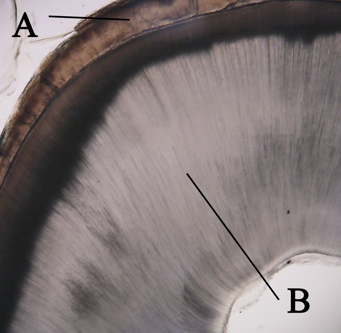
Dentin ( ) (American English) or dentine ( or ) (British English) () is a calcified tissue of the body and, along with enamel, cementum, and pulp, is one of the four major components of teeth. It is usually covered by enamel on the crown and cementum on the root and surrounds the entire pulp. By volume, 45% of dentin consists of the mineral hydroxyapatite, 33% is organic material, and 22% is water. Yellow in appearance, it greatly affects the color of a tooth due to the translucency of enamel. Dentin, which is less mineralized and less brittle than enamel, is necessary for the support of enamel. Dentin rates approximately 3 on the Mohs scale of mineral hardness. There are two main characteristics which distinguish dentin from enamel: firstly, dentin forms throughout life; secondly, dentin is sensitive and can become hypersensitive to changes in temperature due to the sensory function of odontoblasts, especially when enamel recedes and dentin channels become exposed. Devel ... [...More Info...] [...Related Items...] OR: [Wikipedia] [Google] [Baidu] |
Dental Intrusion
Dental intrusion is an apical displacement of the tooth into the alveolar bone. This injury is accompanied by extensive damage to periodontal ligament, cementum, disruption of the neurovascular supply to the pulp, and communication or fracture of the alveolar socket. Intrusive traumas have been found to comprise 0.3-1.9% of the traumas affecting permanent dentition. Diagnosis In most cases of intrusion with fully erupted permanent dentition, diagnosis can be made by comparing incisal height of teeth next to the injured one. In cases with mixed dentition, a percussion test must be performed as an intruded tooth can mimic an erupting tooth. Clinical and radiographical presentation Clinical findings show shortened crown length to various degree and up to no visible crown in severe cases. Tooth is immobile, and percussion gives high, metallic sound. Bleeding around crown margins can be observed. Radiographical findings shows dislocation of root in an apical direction, and period ... [...More Info...] [...Related Items...] OR: [Wikipedia] [Google] [Baidu] |
Contact Sports
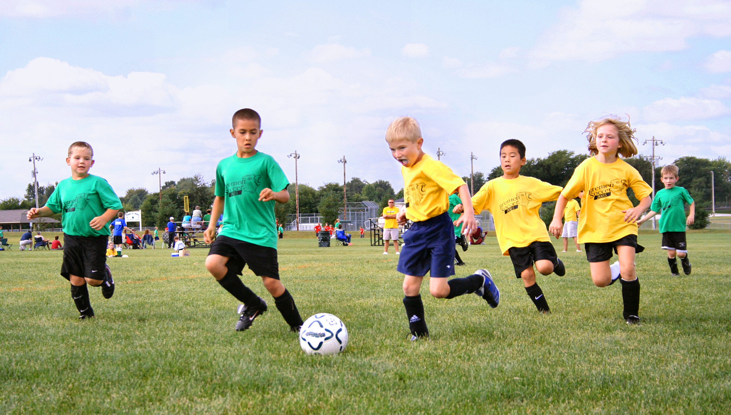
A contact sport is any sport where physical contact between competitors, or their environment, is an integral part of the game. For example, gridiron football. Contact may come about as the result of intentional or incidental actions by the players in the course of play. This is in contrast to noncontact sports where players often have no opportunity to make contact with each other and the laws of the game may expressly forbid contact. In contact sports some forms of contact are encouraged as a critical aspect of the game such as tackling, while others are incidental such as when shielding the ball or contesting an aerial challenge. As the types of contact between players is not equal between all sports they define the types of contact that is deemed acceptable and fall within the laws of the game, while outlawing other types of physical contact that might be considered expressly dangerous or risky such as a high tackle or spear tackle, or against the spirit of the game such as s ... [...More Info...] [...Related Items...] OR: [Wikipedia] [Google] [Baidu] |
Sports
Sport is a physical activity or game, often competitive and organized, that maintains or improves physical ability and skills. Sport may provide enjoyment to participants and entertainment to spectators. The number of participants in a particular sport can vary from hundreds of people to a single individual. Sport competitions may use a team or single person format, and may be open, allowing a broad range of participants, or closed, restricting participation to specific groups or those invited. Competitions may allow a "tie" or "draw", in which there is no single winner; others provide tie-breaking methods to ensure there is only one winner. They also may be arranged in a tournament format, producing a champion. Many sports leagues make an annual champion by arranging games in a regular sports season, followed in some cases by playoffs. Sport is generally recognised as system of activities based in physical athleticism or physical dexterity, with major competi ... [...More Info...] [...Related Items...] OR: [Wikipedia] [Google] [Baidu] |
Enamel Hypocalcification
Enamel is the outermost layer of the tooth which serves as a protective layer from physical, thermal, and chemical damage. Ameloblasts are the cells that produce the enamel. Their life cycle, known as amelogenesis, is divided into six stages: morphogenetic, organizing, formative, maturative, protective, and desmolytic. Enamel mineralization occurs during the maturation stage. Hence, defects in the maturation stage result in hypocalcification or hypomineralization. Enamel hypocalcification is the inadequate deposition of inorganic ions, resulting in the appearance of translucency, white-chalky spots, and yellow-brown discoloration on the surface of the tooth associated with increased sensitivity and a higher risk of developing dental caries. Enamel hypocalcification is a multifactorial disease that targets both primary and permanent dentition and is influenced by local, systemic, environmental, and genetic effects. For instance, trauma, infection, radiation, fluorosis, amelogenesis im ... [...More Info...] [...Related Items...] OR: [Wikipedia] [Google] [Baidu] |
Root Apex (dental)
In dental anatomy, the apical foramen, literally translated "small opening of the apex," is the tooth's natural opening, found at the root's very tip—that is, the root apex — whereby an artery, vein, and nerve enter the tooth and commingle with the tooth's internal soft tissue, called pulp. Additionally, the apical foramen is the point where the pulp meets the periodontal tissues, the connective tissues that surround and support the tooth. The foramen is located 0.5mm to 1.5mm from the apex of the tooth. Each tooth has an apical foramen. Characteristics The average size of the orifice is 0.3 to 0.4 mm in diameter. There can be two or more foramina separated by a portion of dentin and cementum or by cementum only. If more than one foramen is present on each root, the largest one is designated as the apical foramen and the rest are considered accessory foramina. Apical delta Apical delta refers to the branching pattern of small accessory canals and minor foramina seen a ... [...More Info...] [...Related Items...] OR: [Wikipedia] [Google] [Baidu] |
Head Facial Nerve Branches TZBMC
A head is the part of an organism which usually includes the ears, brain, forehead, cheeks, chin, eyes, nose, and mouth, each of which aid in various sensory functions such as sight, hearing, smell, and taste. Some very simple animals may not have a head, but many bilaterally symmetric forms do, regardless of size. Heads develop in animals by an evolutionary trend known as cephalization. In bilaterally symmetrical animals, nervous tissue concentrate at the anterior region, forming structures responsible for information processing. Through biological evolution, sense organs and feeding structures also concentrate into the anterior region; these collectively form the head. Human head The human head is an anatomical unit that consists of the skull, hyoid bone and cervical vertebrae. The skull consists of the brain case which encloses the cranial cavity, and the facial skeleton, which includes the mandible. There are eight bones in the brain case and fourteen in the facia ... [...More Info...] [...Related Items...] OR: [Wikipedia] [Google] [Baidu] |
Occlusion (dentistry)
Occlusion, in a dental context, means simply the contact between teeth. More technically, it is the relationship between the maxillary (upper) and mandibular (lower) teeth when they approach each other, as occurs during chewing or at rest. Static occlusion refers to contact between teeth when the jaw is closed and stationary, while dynamic occlusion refers to occlusal contacts made when the jaw is moving. The masticatory system also involves the periodontium, the TMJ (and other skeletal components) and the neuromusculature, therefore the tooth contacts should not be looked at in isolation, but in relation to the overall masticatory system. Anatomy of Masticatory System One cannot fully understand occlusion without an in depth understanding of the anatomy including that of the teeth, TMJ, musculature surrounding this and the skeletal components. The Dentition and Surrounding Structures The human dentition consists of 32 permanent teeth and these are distributed between ... [...More Info...] [...Related Items...] OR: [Wikipedia] [Google] [Baidu] |
Mandibular Fracture
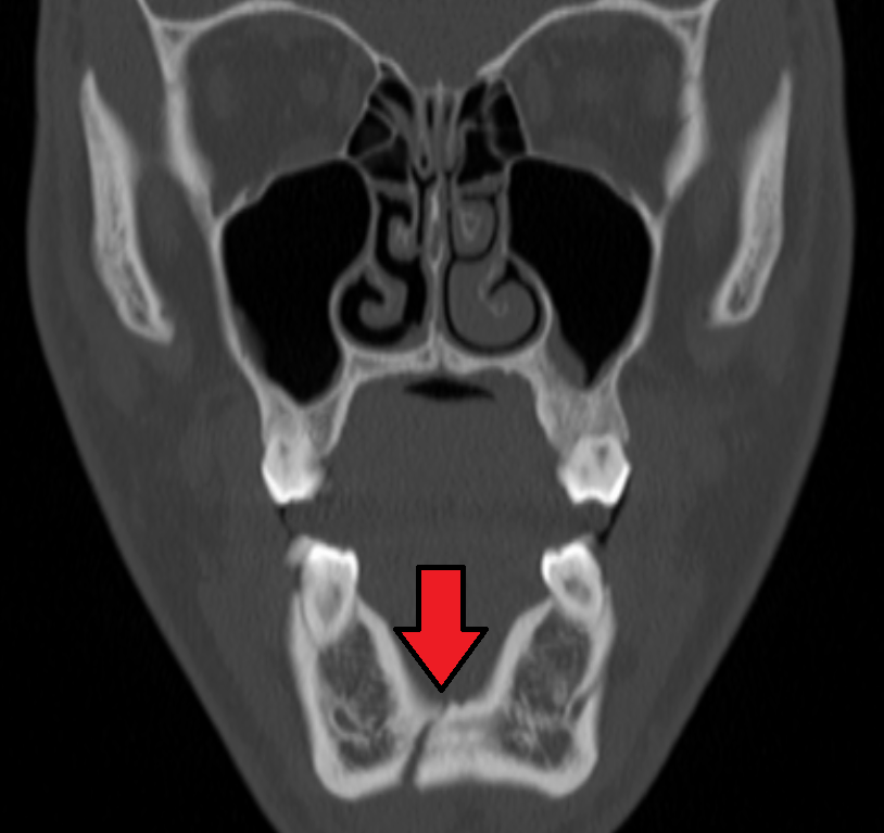
Mandibular fracture, also known as fracture of the jaw, is a break through the mandibular bone. In about 60% of cases the break occurs in two places. It may result in a decreased ability to fully open the mouth. Often the teeth will not feel properly aligned or there may be bleeding of the gums. Mandibular fractures occur most commonly among males in their 30s. Mandibular fractures are typically the result of trauma. This can include a fall onto the chin or a hit from the side. Rarely they may be due to osteonecrosis or tumors in the bone. The most common area of fracture is at the condyle (36%), body (21%), angle (20%) and symphysis (14%). Rarely the fracture may occur at the ramus (3%) or coronoid process (2%). While a diagnosis can occasionally be made with plain X-ray, modern CT scans are more accurate. Immediate surgery is not necessarily required. Occasionally people may go home and follow up for surgery in the next few days. A number of surgical techniques may b ... [...More Info...] [...Related Items...] OR: [Wikipedia] [Google] [Baidu] |
Orbital Blowout Fracture
An orbital blowout fracture is a traumatic deformity of the orbital floor or medial wall that typically results from the impact of a blunt object larger than the orbit (anatomy), orbital aperture, or eye socket. Most commonly this results in a herniation of orbital contents through the orbital fractures. The proximity of maxillary and ethmoidal sinus increases the susceptibility of the floor and medial wall for the orbital blowout fracture in these anatomical sites. Most commonly, the inferior orbital wall, or the floor, is likely to collapse, because the bones of the roof and lateral walls are robust. Although the bone forming the medial wall is the thinnest, it is buttressed by the bone separating the ethmoidal air cells. The comparatively thin bone of the floor of the orbit and roof of the maxillary sinus has no support and so the inferior wall collapses mostly. Therefore, medial wall blowout fractures are the second-most common, and superior wall, or roof and lateral wall, blowou ... [...More Info...] [...Related Items...] OR: [Wikipedia] [Google] [Baidu] |
Zygomaticomaxillary Complex Fracture
The zygomaticomaxillary complex fracture, also known as a quadripod fracture, quadramalar fracture, and formerly referred to as a tripod fracture or trimalar fracture, has four components, three of which are directly related to connections between the zygoma and the face, and the fourth being the orbital floor. Its specific locations are the lateral orbital wall (at its superior junction with the zygomaticofrontal suture or its inferior junction with the zygomaticosphenoid suture at the sphenoid greater wing), separation of the maxilla and zygoma at the anterior maxilla (near the zygomaticomaxillary suture), the zygomatic arch, and the orbital floor near the infraorbital canal. Signs and symptoms On physical exam, the fracture appears as a loss of cheek projection with increased width of the face. In most cases, there is loss of sensation in the cheek and upper lip due to infraorbital nerve injury. Facial bruising, periorbital ecchymosis, soft tissue gas, swelling, trismus, alte ... [...More Info...] [...Related Items...] OR: [Wikipedia] [Google] [Baidu] |


