|
Deep Femoral Vein
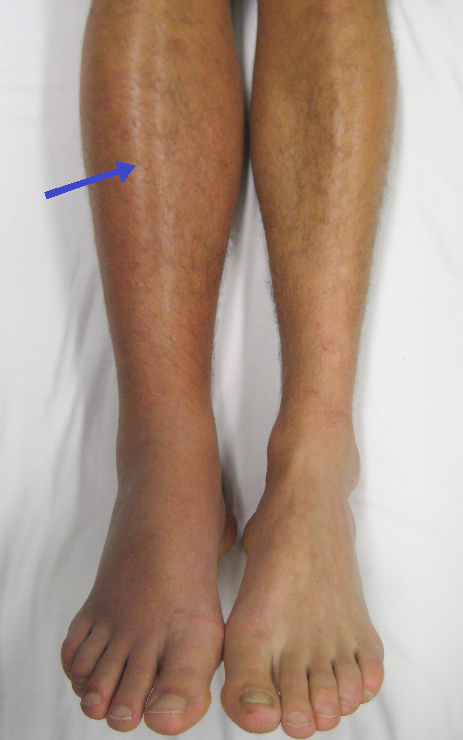
The deep femoral vein, deep vein of the thigh or profunda femoris vein is a large deep vein in the thigh. It collects blood from the inner thigh, passing superiorly and medially alongside the deep femoral artery before emptying into the femoral vein. Anatomy Fate The deep femoral vein drains into the femoral vein at approximately the level of the inferior-most portion of the ischial tuberosity. Function The deep femoral vein drains the inner thigh. It contributes the largest volume of blood entering the femoral vein. Clinical significance The deep femoral vein is commonly affected by phlebitis which can be a dangerous condition in the case of a thrombus, or blood clot, forming, as the thrombus may dislodge and travel to the lungs, causing pulmonary embolism. Risk factors for deep vein thrombosis include prolonged bed rest following surgery, immobility due to disability or fracture, an excessively sedentary lifestyle Sedentary lifestyle is a Lifestyle (social scienc ... [...More Info...] [...Related Items...] OR: [Wikipedia] [Google] [Baidu] |
Femoral Vein
In the human body, the femoral vein is the vein that accompanies the femoral artery in the femoral sheath. It is a deep vein that begins at the adductor hiatus (an opening in the adductor magnus muscle) as the continuation of the popliteal vein. The great saphenous vein (a superficial vein), and the deep femoral vein drain into the femoral vein in the femoral triangle when it becomes known as the common femoral vein. It ends at the inferior margin of the inguinal ligament where it becomes the external iliac vein. Its major tributaries are the deep femoral vein, and the great saphenous vein. The femoral vein contains valves. Structure The femoral vein bears valves which are mostly bicuspid and whose number is variable between individuals and often between left and right leg. Course The femoral vein continues into the thigh as the continuation from the popliteal vein at the back of the knee. It drains blood from the deep thigh muscles and thigh bone. Proximal to th ... [...More Info...] [...Related Items...] OR: [Wikipedia] [Google] [Baidu] |
Profunda Femoris Artery
The deep femoral artery also known as the deep artery of the thigh, or profunda femoris artery, is a large branch of the femoral artery. It travels more deeply ("profoundly") than the rest of the femoral artery. It gives rise to the lateral circumflex femoral artery and medial circumflex femoral artery, and the perforating arteries, terminating within the thigh. Structure Origin The deep femoral artery branches off the posterolateral side of the femoral artery soon after its origin. Course It travels down the thigh closer to the femur than the femoral artery. It runs between the pectineus muscle and the adductor longus muscle. It runs on the posterior side of adductor longus muscle. It pierces the adductor magnus muscle, and may be known as the fourth perforating artery as it continues. The deep femoral artery does not leave the thigh; terminating as perforating tissue branches within the thigh. Branches The deep femoral artery gives off the following branches: * ... [...More Info...] [...Related Items...] OR: [Wikipedia] [Google] [Baidu] |
Deep Vein
A deep vein is a vein that is deep in the body. This contrasts with superficial veins that are close to the body's surface. Deep veins are almost always beside an artery with the same name (e.g. the femoral vein is beside the femoral artery). Collectively, they carry the vast majority of the blood. Occlusion of a deep vein can be life-threatening and is most often caused by thrombosis. Occlusion of a deep vein by thrombosis is called ''deep vein thrombosis''. Because of their location deep within the body, operation on these veins can be difficult. List *Internal jugular vein Upper limb * Brachial vein * Axillary vein *Subclavian vein Lower limb *Common femoral vein *Femoral vein * Profunda femoris vein * Popliteal vein * Peroneal vein * Anterior tibial vein *Posterior tibial vein The posterior tibial veins are veins of the leg in humans. They drain the posterior compartment of the leg and the plantar surface of the foot to the popliteal vein. Structure The poste ... [...More Info...] [...Related Items...] OR: [Wikipedia] [Google] [Baidu] |
Thigh
In anatomy, the thigh is the area between the hip (pelvis) and the knee. Anatomically, it is part of the lower limb. The single bone in the thigh is called the femur. This bone is very thick and strong (due to the high proportion of bone tissue), and forms a ball and socket joint at the hip, and a modified hinge joint at the knee. Structure Bones The femur is the only bone in the thigh and serves as an attachment site for all thigh muscles. The head of the femur articulates with the acetabulum in the pelvic bone forming the hip joint, while the distal part of the femur articulates with the tibia and patella forming the knee. By most measures, the femur is the strongest and longest bone in the body. The femur is categorised as a long bone and comprises a diaphysis, the shaft (or body) and two epiphyses, the lower extremity and the upper extremity of femur, that articulate with adjacent bones in the hip and knee. Muscular compartments In cross-section, the thigh is d ... [...More Info...] [...Related Items...] OR: [Wikipedia] [Google] [Baidu] |
Deep Femoral Artery
The deep femoral artery also known as the deep artery of the thigh, or profunda femoris artery, is a large branch of the femoral artery. It travels more deeply ("profoundly") than the rest of the femoral artery. It gives rise to the lateral circumflex femoral artery and medial circumflex femoral artery, and the perforating arteries, terminating within the thigh. Structure Origin The deep femoral artery branches off the posterolateral side of the femoral artery soon after its origin. Course It travels down the thigh closer to the femur than the femoral artery. It runs between the pectineus muscle and the adductor longus muscle. It runs on the posterior side of adductor longus muscle. It pierces the adductor magnus muscle, and may be known as the fourth perforating artery as it continues. The deep femoral artery does not leave the thigh; terminating as perforating tissue branches within the thigh. Branches The deep femoral artery gives off the following branches: * La ... [...More Info...] [...Related Items...] OR: [Wikipedia] [Google] [Baidu] |
Femoral Vein
In the human body, the femoral vein is the vein that accompanies the femoral artery in the femoral sheath. It is a deep vein that begins at the adductor hiatus (an opening in the adductor magnus muscle) as the continuation of the popliteal vein. The great saphenous vein (a superficial vein), and the deep femoral vein drain into the femoral vein in the femoral triangle when it becomes known as the common femoral vein. It ends at the inferior margin of the inguinal ligament where it becomes the external iliac vein. Its major tributaries are the deep femoral vein, and the great saphenous vein. The femoral vein contains valves. Structure The femoral vein bears valves which are mostly bicuspid and whose number is variable between individuals and often between left and right leg. Course The femoral vein continues into the thigh as the continuation from the popliteal vein at the back of the knee. It drains blood from the deep thigh muscles and thigh bone. Proximal to th ... [...More Info...] [...Related Items...] OR: [Wikipedia] [Google] [Baidu] |
Ischial Tuberosity
The ischial tuberosity (or tuberosity of the ischium, tuber ischiadicum), also known colloquially as the sit bones or sitz bones, or as a pair the sitting bones, is a large posterior bony protuberance on the superior ramus of the ischium. It marks the lateral boundary of the pelvic outlet. When sitting, the weight is frequently placed upon the ischial tuberosity. The gluteus maximus provides cover in the upright posture, but leaves it free in the seated position.Platzer (2004), p 236 The distance between a cyclist's ischial tuberosities is one of the factors in the choice of a bicycle saddle. Divisions The tuberosity is divided into two portions: a lower, rough, somewhat triangular part, and an upper, smooth, quadrilateral portion. * The ''lower portion'' is subdivided by a prominent longitudinal ridge, passing from base to apex, into two parts: ** The outer gives attachment to the adductor magnus ** The inner to the sacrotuberous ligament * The ''upper portion'' is subdiv ... [...More Info...] [...Related Items...] OR: [Wikipedia] [Google] [Baidu] |
Phlebitis
Phlebitis (or venitis) is inflammation of a vein, usually in the legs. It most commonly occurs in superficial veins. Phlebitis often occurs in conjunction with thrombosis (clotting inside blood vessels) and is then called thrombophlebitis or superficial thrombophlebitis. Unlike deep vein thrombosis, the probability that superficial thrombophlebitis will cause a clot to break up and be transported in pieces to the lung is very low. Signs and symptoms * Localized redness and swelling * Pain or burning along the length of the vein * Vein being hard and cord-like There is usually a slow onset of a tender red area along the superficial veins on the skin. A long, thin red area may be seen as the inflammation follows a superficial vein. This area may feel hard, warm, and tender. The skin around the vein may be itchy and swollen. The area may begin to throb or burn. Symptoms may be worse when the leg is lowered, especially when first getting out of bed in the morning. A low-grade ... [...More Info...] [...Related Items...] OR: [Wikipedia] [Google] [Baidu] |
Thrombus
A thrombus ( thrombi) is a solid or semisolid aggregate from constituents of the blood (platelets, fibrin, red blood cells, white blood cells) within the circulatory system during life. A blood clot is the final product of the blood coagulation step in hemostasis in or out of the circulatory system. There are two components to a thrombus: aggregated platelets and red blood cells that form a plug, and a mesh of cross-linked fibrin protein. The substance making up a thrombus is sometimes called cruor. A thrombus is a healthy response to injury intended to stop and prevent further bleeding, but can be harmful in thrombosis, when a clot obstructs blood flow through a healthy blood vessel in the circulatory system. In the microcirculation consisting of the very small and smallest blood vessels the capillaries, tiny thrombi known as microclots can obstruct the flow of blood in the capillaries. This can cause a number of problems particularly affecting the pulmonary alveolus, alveoli ... [...More Info...] [...Related Items...] OR: [Wikipedia] [Google] [Baidu] |
Pulmonary Embolism
Pulmonary embolism (PE) is a blockage of an pulmonary artery, artery in the lungs by a substance that has moved from elsewhere in the body through the bloodstream (embolism). Symptoms of a PE may include dyspnea, shortness of breath, chest pain particularly upon breathing in, and coughing up blood. Symptoms of a deep vein thrombosis, blood clot in the leg may also be present, such as a erythema, red, warm, swollen, and painful leg. Signs of a PE include low blood oxygen saturation, oxygen levels, tachypnea, rapid breathing, tachycardia, rapid heart rate, and sometimes a mild fever. Severe cases can lead to Syncope (medicine), passing out, shock (circulatory), abnormally low blood pressure, obstructive shock, and cardiac arrest, sudden death. PE usually results from a blood clot in the leg that travels to the lung. The risk of blood clots is increased by advanced age, cancer, prolonged bed rest and immobilization, smoking, stroke, long-haul travel over 4 hours, certain genetics, ... [...More Info...] [...Related Items...] OR: [Wikipedia] [Google] [Baidu] |
Deep Vein Thrombosis
Deep vein thrombosis (DVT) is a type of venous thrombosis involving the formation of a blood clot in a deep vein, most commonly in the legs or pelvis. A minority of DVTs occur in the arms. Symptoms can include pain, swelling, redness, and enlarged veins in the affected area, but some DVTs have no symptoms. The most common life-threatening concern with DVT is the potential for a clot to embolize (detach from the veins), travel as an embolus through the right side of the heart, and become lodged in a pulmonary artery that supplies blood to the lungs. This is called a pulmonary embolism (PE). DVT and PE comprise the cardiovascular disease of venous thromboembolism (VTE). About two-thirds of VTE manifests as DVT only, with one-third manifesting as PE with or without DVT. The most frequent long-term DVT Complication (medicine), complication is post-thrombotic syndrome, which can cause pain, swelling, a sensation of heaviness, itching, and in severe cases, ulcers. Recurrent VTE o ... [...More Info...] [...Related Items...] OR: [Wikipedia] [Google] [Baidu] |
Sedentary Lifestyle
Sedentary lifestyle is a Lifestyle (social sciences), lifestyle type, in which one is physically inactive and does little or no physical movement and/or exercise. A person living a sedentary lifestyle is often sitting or lying down while engaged in an activity like socializing, Social aspects of television, watching TV, Gameplay, playing video games, reading or Problematic smartphone use, using a mobile phone or computer for much of the day. A sedentary lifestyle contributes to poor health quality, diseases as well as many preventable causes of death. Sitting time is a common measure of a sedentary lifestyle. A global review representing 47% of the global adult population found that the average person sits down for 4.7 to 6.5 hours a day with the average going up every year. The Centers for Disease Control and Prevention, CDC found that 25.3% of all American adults are physically inactive. Screen time is a term for the amount of time a person spends looking at a screen such a ... [...More Info...] [...Related Items...] OR: [Wikipedia] [Google] [Baidu] |


