|
Cutis (anatomy)
Cutis, often termed the "true skin", is composed of the epidermis and the dermis. The dermis contains blood vessels, sweat glands, sebaceous glands, and hair follicles. The epidermis and the dermis contain sensory nerve endings to detect changes in the environment. The cutis is the layer located above the subcutis The subcutaneous tissue (), also called the hypodermis, hypoderm (), subcutis, or superficial fascia, is the lowermost layer of the integumentary system in vertebrates. The types of cells found in the layer are fibroblasts, adipose cells, and ma .... Pathology Aplasia cutis congenita results in thin, transparent skin usually on the head. The disease is defined as a congenital absence of skin. References Skin anatomy {{anatomy-stub ... [...More Info...] [...Related Items...] OR: [Wikipedia] [Google] [Baidu] |
Epidermis
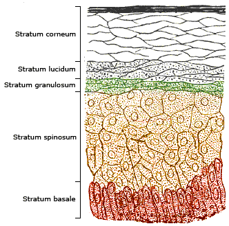
The epidermis is the outermost of the three layers that comprise the skin, the inner layers being the dermis and Subcutaneous tissue, hypodermis. The epidermal layer provides a barrier to infection from environmental pathogens and regulates the amount of water released from the body into the atmosphere through transepidermal water loss. The epidermis is composed of stratified squamous epithelium, multiple layers of flattened cells that overlie a base layer (stratum basale) composed of Epithelium#Cell types, columnar cells arranged perpendicularly. The layers of cells develop from stem cells in the basal layer. The thickness of the epidermis varies from 31.2μm for the penis to 596.6μm for the Sole (foot), sole of the foot with most being roughly 90μm. Thickness does not vary between the sexes but becomes thinner with age. The human epidermis is an example of epithelium, particularly a stratified squamous epithelium. The word epidermis is derived through Latin , itself and . ... [...More Info...] [...Related Items...] OR: [Wikipedia] [Google] [Baidu] |
Dermis
The dermis or corium is a layer of skin between the epidermis (skin), epidermis (with which it makes up the cutis (anatomy), cutis) and subcutaneous tissues, that primarily consists of dense irregular connective tissue and cushions the body from stress and strain. It is divided into two layers, the superficial area adjacent to the epidermis called the papillary region and a deep thicker area known as the reticular dermis.James, William; Berger, Timothy; Elston, Dirk (2005). ''Andrews' Diseases of the Skin: Clinical Dermatology'' (10th ed.). Saunders. Pages 1, 11–12. . The dermis is tightly connected to the epidermis through a basement membrane. Structural components of the dermis are collagen, elastic fibers, and Ground substance, extrafibrillar matrix.Marks, James G; Miller, Jeffery (2006). ''Lookingbill and Marks' Principles of Dermatology'' (4th ed.). Elsevier Inc. Page 8–9. . It also contains mechanoreceptors that provide the sense of touch and thermoreceptors that provide ... [...More Info...] [...Related Items...] OR: [Wikipedia] [Google] [Baidu] |
Blood Vessel
Blood vessels are the tubular structures of a circulatory system that transport blood throughout many Animal, animals’ bodies. Blood vessels transport blood cells, nutrients, and oxygen to most of the Tissue (biology), tissues of a Body (biology), body. They also take waste and carbon dioxide away from the tissues. Some tissues such as cartilage, epithelium, and the lens (anatomy), lens and cornea of the eye are not supplied with blood vessels and are termed ''avascular''. There are five types of blood vessels: the arteries, which carry the blood away from the heart; the arterioles; the capillaries, where the exchange of water and chemicals between the blood and the tissues occurs; the venules; and the veins, which carry blood from the capillaries back towards the heart. The word ''vascular'', is derived from the Latin ''vas'', meaning ''vessel'', and is mostly used in relation to blood vessels. Etymology * artery – late Middle English; from Latin ''arteria'', from Gree ... [...More Info...] [...Related Items...] OR: [Wikipedia] [Google] [Baidu] |
Sweat Gland
Sweat glands, also known as sudoriferous or sudoriparous glands, , are small tubular structures of the skin that produce sweat. Sweat glands are a type of exocrine gland, which are glands that produce and secrete substances onto an epithelial surface by way of a duct. There are two main types of sweat glands that differ in their structure, function, secretory product, mechanism of excretion, anatomic distribution, and distribution across species: * Eccrine sweat glands are distributed almost all over the human body, in varying densities, with the highest density in palms and soles, then on the head, but much less on the trunk and the extremities. Their water-based secretion represents a primary form of cooling in humans. * Apocrine sweat glands are mostly limited to the axillae (armpits) and perineal area in humans. They are not significant for cooling in humans, but are the sole effective sweat glands in hoofed animals, such as the camels, donkeys, horses, and cattle. Ceru ... [...More Info...] [...Related Items...] OR: [Wikipedia] [Google] [Baidu] |
Sebaceous Gland
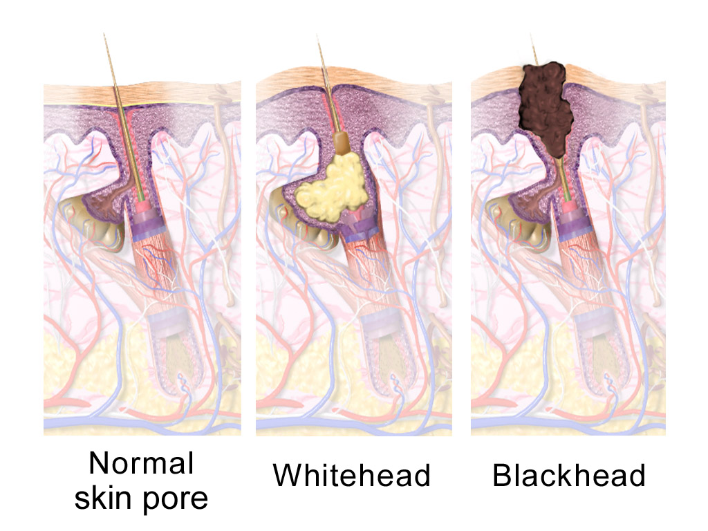
A sebaceous gland or oil gland is a microscopic exocrine gland in the skin that opens into a hair follicle to secrete an oily or waxy matter, called sebum, which lubricates the hair and skin of mammals. In humans, sebaceous glands occur in the greatest number on the face and scalp, but also on all parts of the skin except the palms of the hands and soles of the feet. In the eyelids, meibomian glands, also called tarsal glands, are a type of sebaceous gland that secrete a special type of sebum into tears. Surrounding the female nipples, areolar glands are specialized sebaceous glands for lubricating the nipples. Fordyce spots are benign, visible, sebaceous glands found usually on the lips, gums and inner cheeks, and genitals. Structure Location In humans, sebaceous glands are found throughout all areas of the skin, except the palms of the hands and soles of the feet. There are two types of sebaceous glands: those connected to hair follicles and those that ex ... [...More Info...] [...Related Items...] OR: [Wikipedia] [Google] [Baidu] |
Hair Follicle
The hair follicle is an organ found in mammalian skin. It resides in the dermal layer of the skin and is made up of 20 different cell types, each with distinct functions. The hair follicle regulates hair growth via a complex interaction between hormones, neuropeptides, and immune cells. This complex interaction induces the hair follicle to produce different types of hair as seen on different parts of the body. For example, terminal hairs grow on the scalp and lanugo hairs are seen covering the bodies of fetuses in the uterus and in some newborn babies. The process of hair growth occurs in distinct sequential stages: ''anagen'' is the active growth phase, ''catagen'' is the regression of the hair follicle phase, ''telogen'' is the resting stage, ''exogen'' is the active shedding of hair phase and ''kenogen'' is the phase between the empty hair follicle and the growth of new hair. The function of hair in humans has long been a subject of interest and continues to be an impor ... [...More Info...] [...Related Items...] OR: [Wikipedia] [Google] [Baidu] |
Sensory Nerve
A sensory nerve, or afferent nerve, is a nerve that contains exclusively afferent nerve fibers. Nerves containing also motor fibers are called mixed nerve, mixed. Afferent nerve fibers in a sensory nerve carry sensory system, sensory information toward the central nervous system (CNS) from different sensory receptors of sensory neurons in the peripheral nervous system (PNS). A motor nerve carries information from the CNS to the PNS. Afferent nerve fibers link the sensory neurons throughout the body, in neural pathway, pathways to the relevant processing circuits in the central nervous system. Afferent nerve fibers are often paired with efferent nerve fibers from the motor neurons (that travel from the CNS to the PNS), in mixed nerves. Stimuli cause nerve impulses in the receptors and alter the potentials, which is known as sensory transduction. Spinal cord entry Afferent nerve fibers leave the sensory neuron from the dorsal root ganglion, dorsal root ganglia of the spinal cor ... [...More Info...] [...Related Items...] OR: [Wikipedia] [Google] [Baidu] |
Subcutis
The subcutaneous tissue (), also called the hypodermis, hypoderm (), subcutis, or superficial fascia, is the lowermost layer of the integumentary system in vertebrates. The types of cells found in the layer are fibroblasts, adipose cells, and macrophages. The subcutaneous tissue is derived from the mesoderm, but unlike the dermis, it is not derived from the mesoderm's Dermatome (anatomy), dermatome region. It consists primarily of loose connective tissue and contains larger blood vessels and nerves than those found in the dermis. It is a major site of fat storage in the body. In arthropods, a hypodermis can refer to an epidermal layer of cells that secretes the chitinous cuticle. The term also refers to a layer of cells lying immediately below the Epidermis (botany), epidermis of plants. Structure * Fibrous bands anchoring the skin to the deep fascia * Collagen and elastin fibers attaching it to the dermis * Fat is absent from the eyelids, clitoris, penis, much of Pinna (anatomy ... [...More Info...] [...Related Items...] OR: [Wikipedia] [Google] [Baidu] |


