|
Cross Modal Plasticity
Cross modal plasticity is the adaptive reorganization of neurons to integrate the function of two or more sensory systems. Cross modal plasticity is a type of neuroplasticity and often occurs after sensory deprivation due to disease or brain damage. The reorganization of the neural network is greatest following long-term sensory deprivation, such as congenital blindness or pre-lingual deafness. For example, individuals with auditory deficits may experience strengthened visual processing compared to hearing individuals. In these instances, cross modal plasticity can strengthen other sensory systems to compensate for the lack of vision or hearing. This strengthening is due to new connections that are formed to brain cortices that no longer receive sensory input. It is important to note that cross modal plasticity is not only limited to auditory and visual systems but can cause reorganization in tactile and olfactory systems too. Plasticity after visual deficit In people who are bl ... [...More Info...] [...Related Items...] OR: [Wikipedia] [Google] [Baidu] |
Two Streams Hypothesis
The two-streams hypothesis is a model of the neural processing of vision as well as hearing. The hypothesis, given its initial characterisation in a paper by David Milner and Melvyn A. Goodale in 1992, argues that humans possess two distinct visual systems. Recently there seems to be evidence of two distinct auditory systems as well. As visual information exits the occipital lobe, and as sound leaves the phonological network, it follows two main pathways, or "streams". The ventral stream (also known as the "what pathway") leads to the temporal lobe, which is involved with object and visual identification and recognition. The dorsal stream (or, "where pathway") leads to the parietal lobe, which is involved with processing the object's spatial location relative to the viewer and with speech repetition. History Several researchers had proposed similar ideas previously. The authors themselves credit the inspiration of work on blindsight by Weiskrantz, and previous neuroscientific ... [...More Info...] [...Related Items...] OR: [Wikipedia] [Google] [Baidu] |
Neuroimaging
Neuroimaging is the use of quantitative (computational) techniques to study the neuroanatomy, structure and function of the central nervous system, developed as an objective way of scientifically studying the healthy human brain in a non-invasive manner. Increasingly it is also being used for quantitative research studies of brain disease and psychiatric illness. Neuroimaging is highly multidisciplinary involving neuroscience, computer science, psychology and statistics, and is not a medical specialty. Neuroimaging is sometimes confused with neuroradiology. Neuroradiology is a medical specialty that uses non-statistical brain imaging in a clinical setting, practiced by radiologists who are medical practitioners. Neuroradiology primarily focuses on recognizing brain lesions, such as vascular diseases, strokes, tumors, and inflammatory diseases. In contrast to neuroimaging, neuroradiology is qualitative (based on subjective impressions and extensive clinical training) but sometime ... [...More Info...] [...Related Items...] OR: [Wikipedia] [Google] [Baidu] |
Retinal Ganglion Cells
A retinal ganglion cell (RGC) is a type of neuron located near the inner surface (the ganglion cell layer) of the retina of the eye. It receives visual information from photoreceptors via two intermediate neuron types: bipolar cells and retina amacrine cells. Retina amacrine cells, particularly narrow field cells, are important for creating functional subunits within the ganglion cell layer and making it so that ganglion cells can observe a small dot moving a small distance. Retinal ganglion cells collectively transmit image-forming and non-image forming visual information from the retina in the form of action potential to several regions in the thalamus, hypothalamus, and mesencephalon, or midbrain. Retinal ganglion cells vary significantly in terms of their size, connections, and responses to visual stimulation but they all share the defining property of having a long axon that extends into the brain. These axons form the optic nerve, optic chiasm, and optic tract. A small p ... [...More Info...] [...Related Items...] OR: [Wikipedia] [Google] [Baidu] |
Posterior Parietal Cortex
The posterior parietal cortex (the portion of parietal neocortex posterior to the primary somatosensory cortex) plays an important role in planned movements, spatial reasoning, and attention. Damage to the posterior parietal cortex can produce a variety of sensorimotor deficits, including deficits in the perception and memory of spatial relationships, inaccurate reaching and grasping, in the control of eye movement, and inattention. The two most striking consequences of PPC damage are apraxia and hemispatial neglect. Anatomy The posterior parietal cortex is located just behind the central sulcus, between the visual cortex, the caudal pole and the somatosensory cortex. The posterior parietal cortex receives input from the three sensory systems that play roles in the localization of the body and external objects in space: the visual system, the auditory system, and the somatosensory system. In turn, much of the output of the posterior parietal cortex goes to areas of frontal moto ... [...More Info...] [...Related Items...] OR: [Wikipedia] [Google] [Baidu] |
Auditory System
The auditory system is the sensory system for the sense of hearing. It includes both the ear, sensory organs (the ears) and the auditory parts of the sensory system. System overview The outer ear funnels sound vibrations to the eardrum, increasing the sound pressure in the middle frequency range. The Middle ear, middle-ear ossicles further amplify the vibration pressure roughly 20 times. The base of the stapes couples vibrations into the cochlea via the oval window, which vibrates the perilymph liquid (present throughout the inner ear) and causes the round window to bulb out as the oval window bulges in. Vestibular duct, Vestibular and tympanic ducts are filled with perilymph, and the smaller cochlear duct between them is filled with endolymph, a fluid with a very different ion concentration and voltage. Vestibular duct perilymph vibrations bend organ of Corti outer cells (4 lines) causing prestin to be released in cell tips. This causes the cells to be chemically elongated an ... [...More Info...] [...Related Items...] OR: [Wikipedia] [Google] [Baidu] |
Ears
In vertebrates, an ear is the organ that enables hearing and (in mammals) body balance using the vestibular system. In humans, the ear is described as having three parts: the outer ear, the middle ear and the inner ear. The outer ear consists of the auricle and the ear canal. Since the outer ear is the only visible portion of the ear, the word "ear" often refers to the external part (auricle) alone. The middle ear includes the tympanic cavity and the three ossicles. The inner ear sits in the bony labyrinth, and contains structures which are key to several senses: the semicircular canals, which enable balance and eye tracking when moving; the utricle and saccule, which enable balance when stationary; and the cochlea, which enables hearing. The ear canal is cleaned via earwax, which naturally migrates to the auricle. The ear develops from the first pharyngeal pouch and six small swellings that develop in the early embryo called otic placodes, which are derived from ... [...More Info...] [...Related Items...] OR: [Wikipedia] [Google] [Baidu] |
Sign Language
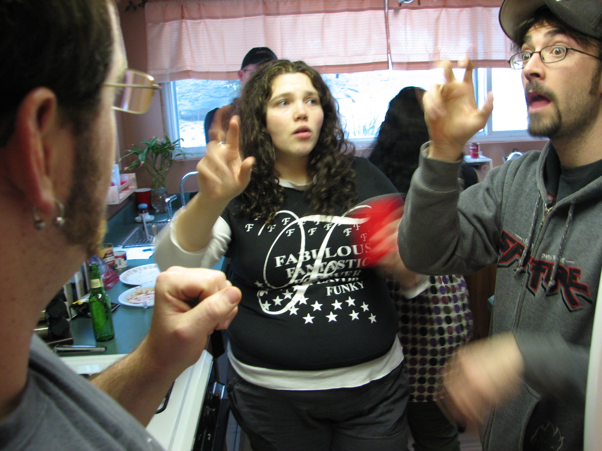
Sign languages (also known as signed languages) are languages that use the visual-manual modality to convey meaning, instead of spoken words. Sign languages are expressed through manual articulation in combination with #Non-manual elements, non-manual markers. Sign languages are full-fledged natural languages with their own grammar and lexicon. Sign languages are not universal and are usually not mutual intelligibility, mutually intelligible, although there are similarities among different sign languages. Linguists consider both spoken and signed communication to be types of natural language, meaning that both emerged through an abstract, protracted aging process and evolved over time without meticulous planning. This is supported by the fact that there is substantial overlap between the neural substrates of sign and spoken language processing, despite the obvious differences in modality. Sign language should not be confused with body language, a type of non verbal communicati ... [...More Info...] [...Related Items...] OR: [Wikipedia] [Google] [Baidu] |
Primary Auditory Cortex
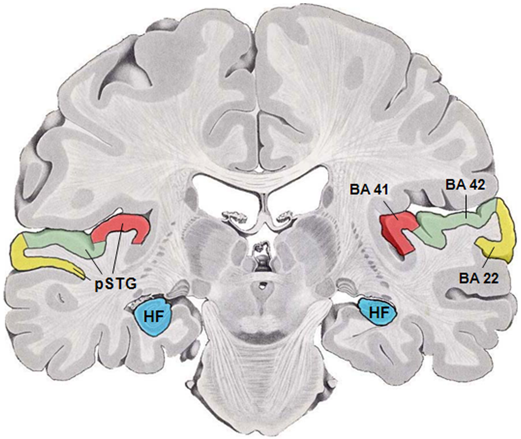
The auditory cortex is the part of the temporal lobe that processes auditory information in humans and many other vertebrates. It is a part of the auditory system, performing basic and higher functions in hearing, such as possible relations to language switching.Cf. Pickles, James O. (2012). ''An Introduction to the Physiology of Hearing'' (4th ed.). Bingley, UK: Emerald Group Publishing Limited, p. 238. It is located bilaterally, roughly at the upper sides of the temporal lobes – in humans, curving down and onto the medial surface, on the superior temporal plane, within the lateral sulcus and comprising parts of the transverse temporal gyri, and the superior temporal gyrus, including the planum polare and planum temporale (roughly Brodmann areas 41 and 42, and partially 22). The auditory cortex takes part in the spectrotemporal, meaning involving time and frequency, analysis of the inputs passed on from the ear. The cortex then filters and passes on the information to t ... [...More Info...] [...Related Items...] OR: [Wikipedia] [Google] [Baidu] |
FMRI
Functional magnetic resonance imaging or functional MRI (fMRI) measures brain activity by detecting changes associated with blood flow. This technique relies on the fact that cerebral blood flow and neuronal activation are coupled. When an area of the brain is in use, blood flow to that region also increases. The primary form of fMRI uses the blood-oxygen-level dependent (BOLD) contrast, discovered by Seiji Ogawa in 1990. This is a type of specialized brain and body scan used to map neural activity in the brain or spinal cord of humans or other animals by imaging the change in blood flow ( hemodynamic response) related to energy use by brain cells. Since the early 1990s, fMRI has come to dominate brain mapping research because it does not involve the use of injections, surgery, the ingestion of substances, or exposure to ionizing radiation. This measure is frequently corrupted by noise from various sources; hence, statistical procedures are used to extract the underlying signa ... [...More Info...] [...Related Items...] OR: [Wikipedia] [Google] [Baidu] |
Prelingual Deafness
Prelingual deafness refers to deafness that occurs before learning speech or language. Speech and language typically begin to develop very early with infants saying their first words by age one. Therefore, prelingual deafness is considered to occur before the age of one, where a baby is either born deaf (known as congenital deafness) or loses hearing before the age of one. This hearing loss may occur for a variety of reasons and impacts cognitive, social, and language development. Statistics There are approximately 12,000 children with hearing loss in the United States. Profound hearing loss occurs in somewhere between 4 and 11 per every 10,000 children. In 2017, according to the CDC, of the 3,742,608 babies screened, 3,896 were diagnosed with hearing loss before the age of three months or 1.7 babies per 1,000 births were diagnosed with hearing loss in the United States. Causes Prelingual hearing loss can be considered congenital, present at birth, or acquired, occurring after birt ... [...More Info...] [...Related Items...] OR: [Wikipedia] [Google] [Baidu] |
Sense Of Touch
The somatosensory system, or somatic sensory system is a subset of the sensory nervous system. The main functions of the somatosensory system are the perception of external stimuli, the perception of internal stimuli, and the regulation of body position and balance (proprioception). It is believed to act as a pathway between the different sensory modalities within the body. As of 2024 debate continued on the underlying mechanisms, correctness and validity of the somatosensory system model, and whether it impacts emotions in the body. The somatosensory system has been thought of as having two subdivisions; *one for the detection of mechanosensory information related to touch. Mechanosensory information includes that of light touch, vibration, pressure and tension in the skin. Much of this information belongs to the sense of touch which is a general somatic sense in contrast to the special senses of sight, smell, taste, hearing, and balance. * one for the nociception det ... [...More Info...] [...Related Items...] OR: [Wikipedia] [Google] [Baidu] |


