|
Coronary Catheterization
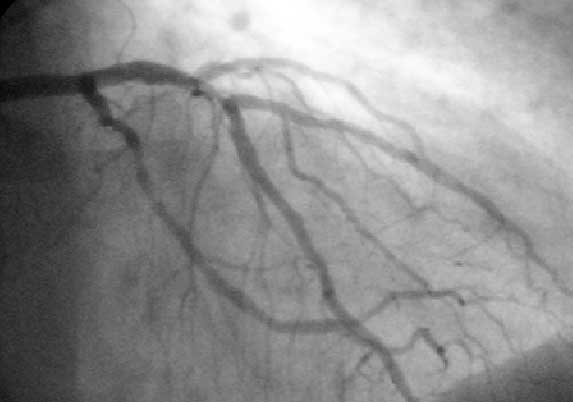
A coronary catheterization is a minimally invasive procedure to access the coronary circulation and blood filled chambers of the heart using a catheter. It is performed for both diagnostic and interventional (treatment) purposes. Coronary catheterization is one of the several cardiology diagnostic tests and procedures. Specifically, through the injection of a liquid radiocontrast agent and illumination with X-rays, angiocardiography allows the recognition of Vascular occlusion, occlusion, stenosis, restenosis, thrombosis or aneurysmal enlargement of the coronary artery lumen (anatomy), lumens; heart chamber size; heart muscle contraction performance; and some aspects of heart valve function. Important internal heart and lung blood pressures, not measurable from outside the body, can be accurately measured during the test. The relevant problems that the test deals with most commonly occur as a result of advanced atherosclerosis – atheroma activity within the wall of the coronary ... [...More Info...] [...Related Items...] OR: [Wikipedia] [Google] [Baidu] |
Minimally Invasive
Minimally invasive procedures (also known as minimally invasive surgeries) encompass surgical techniques that limit the size of incisions needed, thereby reducing wound healing time, associated pain, and risk of infection. Surgery by definition is invasive, and many operations requiring incisions of some size are referred to as ''open surgery''. Incisions made during open surgery can sometimes leave large wounds that may be painful and take a long time to heal. Advancements in medical technologies have enabled the development and regular use of minimally invasive procedures. For example, endovascular aneurysm repair, a minimally invasive surgery, has become the most common method of repairing abdominal aortic aneurysms in the US as of 2003. The procedure involves much smaller incisions than the corresponding open surgery procedure of open aortic surgery. Interventional radiologists were the forerunners of minimally invasive procedures. Using imaging techniques, radiologist ... [...More Info...] [...Related Items...] OR: [Wikipedia] [Google] [Baidu] |
Lung
The lungs are the primary Organ (biology), organs of the respiratory system in many animals, including humans. In mammals and most other tetrapods, two lungs are located near the Vertebral column, backbone on either side of the heart. Their function in the respiratory system is to extract oxygen from the atmosphere and transfer it into the bloodstream, and to release carbon dioxide from the bloodstream into the atmosphere, in a process of gas exchange. Respiration is driven by different muscular systems in different species. Mammals, reptiles and birds use their musculoskeletal systems to support and foster breathing. In early tetrapods, air was driven into the lungs by the pharyngeal muscles via buccal pumping, a mechanism still seen in amphibians. In humans, the primary muscle that drives breathing is the Thoracic diaphragm, diaphragm. The lungs also provide airflow that makes Animal communication#Auditory, vocalisation including speech possible. Humans have two lungs, a ri ... [...More Info...] [...Related Items...] OR: [Wikipedia] [Google] [Baidu] |
Werner Forssmann
Werner Theodor Otto Forßmann (Forssmann in English; ; 29 August 1904 – 1 June 1979) was a German researcher and physician from Germany who shared the 1956 Nobel Prize in Medicine (with Andre Frederic Cournand and Dickinson W. Richards) for developing a procedure that allowed cardiac catheterization. In 1929, he put himself under local anesthesia and inserted a catheter into a vein of his arm. Not knowing if the catheter might pierce a vein, he put his life at risk. Forssmann was nevertheless successful; he safely passed the catheter into his heart. Early life Forssmann was born in Berlin on 29 August 1904. Upon graduating from , he entered the University of Berlin to study medicine, passing the State Examination in 1929. Career He hypothesized that a catheter could be inserted directly into the heart, for such applications as directly delivering drugs, injecting Radiodensity, radiopaque dyes, or measuring blood pressure. The fear at the time was that such an intrusion into ... [...More Info...] [...Related Items...] OR: [Wikipedia] [Google] [Baidu] |
Cerebral Angiography
Cerebral angiography is a form of angiography which provides images of blood vessels in and around the brain, thereby allowing detection of abnormalities such as arteriovenous malformations and aneurysms. It was pioneered in 1927 by the Portuguese neurologist Egas Moniz at the University of Lisbon, who also helped develop thorotrast for use in the procedure. Typically a catheter is inserted into a large artery (such as the femoral artery) and threaded through the circulatory system to the Common carotid artery, carotid artery, where a contrast agent is injected. A series of radiographs are taken as the contrast agent spreads through the brain's arterial system, then a second series as it reaches the venous system. For some applications, cerebral angiography may yield better images than less invasive methods such as computed tomography angiography and magnetic resonance angiography. In addition, cerebral angiography allows certain treatments to be performed immediately, based on ... [...More Info...] [...Related Items...] OR: [Wikipedia] [Google] [Baidu] |
University Of Lisbon
The University of Lisbon (ULisboa; ) is a public university, public research university in Lisbon, and Portugal's largest university. It was founded in 1911, but the university's present structure dates to the 2013 merger of the former University of Lisbon (1911–2013) and the Technical University of Lisbon, Technical University of Lisbon (1930–2013). History University of Coimbra, the first Portuguese university, was established in Lisbon between 1288 and 1290, when Denis of Portugal, Dinis I promulgated the letter ''Scientiae thesaurus mirabili'', granting several privileges to the students of the ''studium generale'' in Lisbon, proving that it was already founded on that date. There was an active participation in this educational activity by the Portuguese Crown and its king, through its commitment of part of the subsidy of the same, as by the fixed incomes of the Church. This institution moved several times between Lisbon and Coimbra, where it settled permanently in 1537. ... [...More Info...] [...Related Items...] OR: [Wikipedia] [Google] [Baidu] |
IVUS
Intravascular ultrasound (IVUS) or intravascular echocardiography is a medical imaging methodology using a specially designed catheter with a miniaturized ultrasound probe attached to the distal end of the catheter. The proximal end of the catheter is attached to computerized ultrasound equipment. It allows the application of ultrasound technology, such as piezoelectric transducer or CMUT, to see from inside blood vessels out through the surrounding blood column, visualizing the endothelium (inner wall) of blood vessels. The arteries of the heart (the coronary arteries) are the most frequent imaging target for IVUS. IVUS is used in the coronary arteries to determine the amount of atheromatous plaque built up at any particular point in the epicardial coronary artery. Intravascular ultrasound provides a unique method to study the regression or progression of atherosclerotic lesions in vivo. The progressive accumulation of plaque within the artery wall over decades leads to the ... [...More Info...] [...Related Items...] OR: [Wikipedia] [Google] [Baidu] |
Myocardial Infarction
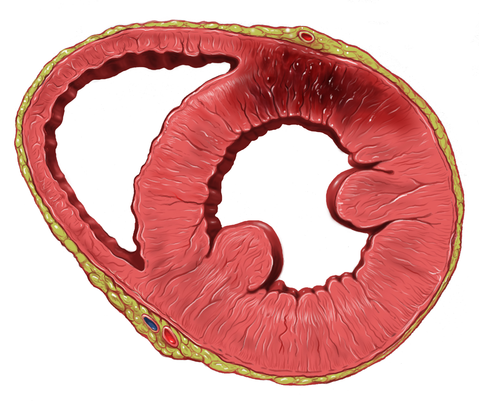
A myocardial infarction (MI), commonly known as a heart attack, occurs when Ischemia, blood flow decreases or stops in one of the coronary arteries of the heart, causing infarction (tissue death) to the heart muscle. The most common symptom is retrosternal Angina, chest pain or discomfort that classically radiates to the left shoulder, arm, or jaw. The pain may occasionally feel like heartburn. This is the dangerous type of acute coronary syndrome. Other symptoms may include shortness of breath, nausea, presyncope, feeling faint, a diaphoresis, cold sweat, Fatigue, feeling tired, and decreased level of consciousness. About 30% of people have atypical symptoms. Women more often present without chest pain and instead have neck pain, arm pain or feel tired. Among those over 75 years old, about 5% have had an MI with little or no history of symptoms. An MI may cause heart failure, an Cardiac arrhythmia, irregular heartbeat, cardiogenic shock or cardiac arrest. Most MIs occur d ... [...More Info...] [...Related Items...] OR: [Wikipedia] [Google] [Baidu] |
Angina Pectoris
Angina, also known as angina pectoris, is chest pain or pressure, usually caused by insufficient blood flow to the heart muscle (myocardium). It is most commonly a symptom of coronary artery disease. Angina is typically the result of partial obstruction or spasm of the arteries that supply blood to the heart muscle. The main mechanism of coronary artery obstruction is atherosclerosis as part of coronary artery disease. Other causes of angina include abnormal heart rhythms, heart failure and, less commonly, anemia. The term derives , and can therefore be translated as "a strangling feeling in the chest". An urgent medical assessment is suggested to rule out serious medical conditions. There is a relationship between severity of angina and degree of oxygen deprivation in the heart muscle. However, the severity of angina does not always match the degree of oxygen deprivation to the heart or the risk of a heart attack (myocardial infarction). Some people may experience s ... [...More Info...] [...Related Items...] OR: [Wikipedia] [Google] [Baidu] |
Coronary Artery
The coronary arteries are the arterial blood vessels of coronary circulation, which transport oxygenated blood to the heart muscle. The heart requires a continuous supply of oxygen to function and survive, much like any other tissue or organ of the body. The coronary arteries wrap around the entire heart. The two main branches are the left coronary artery and right coronary artery. The arteries can additionally be categorized based on the area of the heart for which they provide circulation. These categories are called ''epicardial'' (above the epicardium, or the outermost tissue of the heart) and ''microvascular'' (close to the endocardium, or the innermost tissue of the heart). Reduced function of the coronary arteries can lead to decreased flow of oxygen and nutrients to the heart. Not only does this affect supply to the heart muscle itself, but it also can affect the ability of the heart to pump blood throughout the body. Therefore, any disorder or disease of the coronar ... [...More Info...] [...Related Items...] OR: [Wikipedia] [Google] [Baidu] |
Heart Arrhythmia
Arrhythmias, also known as cardiac arrhythmias, are irregularities in the cardiac cycle, heartbeat, including when it is too fast or too slow. Essentially, this is anything but normal sinus rhythm. A resting heart rate that is too fast – above 100 beats per minute in adults – is called tachycardia, and a resting heart rate that is too slow – below 60 beats per minute – is called bradycardia. Some types of arrhythmias have no symptoms. Symptoms, when present, may include palpitations or feeling a pause between heartbeats. In more serious cases, there may be presyncope, lightheadedness, Syncope (medicine), passing out, shortness of breath, chest pain, or decreased level of consciousness. While most cases of arrhythmia are not serious, some predispose a person to complications such as stroke or heart failure. Others may result in cardiac arrest, sudden death. Arrhythmias are often categorized into four groups: premature heart beat, extra beats, supraventricular tachycard ... [...More Info...] [...Related Items...] OR: [Wikipedia] [Google] [Baidu] |
Artery
An artery () is a blood vessel in humans and most other animals that takes oxygenated blood away from the heart in the systemic circulation to one or more parts of the body. Exceptions that carry deoxygenated blood are the pulmonary arteries in the pulmonary circulation that carry blood to the lungs for oxygenation, and the umbilical arteries in the fetal circulation that carry deoxygenated blood to the placenta. It consists of a multi-layered artery wall wrapped into a tube-shaped channel. Arteries contrast with veins, which carry deoxygenated blood back towards the heart; or in the pulmonary and fetal circulations carry oxygenated blood to the lungs and fetus respectively. Structure The anatomy of arteries can be separated into gross anatomy, at the macroscopic scale, macroscopic level, and histology, microanatomy, which must be studied with a microscope. The arterial system of the human body is divided into systemic circulation, systemic arteries, carrying blood from the ... [...More Info...] [...Related Items...] OR: [Wikipedia] [Google] [Baidu] |


