|
Coronary Artery Aneurysm
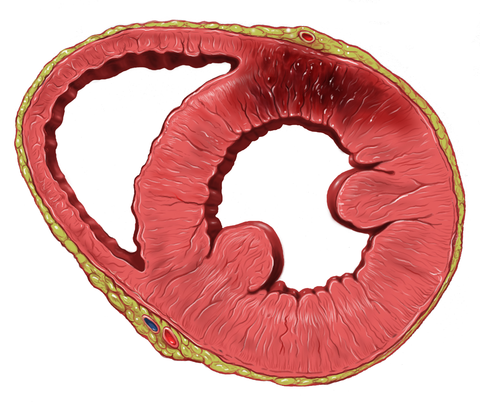
Coronary artery aneurysm is an abnormal dilatation of part of the coronary artery. This rare disorder occurs in about 0.3–4.9% of patients who undergo coronary angiography. Signs and symptoms The majority of individuals suffering from coronary artery aneurysms do not exhibit any symptoms; the development of complications or concurrent atherosclerotic coronary artery disease is what causes clinical manifestations to occur. The most common complications include coronary spasm, distal embolization, aneurysm rupture, local thrombosis, and compression of surrounding structures due to massive enlargement of coronary artery aneurysm. Causes Acquired causes include atherosclerosis in adults, Kawasaki disease in children and coronary catheterization. With the invention of drug eluting stents, there has been more cases implying stents lead to coronary aneurysms. The pathophysiology, although not completely understood, might be comparable to that of aneurysms of larger vessels. This ... [...More Info...] [...Related Items...] OR: [Wikipedia] [Google] [Baidu] |
Coronary Angiography , part of a mammalian eye.
{{Set index article ...
Coronary () may, as shorthand in English, be used to mean: * Coronary circulation, the system of arteries and veins in mammals ** Coronary artery disease ** Coronary occlusion ** A myocardial infarction, a heart attack As adjective * Referring to the work of a Coroner, a person entitled to investigate deaths * Referring to a stellar corona, the outermost atmosphere of a star * Mistakenly to a Cornea The cornea is the transparency (optics), transparent front part of the eyeball which covers the Iris (anatomy), iris, pupil, and Anterior chamber of eyeball, anterior chamber. Along with the anterior chamber and Lens (anatomy), lens, the cornea ... [...More Info...] [...Related Items...] OR: [Wikipedia] [Google] [Baidu] |
Marfan Syndrome
Marfan syndrome (MFS) is a multi-systemic genetic disorder that affects the connective tissue. Those with the condition tend to be tall and thin, with dolichostenomelia, long arms, legs, Arachnodactyly, fingers, and toes. They also typically have hypermobility (joints), exceptionally flexible joints and scoliosis, abnormally curved spines. The most serious complications involve the heart and aorta, with an increased risk of mitral valve prolapse and aortic aneurysm. The lungs, eyes, bones, and the dura mater, covering of the spinal cord are also commonly affected. The severity of the symptoms is variable. MFS is caused by a mutation in ''FBN1'', one of the genes that make fibrillin, which results in abnormal connective tissue. It is an autosomal dominant disorder. In about 75% of cases, it is inherited from a parent with the condition, while in about 25% it is a new mutation. Diagnosis is often based on the Ghent criteria, family history and genetic testing (DNA analysis). Th ... [...More Info...] [...Related Items...] OR: [Wikipedia] [Google] [Baidu] |
Coronary Artery Ectasia
Coronary artery ectasia is a rare disease that occurs in only 0.3-4.9% of people in North America. Coronary artery ectasia is characterized by the enlargement of a coronary artery to 1.5 times or more than its normal diameter.Lin, C.T., Chen, C.W., Lin, T.W., and Lin, C.L (2008) Coronary artery ectasia, ''Tzu Chi Med'', 20(14), 270-274 The disease is commonly asymptomatic and is normally discovered when performing tests for other conditions such as coronary artery disease, stable angina and other acute coronary syndromes.Hsu, P.C., Su, H.M., Lee, H.C., Juo, S.H., Lin, T.H., Voon, W.C., Lai, W.T., and Sheu, S.H.(2014)Coronary artery collateral circulation in patients of coronary ectasia with significant coronary artery disease.''PLOS ONE'',9(1), doi: 10.13.71Antoniadia, A.P., Chatzizisis, Y.S., and Giamoglou, G.P. (2008) Pathogenic mechanisms of coronary ectasia. ''Int J Cardio'', 130, 335-343 Coronary artery ectasia occurs 4 times more frequently in males than in females and in peop ... [...More Info...] [...Related Items...] OR: [Wikipedia] [Google] [Baidu] |
Aneurysm
An aneurysm is an outward :wikt:bulge, bulging, likened to a bubble or balloon, caused by a localized, abnormal, weak spot on a blood vessel wall. Aneurysms may be a result of a hereditary condition or an acquired disease. Aneurysms can also be a wikt:Special:Search/nidus, nidus (starting point) for clot formation (thrombosis) and Embolism, embolization. As an aneurysm increases in size, the risk of rupture increases, which could lead to uncontrolled bleeding. Although they may occur in any blood vessel, particularly lethal examples include aneurysms of the circle of Willis in the brain, aortic aneurysms affecting the thoracic aorta, and abdominal aortic aneurysms. Aneurysms can arise in the heart itself following a Myocardial infarction, heart attack, including both Ventricular aneurysm, ventricular and atrial septal aneurysms. There are congenital atrial septal defect, atrial septal aneurysms, a rare heart defect. Etymology The word is from Greek language, Greek: ἀνεύρ� ... [...More Info...] [...Related Items...] OR: [Wikipedia] [Google] [Baidu] |
Myocardial Infarction
A myocardial infarction (MI), commonly known as a heart attack, occurs when Ischemia, blood flow decreases or stops in one of the coronary arteries of the heart, causing infarction (tissue death) to the heart muscle. The most common symptom is retrosternal Angina, chest pain or discomfort that classically radiates to the left shoulder, arm, or jaw. The pain may occasionally feel like heartburn. This is the dangerous type of acute coronary syndrome. Other symptoms may include shortness of breath, nausea, presyncope, feeling faint, a diaphoresis, cold sweat, Fatigue, feeling tired, and decreased level of consciousness. About 30% of people have atypical symptoms. Women more often present without chest pain and instead have neck pain, arm pain or feel tired. Among those over 75 years old, about 5% have had an MI with little or no history of symptoms. An MI may cause heart failure, an Cardiac arrhythmia, irregular heartbeat, cardiogenic shock or cardiac arrest. Most MIs occur d ... [...More Info...] [...Related Items...] OR: [Wikipedia] [Google] [Baidu] |
Arrhythmia
Arrhythmias, also known as cardiac arrhythmias, are irregularities in the cardiac cycle, heartbeat, including when it is too fast or too slow. Essentially, this is anything but normal sinus rhythm. A resting heart rate that is too fast – above 100 beats per minute in adults – is called tachycardia, and a resting heart rate that is too slow – below 60 beats per minute – is called bradycardia. Some types of arrhythmias have no symptoms. Symptoms, when present, may include palpitations or feeling a pause between heartbeats. In more serious cases, there may be presyncope, lightheadedness, Syncope (medicine), passing out, shortness of breath, chest pain, or decreased level of consciousness. While most cases of arrhythmia are not serious, some predispose a person to complications such as stroke or heart failure. Others may result in cardiac arrest, sudden death. Arrhythmias are often categorized into four groups: premature heart beat, extra beats, supraventricular tachycard ... [...More Info...] [...Related Items...] OR: [Wikipedia] [Google] [Baidu] |
Immunoglobulin Therapy
Immunoglobulin therapy is the use of a mixture of antibodies (normal human immunoglobulin) to treat several health conditions. These conditions include primary immunodeficiency, immune thrombocytopenic purpura, chronic inflammatory demyelinating polyneuropathy, Kawasaki disease, certain cases of HIV/AIDS and measles, Guillain–Barré syndrome, and certain other infections when a more specific immunoglobulin is not available. Depending on the formulation it can be given by injection into muscle, a vein, or under the skin. The effects last a few weeks. Common side effects include pain at the site of injection, muscle pain, and allergic reactions. Other severe side effects include kidney problems, anaphylaxis, blood clots, and red blood cell breakdown. Use is not recommended in people with some types of IgA deficiency. Use appears to be relatively safe during pregnancy. Human immunoglobulin is made from human blood plasma. It contains antibodies against many viruses. Huma ... [...More Info...] [...Related Items...] OR: [Wikipedia] [Google] [Baidu] |
Angiography
Angiography or arteriography is a medical imaging technique used to visualize the inside, or lumen, of blood vessels and organs of the body, with particular interest in the arteries, veins, and the heart chambers. Modern angiography is performed by injecting a radio-opaque contrast agent into the blood vessel and imaging using X-ray based techniques such as fluoroscopy. With time-of-flight (TOF) magnetic resonance it is no longer necessary to use a contrast. The word itself comes from the Greek words ἀνγεῖον ''angeion'' 'vessel' and γράφειν ''graphein'' 'to write, record'. The film or image of the blood vessels is called an ''angiograph'', or more commonly an ''angiogram''. Though the word can describe both an arteriogram and a venogram, in everyday usage the terms angiogram and arteriogram are often used synonymously, whereas the term venogram is used more precisely. The term angiography has been applied to radionuclide angiography and newer vascular ima ... [...More Info...] [...Related Items...] OR: [Wikipedia] [Google] [Baidu] |
Computerized Tomography
A computed tomography scan (CT scan), formerly called computed axial tomography scan (CAT scan), is a medical imaging technique used to obtain detailed internal images of the body. The personnel that perform CT scans are called radiographers or radiology technologists. CT scanners use a rotating X-ray tube and a row of detectors placed in a gantry (medical), gantry to measure X-ray Attenuation#Radiography, attenuations by different tissues inside the body. The multiple X-ray measurements taken from different angles are then processed on a computer using tomographic reconstruction algorithms to produce Tomography, tomographic (cross-sectional) images (virtual "slices") of a body. CT scans can be used in patients with metallic implants or pacemakers, for whom magnetic resonance imaging (MRI) is Contraindication, contraindicated. Since its development in the 1970s, CT scanning has proven to be a versatile imaging technique. While CT is most prominently used in medical diagnosis, i ... [...More Info...] [...Related Items...] OR: [Wikipedia] [Google] [Baidu] |
Magnetic Resonance Imaging
Magnetic resonance imaging (MRI) is a medical imaging technique used in radiology to generate pictures of the anatomy and the physiological processes inside the body. MRI scanners use strong magnetic fields, magnetic field gradients, and radio waves to form images of the organs in the body. MRI does not involve X-rays or the use of ionizing radiation, which distinguishes it from computed tomography (CT) and positron emission tomography (PET) scans. MRI is a medical application of nuclear magnetic resonance (NMR) which can also be used for imaging in other NMR applications, such as NMR spectroscopy. MRI is widely used in hospitals and clinics for medical diagnosis, staging and follow-up of disease. Compared to CT, MRI provides better contrast in images of soft tissues, e.g. in the brain or abdomen. However, it may be perceived as less comfortable by patients, due to the usually longer and louder measurements with the subject in a long, confining tube, although ... [...More Info...] [...Related Items...] OR: [Wikipedia] [Google] [Baidu] |
Echocardiography
Echocardiography, also known as cardiac ultrasound, is the use of ultrasound to examine the heart. It is a type of medical imaging, using standard ultrasound or Doppler ultrasound. The visual image formed using this technique is called an echocardiogram, a cardiac echo, or simply an echo. Echocardiography is routinely used in the diagnosis, management, and follow-up of patients with any suspected or known heart diseases. It is one of the most widely used diagnostic imaging modalities in cardiology. It can provide a wealth of helpful information, including the size and shape of the heart (internal chamber size quantification), pumping capacity, location and extent of any tissue damage, and assessment of valves. An echocardiogram can also give physicians other estimates of heart function, such as a calculation of the cardiac output, ejection fraction, and diastolic function (how well the heart relaxes). Echocardiography is an important tool in assessing wall motion abnorma ... [...More Info...] [...Related Items...] OR: [Wikipedia] [Google] [Baidu] |
Arterial Wall
An artery () is a blood vessel in humans and most other animals that takes oxygenated blood away from the heart in the systemic circulation to one or more parts of the body. Exceptions that carry deoxygenated blood are the pulmonary arteries in the pulmonary circulation that carry blood to the lungs for oxygenation, and the umbilical arteries in the fetal circulation that carry deoxygenated blood to the placenta. It consists of a multi-layered artery wall wrapped into a tube-shaped channel. Arteries contrast with veins, which carry deoxygenated blood back towards the heart; or in the pulmonary and fetal circulations carry oxygenated blood to the lungs and fetus respectively. Structure The anatomy of arteries can be separated into gross anatomy, at the macroscopic level, and microanatomy, which must be studied with a microscope. The arterial system of the human body is divided into systemic arteries, carrying blood from the heart to the whole body, and pulmonary arteries, c ... [...More Info...] [...Related Items...] OR: [Wikipedia] [Google] [Baidu] |


