|
Contrahentes
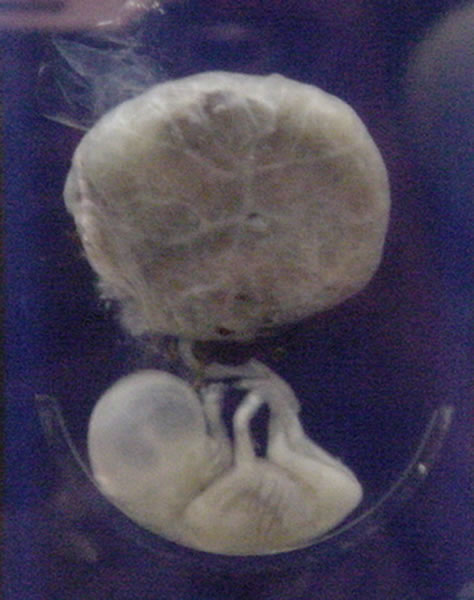
The contrahentes (singular contrahens) are muscles widely present in the hands of mammals, including monkeys. They are on the palmar/plantar side. There is one each for digits I, II, IV and V, but not III. They pull the fingers/toes down and together. Human anatomy In humans, the adductor pollicis muscle in the hand and the adductor hallucis in the foot are well-developed remnants of the first contrahens, though they have lost the insertion on the distal phalanx of the thumb or big toe. The other contrahentes only appear as rare atavistic abnormalities. In other mammals, the contrahentes may have their origin either on the carpus or the metacarpus, which suggests that the palmar interossei muscles also contain elements of the contrahentes. They appear in the human fetus as a layer of flesh that mostly disappears. In other animals The contrahens of the fourth digit is absent in dogs but present in cats and rabbits. In primates, the contrahentes vary in number between zero and fo ... [...More Info...] [...Related Items...] OR: [Wikipedia] [Google] [Baidu] |
Thumb
The thumb is the first digit of the hand, next to the index finger. When a person is standing in the medical anatomical position (where the palm is facing to the front), the thumb is the outermost digit. The Medical Latin English noun for thumb is ''pollex'' (compare ''hallux'' for big toe), and the corresponding adjective for thumb is ''pollical''. Definition Thumb and fingers The English word ''finger'' has two senses, even in the context of appendages of a single typical human hand: 1) Any of the five terminal members of the hand. 2) Any of the four terminal members of the hand, other than the thumb. Linguistically, it appears that the original sense was the first of these two: (also rendered as ) was, in the inferred Proto-Indo-European language, a suffixed form of (or ), which has given rise to many Indo-European-family words (tens of them defined in English dictionaries) that involve, or stem from, concepts of fiveness. The thumb shares the following with each of ... [...More Info...] [...Related Items...] OR: [Wikipedia] [Google] [Baidu] |
Adductor Pollicis Muscle
In human anatomy, the adductor pollicis muscle is a muscle in the hand that functions to adduct the thumb. It has two heads: transverse and oblique. It is a fleshy, flat, triangular, and fan-shaped muscle deep in the thenar compartment beneath the long flexor tendons and the lumbrical muscles at the center of the palm. It overlies the metacarpal bones and the interosseous muscles. Structure Oblique head The oblique head (Latin: ''adductor obliquus pollicis'') arises by several slips from the capitate bone, the bases of the second and third metacarpals, the intercarpal ligaments, and the sheath of the tendon of the flexor carpi radialis. Gray's Anatomy 1918. (See infobox) From this origin the greater number of fibers pass obliquely downward and converge to a tendon, which, uniting with the tendons of the medial portion of the flexor pollicis brevis and the transverse head of the adductor pollicis, is inserted into the ulnar side of the base of the proximal phalanx ... [...More Info...] [...Related Items...] OR: [Wikipedia] [Google] [Baidu] |
Muscle
Muscle is a soft tissue, one of the four basic types of animal tissue. There are three types of muscle tissue in vertebrates: skeletal muscle, cardiac muscle, and smooth muscle. Muscle tissue gives skeletal muscles the ability to muscle contraction, contract. Muscle tissue contains special Muscle contraction, contractile proteins called actin and myosin which interact to cause movement. Among many other muscle proteins, present are two regulatory proteins, troponin and tropomyosin. Muscle is formed during embryonic development, in a process known as myogenesis. Skeletal muscle tissue is striated consisting of elongated, multinucleate muscle cells called muscle fibers, and is responsible for movements of the body. Other tissues in skeletal muscle include tendons and perimysium. Smooth and cardiac muscle contract involuntarily, without conscious intervention. These muscle types may be activated both through the interaction of the central nervous system as well as by innervation ... [...More Info...] [...Related Items...] OR: [Wikipedia] [Google] [Baidu] |
Proximal Phalanges
The phalanges (: phalanx ) are digital bones in the hands and feet of most vertebrates. In primates, the thumbs and big toes have two phalanges while the other digits have three phalanges. The phalanges are classed as long bones. Structure The phalanges are the bones that make up the fingers of the hand and the toes of the foot. There are 56 phalanges in the human body, with fourteen on each hand and foot. Three phalanges are present on each finger and toe, with the exception of the thumb and big toe, which possess only two. The middle and far phalanges of the fifth toes are often fused together (symphalangism). The phalanges of the hand are commonly known as the finger bones. The phalanges of the foot differ from the hand in that they are often shorter and more compressed, especially in the proximal phalanges, those closest to the torso. A phalanx is named according to whether it is proximal, middle, or distal and its associated finger or toe. The proximal phalan ... [...More Info...] [...Related Items...] OR: [Wikipedia] [Google] [Baidu] |
Hand
A hand is a prehensile, multi-fingered appendage located at the end of the forearm or forelimb of primates such as humans, chimpanzees, monkeys, and lemurs. A few other vertebrates such as the Koala#Characteristics, koala (which has two thumb#Opposition and apposition, opposable thumbs on each "hand" and fingerprints extremely similar to human fingerprints) are often described as having "hands" instead of paws on their front limbs. The raccoon is usually described as having "hands" though opposable thumbs are lacking. Some evolutionary anatomists use the term ''hand'' to refer to the appendage of digits on the forelimb more generally—for example, in the context of whether the three Digit (anatomy), digits of the bird hand involved the same Homology (biology), homologous loss of two digits as in the dinosaur hand. The human hand usually has five digits: Finger numbering#Four-finger system, four fingers plus one thumb; however, these are often referred to collectively as Finger ... [...More Info...] [...Related Items...] OR: [Wikipedia] [Google] [Baidu] |
Muscles Of The Upper Limb
Muscle is a soft tissue, one of the four basic types of animal tissue. There are three types of muscle tissue in vertebrates: skeletal muscle, cardiac muscle, and smooth muscle. Muscle tissue gives skeletal muscles the ability to contract. Muscle tissue contains special contractile proteins called actin and myosin which interact to cause movement. Among many other muscle proteins, present are two regulatory proteins, troponin and tropomyosin. Muscle is formed during embryonic development, in a process known as myogenesis. Skeletal muscle tissue is striated consisting of elongated, multinucleate muscle cells called muscle fibers, and is responsible for movements of the body. Other tissues in skeletal muscle include tendons and perimysium. Smooth and cardiac muscle contract involuntarily, without conscious intervention. These muscle types may be activated both through the interaction of the central nervous system as well as by innervation from peripheral plexus or endocrine ... [...More Info...] [...Related Items...] OR: [Wikipedia] [Google] [Baidu] |
Distal Interphalangeal Joint
Distal interphalangeal joints are the articulations between the phalanges of the hand or foot. This term therefore includes: * Interphalangeal joints of the hand * Interphalangeal joints of the foot The interphalangeal joints of the foot are the joints between the phalanx bones of the toes in the feet. Since the great toe only has two phalanx bones ( proximal and distal phalanges), it only has one interphalangeal joint, which is often ab ... {{Set index article Joints ... [...More Info...] [...Related Items...] OR: [Wikipedia] [Google] [Baidu] |
Interphalangeal Articulations Of Hand
The interphalangeal joints of the hand are the hinge joints between the phalanges of the fingers that provide flexion towards the palm of the hand. There are two sets in each finger (except in the thumb, which has only one joint): * "proximal interphalangeal joints" (PIJ or PIP), those between the first (also called proximal) and second (intermediate) phalanges * "distal interphalangeal joints" (DIJ or DIP), those between the second (intermediate) and third (distal) phalanges Anatomically, the proximal and distal interphalangeal joints are very similar. There are some minor differences in how the palmar plates are attached proximally and in the segmentation of the flexor tendon sheath, but the major differences are the smaller dimension and reduced mobility of the distal joint. Joint structure The PIP joint exhibits great lateral stability. Its transverse diameter is greater than its antero-posterior diameter and its thick collateral ligaments are tight in all positions dur ... [...More Info...] [...Related Items...] OR: [Wikipedia] [Google] [Baidu] |
Metacarpophalangeal Joint
The metacarpophalangeal joints (MCP) are situated between the metacarpal bones and the proximal phalanges of the fingers. These joints are of the condyloid kind, formed by the reception of the rounded heads of the metacarpal bones into shallow cavities on the proximal ends of the proximal phalanges. Being condyloid, they allow the movements of flexion, extension, abduction, adduction and circumduction (see anatomical terms of motion) at the joint. Structure Ligaments Each joint has: * palmar ligaments of metacarpophalangeal articulations * collateral ligaments of metacarpophalangeal articulations Dorsal surfaces The dorsal surfaces of these joints are covered by the expansions of the Extensor tendons, together with some loose areolar tissue which connects the deep surfaces of the tendons to the bones. Function The movements which occur in these joints are flexion, extension, adduction, abduction, and circumduction; the movements of abduction and adduction are very ... [...More Info...] [...Related Items...] OR: [Wikipedia] [Google] [Baidu] |
Tarsier
Tarsiers ( ) are haplorhine primates of the family Tarsiidae, which is the lone extant family within the infraorder Tarsiiformes. Although the group was prehistorically more globally widespread, all of the existing species are restricted to Maritime Southeast Asia, predominantly in Brunei, Indonesia, Malaysia and the Philippines.They are found primarily in forested habitats, especially forests that have liana, since the vine gives tarsiers vertical support when climbing trees. Evolutionary history Fossil record Fossils of tarsiiform primates have been found in Asia, Europe, and North America (with disputed fossils from Northern Africa), but extant tarsiers are restricted to several Southeast Asian islands. The fossil record indicates that their dentition has not changed much, except in size, over the past 45 million years. Within the family Tarsiidae, there are two extinct genera—''Xanthorhysis'' and '' Afrotarsius''; however, the placement of ''Afrotarsius'' is not certai ... [...More Info...] [...Related Items...] OR: [Wikipedia] [Google] [Baidu] |
Palmar Interossei Muscles
In human anatomy, the palmar or volar interossei (interossei volares in older literature) are four muscles, one on the thumb that is occasionally missing, and three small, unipennate, central muscles in the hand that lie between the Metacarpus, metacarpal bones and are attached to the Index finger, index, Ring finger, ring, and Little finger, little fingers. They are smaller than the dorsal interossei of the hand. Structure All palmar interossei originate along the shaft of the metacarpal bone of the digit on which they act. They are inserted into the base of the Phalanx bone, proximal phalanx and the extensor expansion of the Extensor digitorum muscle, extensor digitorum of the same digit. Pollical palmar interosseous The first palmar interosseous is located at the thumb's medial side. Passing between the first dorsal interosseous and the oblique head of Adductor pollicis muscle, adductor pollicis, it is inserted on the base of the thumb's proximal phalanx together with Adductor ... [...More Info...] [...Related Items...] OR: [Wikipedia] [Google] [Baidu] |
Human Fetus
A fetus or foetus (; : fetuses, foetuses, rarely feti or foeti) is the unborn offspring of a viviparous animal that develops from an embryo. Following the embryonic stage, the fetal stage of development takes place. Prenatal development is a continuum, with no clear defining feature distinguishing an embryo from a fetus. However, in general a fetus is characterized by the presence of all the major body organs, though they will not yet be fully developed and functional, and some may not yet be situated in their final anatomical location. In human prenatal development, fetal development begins from the ninth week after fertilization (which is the eleventh week of gestational age) and continues until the birth of a newborn. Etymology The word ''fetus'' (plural ''fetuses'' or rarely, the solecism '' feti''''Oxford English Dictionary'', 2013''s.v.'' 'fetus') comes from Latin '' fētus'' 'offspring, bringing forth, hatching of young'. The Latin plural ''fetūs'' is not used in ... [...More Info...] [...Related Items...] OR: [Wikipedia] [Google] [Baidu] |


