|
Congenital Cartilaginous Rest Of The Neck
Congenital cartilaginous rest of the neck (CCRN) is a minor and very rare congenital cutaneous condition characterized by branchial arch remnants that are considered to be the cervical variant of accessory tragus. It resembles a rudimentary Pinna (anatomy), pinna that in most cases is located in the lower anterior part of the neck. Diagnosis CCRN histopathology indicates the presence of elastic cartilage enclosed by various skin structures such as eccrine glands, adipose tissue, and Sebaceous gland, pilosebaceous units. To assess the extent of the lesion as well as look for any underlying Sinus tract, sinus tracts, ultrasonography alongside computed tomography (CT) scans can be used. Alternative diagnoses for CCRN consist of thyroglossal duct cyst, Hair follicle nevus, hair follicle naevus, fibroepithelial polyp, and branchial cleft cyst. Thyroglossal duct cysts are typically found in the midline of the neck, near the hyoid bone, and move with tongue protrusion or swallowing. H ... [...More Info...] [...Related Items...] OR: [Wikipedia] [Google] [Baidu] |
Congenital
A birth defect is an abnormal condition that is present at childbirth, birth, regardless of its cause. Birth defects may result in disability, disabilities that may be physical disability, physical, intellectual disability, intellectual, or developmental disability, developmental. The disabilities can range from mild to severe. Birth defects are divided into two main types: structural disorders in which problems are seen with the shape of a body part and functional disorders in which problems exist with how a body part works. Functional disorders include metabolic disorder, metabolic and degenerative disease, degenerative disorders. Some birth defects include both structural and functional disorders. Birth defects may result from genetic disorder, genetic or chromosome abnormality, chromosomal disorders, exposure to certain medications or chemicals, or certain vertically transmitted infection, infections during pregnancy. Risk factors include folate deficiency, alcohol drink, d ... [...More Info...] [...Related Items...] OR: [Wikipedia] [Google] [Baidu] |
Hair Follicle Nevus
Hair follicle nevus is a cutaneous condition that presents as a small papule from which fine hairs protrude evenly from the surface. Signs and symptoms Hair follicle nevus usually presents as a single, skin-colored papule or nodule on the face after birth that exhibit no symptoms. Diagnosis Histologically, vellus hair follicle growth with perifollicular fibrous thickening occasionally encircled by a cellular stroma is the hallmark of hair follicle nevus. Smooth muscle fibers and eccrine and sebaceous glands are at times visible. See also * Skin lesion * List of cutaneous conditions Many skin conditions affect the human integumentary system—the organ system covering the entire surface of the Human body, body and composed of Human skin, skin, hair, Nail (anatomy), nails, and related muscle and glands. The major function o ... References Further reading * * External links VisualDx {{Medical resources , ICD11 = {{ICD11, LC01 , ICD10 = {{I ... [...More Info...] [...Related Items...] OR: [Wikipedia] [Google] [Baidu] |
Congenital Smooth Muscle Hamartoma
Congenital smooth muscle hamartoma is typically a skin colored or lightly pigmented patch or plaque with hypertrichosis.James, William; Berger, Timothy; Elston, Dirk (2005). ''Andrews' Diseases of the Skin: Clinical Dermatology''. (10th ed.). Saunders. . Congenital smooth muscle hamartoma was originally reported in 1969 by Sourreil et al. Signs and symptoms Although the clinical presentation of congenital smooth muscle hamartoma varies, it typically takes the form of an irregularly shaped, skin-colored, or slightly hyperpigmented patch or plaque on the trunk or extremities that is accompanied by noticeable vellus hairs. Often, it is located in the lumbosacral region. Causes Congenital smooth muscle hamartoma most likely arises from an abnormal development that occurs during mesodermal maturation, primarily in the arrector pili muscle. It is hypothesized that hypertrichosis results from the CD34 + dermal dendritic cells in the hamartoma stimulating the bulge's epithelial cells ... [...More Info...] [...Related Items...] OR: [Wikipedia] [Google] [Baidu] |
Fistula
In anatomy, a fistula (: fistulas or fistulae ; from Latin ''fistula'', "tube, pipe") is an abnormal connection (i.e. tube) joining two hollow spaces (technically, two epithelialized surfaces), such as blood vessels, intestines, or other hollow organs to each other, often resulting in an abnormal flow of fluid from one space to the other. An anal fistula connects the anal canal to the perianal skin. An anovaginal or rectovaginal fistula is a hole joining the anus or rectum to the vagina. A colovaginal fistula joins the space in the colon to that in the vagina. A urinary tract fistula is an abnormal opening in the urinary tract or an abnormal connection between the urinary tract and another organ. An abnormal communication (i.e. hole or tube) between the bladder and the uterus is called a vesicouterine fistula, while if it is between the bladder and the vagina it is known as a vesicovaginal fistula, and if between the urethra and the vagina: a urethrovaginal fistu ... [...More Info...] [...Related Items...] OR: [Wikipedia] [Google] [Baidu] |
Cyst
A cyst is a closed sac, having a distinct envelope and division compared with the nearby tissue. Hence, it is a cluster of cells that have grouped together to form a sac (like the manner in which water molecules group together to form a bubble); however, the distinguishing aspect of a cyst is that the cells forming the "shell" of such a sac are distinctly abnormal (in both appearance and behaviour) when compared with all surrounding cells for that given location. A cyst may contain air, fluids, or semi-solid material. A collection of pus is called an abscess, not a cyst. Once formed, a cyst may resolve on its own. When a cyst fails to resolve, it may need to be removed surgically, but that would depend upon its type and location. Cancer-related cysts are formed as a defense mechanism for the body following the development of mutations that lead to an uncontrolled cellular division. Once that mutation has occurred, the affected cells divide incessantly and become cancerous, ... [...More Info...] [...Related Items...] OR: [Wikipedia] [Google] [Baidu] |
Blood Vessel
Blood vessels are the tubular structures of a circulatory system that transport blood throughout many Animal, animals’ bodies. Blood vessels transport blood cells, nutrients, and oxygen to most of the Tissue (biology), tissues of a Body (biology), body. They also take waste and carbon dioxide away from the tissues. Some tissues such as cartilage, epithelium, and the lens (anatomy), lens and cornea of the eye are not supplied with blood vessels and are termed ''avascular''. There are five types of blood vessels: the arteries, which carry the blood away from the heart; the arterioles; the capillaries, where the exchange of water and chemicals between the blood and the tissues occurs; the venules; and the veins, which carry blood from the capillaries back towards the heart. The word ''vascular'', is derived from the Latin ''vas'', meaning ''vessel'', and is mostly used in relation to blood vessels. Etymology * artery – late Middle English; from Latin ''arteria'', from Gree ... [...More Info...] [...Related Items...] OR: [Wikipedia] [Google] [Baidu] |
Collagen Fibers
Type I collagen is the most abundant collagen of the human body, consisting of around 90% of the body's total collagen in vertebrates. Due to this, it is also the most abundant protein type found in all vertebrates. Type I forms large, eosinophilic fibers known as collagen fibers, which make up most of the rope-like dense connective tissue in the body. Collagen I itself is created by the combination of both a proalpha1 and a proalpha2 chain created by the COL1alpha1 and COL1alpha2 genes respectively. The Col I gene itself takes up a triple-helical conformation due to its Glycine-X-Y structure, x and y being any type of amino acid. Collagen can also be found in two different isoforms, either as a homotrimer or a heterotrimer, both of which can be found during different periods of development. Heterotrimers, in particular, play an important role in wound healing, and are the dominant isoform found in the body. Type I collagen can be found in a myriad of different places in the b ... [...More Info...] [...Related Items...] OR: [Wikipedia] [Google] [Baidu] |
Hypertrichosis
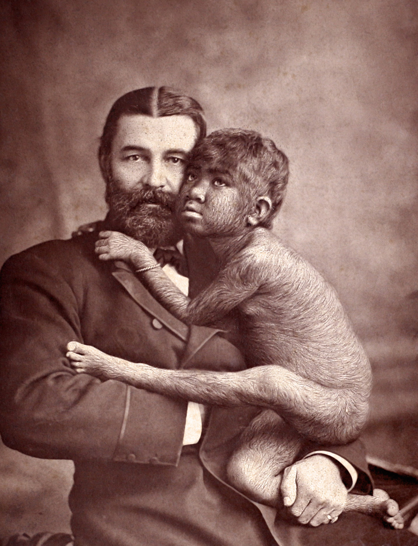
Hypertrichosis (sometimes known as werewolf syndrome) is an abnormal amount of hair growth over the body. The two distinct types of hypertrichosis are generalized hypertrichosis, which occurs over the entire body, and localized hypertrichosis, which is restricted to a certain area. Hypertrichosis can be either congenital (present at birth) or acquired later in life. The excess growth of hair occurs in areas of the skin with the exception of androgen-dependent hair of the pubic area, face, and axillary regions. Several circus sideshow performers in the 19th and early 20th centuries, such as Julia Pastrana, had hypertrichosis. Many of them worked as freaks and were promoted as having distinct human and animal traits. Classification Two methods of classification are used for hypertrichosis. One divides them into either generalized versus localized hypertrichosis, while the other divides them into congenital versus acquired. Congenital Congenital forms of hypertrichosis are ... [...More Info...] [...Related Items...] OR: [Wikipedia] [Google] [Baidu] |
Papule
A papule is a small, well-defined bump in the skin lesion, skin. It may have a rounded, pointed or flat top, and may have a umbilication, dip. It can appear with a Peduncle (anatomy), stalk, be thread-like or look warty. It can be soft or firm and its surface may be rough or smooth. Some have Crust (dermatology), crusts or Scale (dermatology), scales. A papule can be flesh colored, yellow, white, brown, red, blue or purplish. There may be just one or many, and they may occur irregularly in different parts of the body or appear in clusters. It does not contain fluid but may progress to a pustule or vesicle (dermatology), vesicle. A papule is smaller than a Nodule (medicine), nodule; it can be as tiny as a pinhead and is typically less than 1 cm in width, according to some sources, and 0.5 cm according to others. When merged together, it appears as a plaque. A papule's colour might indicate its cause, such as white in Milium (dermatology), milia, red in eczema, yellowish ... [...More Info...] [...Related Items...] OR: [Wikipedia] [Google] [Baidu] |
Hyoid Bone
The hyoid-bone (lingual-bone or tongue-bone) () is a horseshoe-shaped bone situated in the anterior midline of the neck between the chin and the thyroid-cartilage. At rest, it lies between the base of the mandible and the third cervical vertebra. Unlike other bones, the hyoid is only distantly articulated to other bones by muscles or ligaments. It is the only bone in the human body that is not connected to any other bones. The hyoid is anchored by muscles from the anterior, posterior and inferior directions, and aids in tongue movement and swallowing. The hyoid bone provides attachment to the muscles of the floor of the mouth and the tongue above, the larynx below, and the epiglottis and pharynx behind. Its name is derived . Structure The hyoid bone is classed as an irregular bone and consists of a central part called the body, and two pairs of horns, the greater and lesser horns. Body The body of the hyoid bone is the central part of the hyoid bone. *At the fron ... [...More Info...] [...Related Items...] OR: [Wikipedia] [Google] [Baidu] |
Branchial Cleft Cyst
A branchial cleft cyst or simply branchial cyst is a cyst as a swelling in the upper part of neck anterior to sternocleidomastoid. It can, but does not necessarily, have an opening to the skin surface, called a fistula. The cause is usually a developmental abnormality arising in the early prenatal period, typically failure of obliteration of the second, third, and fourth pharyngeal groove, branchial cleft, i.e. failure of fusion of the second pharyngeal arch, branchial arches and epicardial ridge in lower part of the neck. Branchial cleft cysts account for almost 20% of neck masses in children. Less commonly, the cysts can develop from the first, third, or fourth clefts, and their location and the location of associated fistulas differs accordingly. Symptoms and signs Most branchial cleft cysts present in late childhood or early adulthood as a solitary, painless mass, which went previously unnoticed, that has now become Infection, infected (typically after an upper respiratory tract ... [...More Info...] [...Related Items...] OR: [Wikipedia] [Google] [Baidu] |
Fibroepithelial Polyp
A fibroepithelial neoplasm (or tumor) is a biphasic tumor. They consist of epithelial tissue, and stromal or mesenchymal tissue. They may be benign or malignant.Tavassoli, F.A., Devilee, P. (Eds). 2003. World Health Organization Classification of Tumours: Pathology & Genetics: Tumours of the breast and female genital organs. IARC Press: Lyon. Examples include: * Brenner tumor of the ovary * Fibroadenoma of the breast * Phyllodes tumor Phyllodes tumors (from Greek language, Greek: ), are a rare type of Biphasic tumor, biphasic Fibroepithelial neoplasm, fibroepithelial mass that form from the periductal stromal and epithelial cells of the breast. They account for less than 1% of ... of the breast Sometimes fibroepithelial polyps (FEPs) of the vulva may be misdiagnosed as cancers. However not much harm is caused because the treatment of both is excision. The consent for removal must however be completely informed. References External links * - "Premalignant Fibroepithelial ... [...More Info...] [...Related Items...] OR: [Wikipedia] [Google] [Baidu] |


