|
Collectins
Collectins, col-lectins, (collagen-containing C-type lectins) are a part of the innate immune system. They form a sub-family of collagenous Ca2+-dependent lectins of the family of C-type lectins, which are found in animals. Collectins are soluble pattern recognition receptors (PRRs). Their function is to bind to oligosaccharide structure or lipids that are on the surface of microorganisms. Like other PRRs they bind pathogen-associated molecular patterns (PAMPs) and damage-associated molecular patterns (DAMPs) of oligosaccharide origin. Binding of collectins to microorganisms may trigger elimination of microorganisms by aggregation, complement activation, opsonization, activation of phagocytosis, or inhibition of microbial growth. Other functions of collectins are modulation of inflammatory, allergic responses, adaptive immune system and clearance of apoptotic cells. Structure Functionally collectins are trimers. Monomeric subunit consists of four parts: * a cysteine-rich domain a ... [...More Info...] [...Related Items...] OR: [Wikipedia] [Google] [Baidu] |
Conglutinin
Conglutinin is a collectin protein Proteins are large biomolecules and macromolecules that comprise one or more long chains of amino acid residue (biochemistry), residues. Proteins perform a vast array of functions within organisms, including Enzyme catalysis, catalysing metab .... External links * Collectins {{Transmembranereceptor-stub ... [...More Info...] [...Related Items...] OR: [Wikipedia] [Google] [Baidu] |
Collectin Of 46 KDa
Collectin of 46 kDa (CL-46) is a collectin protein. It has two cysteine residues on the N-terminal segment, a hydrophilic loop near the carbohydrate recognition domain's binding site, and a N-glycosylation site in the collagen region. It is expressed in bovine liver and thymus The thymus (: thymuses or thymi) is a specialized primary lymphoid organ of the immune system. Within the thymus, T cells mature. T cells are critical to the adaptive immune system, where the body adapts to specific foreign invaders. The thymus ... glands and binds to pathogens, prompting elimination by macrophages. References Proteins Collectins {{immunology-stub ... [...More Info...] [...Related Items...] OR: [Wikipedia] [Google] [Baidu] |
Collectin Of 43 KDa
Collectin of 43 kDa (CL-43) is a collectin protein that acts as an antigen recognition protein. When an agent, zymosan, was injected into the tunicate Tunicates are marine invertebrates belonging to the subphylum Tunicata ( ). This grouping is part of the Chordata, a phylum which includes all animals with dorsal nerve cords and notochords (including vertebrates). The subphylum was at one time ... ''Styela plicata'' (causing inflammation), secretion of this collectin was tripled within 96 hours. References Collectins Proteins {{Immunology-stub ... [...More Info...] [...Related Items...] OR: [Wikipedia] [Google] [Baidu] |
Collectin Liver 1
Collectin-10, also known as collectin liver 1, is a collectin protein that in humans is encoded by the ''COLEC10'' gene. Its structure is similar to mannan-binding lectin ( MBL). Collectin liver 1 (CL-L1) show very similar carbohydrate selectivity as MBL. Two other discovered collectin Collectins, col-lectins, (collagen-containing C-type lectins) are a part of the innate immune system. They form a sub-family of collagenous Ca2+-dependent lectins of the family of C-type lectins, which are found in animals. Collectins are soluble p ...s include collectin placenta 1 (CL-P1) and collectin kidney 1 (CL-K1). CL-L1's location found to be on chromosome 8 q23-24.1. Research concluded CL-L1 to be a serum protein. References External links * Collectins {{biochem-stub ... [...More Info...] [...Related Items...] OR: [Wikipedia] [Google] [Baidu] |
Surfactant Protein D
Surfactant protein D, also known as SP-D, is a lung surfactant protein part of the collagenous family of lectins called collectin. In humans, SP-D is encoded by the ''SFTPD'' gene and is part of the innate immune system. Each SP-D subunit is composed of an N-terminal domain, a collagenous region, a nucleating neck region, and a C-terminal lectin domain. Three of these subunits assemble to form a homotrimer, which further assemble into a tetrameric complex. Interactions Surfactant protein D has been shown to interact with DMBT1, and hemagglutinin of influenza A virus. Post-translational modification of SP-D i.e. S-nitrosylation switches its function. See also * Pulmonary surfactant Pulmonary surfactant is a surface-active complex of phospholipids and proteins formed by Type II cells, type II alveolar cells. The proteins and lipids that make up the surfactant have both hydrophilic and hydrophobic regions. By adsorption, adso ... * Pulmonary surfactant protein D Refe ... [...More Info...] [...Related Items...] OR: [Wikipedia] [Google] [Baidu] |
Surfactant Protein A
Surfactant protein A is an innate immune system collectin. It is water-soluble and has collagen-like domains similar to SP-D. It is part of the innate immune system and is used to opsonize bacterial cells in the alveoli marking them for phagocytosis by alveolar macrophages. SP-A may also play a role in negative feedback limiting the secretion of pulmonary surfactant. SP-A is not required for pulmonary surfactant to function but does confer immune effects to the organism. During parturition The role of surfactant protein A (SP-A) in childbirth is indicated in studies with mice. Mice which gestate for 19 days typically show signs of SP-A in amniotic fluid at around 16 days. If SP-A is injected into the uterus at 15 days, mice typically deliver early. Inversely, an SP-A inhibitor injection causes notable delays in birth. The presence of surfactant protein A seemed to trigger an inflammatory response in the uterus of the mice, but later studies found an anti-inflammatory response ... [...More Info...] [...Related Items...] OR: [Wikipedia] [Google] [Baidu] |
Mannan-binding Lectin
Mannose-binding lectin (MBL), also called mannan-binding lectin or mannan-binding protein (MBP), is a lectin that is instrumental in innate immunity as an opsonin and via the lectin pathway. Structure MBL has an oligomeric structure (400-700 kDa), built of subunits that contain three presumably identical peptide chains of about 30 kDa each. Although MBL can form several oligomeric forms, there are indications that dimers and trimers are biologically inactive as an opsonin and at least a tetramer form is needed for activation of complement. Genes and polymorphisms Human MBL2 gene is located on chromosome 10q11.2-q21. Mice have two homologous genes, but in human the first of them was lost. A low level expression of an MBL1 pseudogene 1 (MBL1P1) was detected in liver. The pseudogene encodes a truncated 51-amino acid protein that is homologous to the MBLA isoform in rodents and some primates. Structural mutations in exon 1 of the human MBL2 gene, at codon 52 (Arg to Cys, allele D) ... [...More Info...] [...Related Items...] OR: [Wikipedia] [Google] [Baidu] |
C-type Lectin
A C-type lectin (CLEC) is a type of carbohydrate-binding protein known as a lectin. The C-type designation is from their requirement for calcium for binding. Proteins that contain C-type lectin domains have a diverse range of functions including cell-cell adhesion, immune response to pathogens and apoptosis. Classification Drickamer ''et al.'' classified C-type lectins into 7 subgroups (I to VII) based on the order of the various protein domains in each protein. This classification was subsequently updated in 2002, leading to seven additional groups (VIII to XIV). Most recently, three further subgroups were added (XV to XVII). CLECs include: * CLEC1A, CLEC1B * CLEC2A, CLEC2B, CD69 (CLEC2C), CLEC2D, CLEC2L * CLEC3A, CLEC3B * CLEC4A, CLEC4C, CLEC4D, CLEC4E, CLEC4F, CLEC4G, ASGR1 (CLEC4H1), ASGR2 (CLEC4H2), FCER2 (CLEC4J), CD207 (CLEC4K), CD209 (CLEC4L), CLEC4M * CLEC5A * CLEC6A * CLEC7A * OLR1 (CLEC8A) * CLEC9A * CLEC10A * CLEC11A * CLEC12A, CLEC12B * CD302 (CLEC13A), LY75 (CLEC ... [...More Info...] [...Related Items...] OR: [Wikipedia] [Google] [Baidu] |
Carbohydrate Recognition Domain
A carbohydrate () is a biomolecule composed of carbon (C), hydrogen (H), and oxygen (O) atoms. The typical hydrogen-to-oxygen atomic ratio is 2:1, analogous to that of water, and is represented by the empirical formula (where ''m'' and ''n'' may differ). This formula does not imply direct covalent bonding between hydrogen and oxygen atoms; for example, in , hydrogen is covalently bonded to carbon, not oxygen. While the 2:1 hydrogen-to-oxygen ratio is characteristic of many carbohydrates, exceptions exist. For instance, uronic acids and deoxy-sugars like fucose deviate from this precise stoichiometric definition. Conversely, some compounds conforming to this definition, such as formaldehyde and acetic acid, are not classified as carbohydrates. The term is predominantly used in biochemistry, functioning as a synonym for saccharide (), a group that includes sugars, starch, and cellulose. The saccharides are divided into four chemical groups: monosaccharides, disaccharides, oligosac ... [...More Info...] [...Related Items...] OR: [Wikipedia] [Google] [Baidu] |
Gram-positive
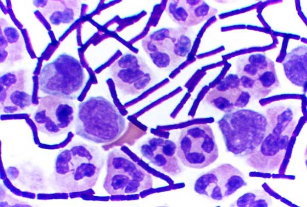
In bacteriology, gram-positive bacteria are bacteria that give a positive result in the Gram stain test, which is traditionally used to quickly classify bacteria into two broad categories according to their type of cell wall. The Gram stain is used by microbiologists to place bacteria into two main categories, gram-positive (+) and gram-negative bacteria, gram-negative (−). Gram-positive bacteria have a thick layer of peptidoglycan within the cell wall, and gram-negative bacteria have a thin layer of peptidoglycan. Gram-positive bacteria retain the crystal violet stain used in the test, resulting in a purple color when observed through an optical microscope. The thick layer of peptidoglycan in the bacterial cell wall retains the Stain (biology), stain after it has been fixed in place by iodine. During the decolorization step, the decolorizer removes crystal violet from all other cells. Conversely, gram-negative bacteria cannot retain the violet stain after the decolorization ... [...More Info...] [...Related Items...] OR: [Wikipedia] [Google] [Baidu] |
Gram-negative
Gram-negative bacteria are bacteria that, unlike gram-positive bacteria, do not retain the crystal violet stain used in the Gram staining method of bacterial differentiation. Their defining characteristic is that their cell envelope consists of a thin peptidoglycan cell wall sandwiched between an inner ( cytoplasmic) membrane and an outer membrane. These bacteria are found in all environments that support life on Earth. Within this category, notable species include the model organism '' Escherichia coli'', along with various pathogenic bacteria, such as '' Pseudomonas aeruginosa'', '' Chlamydia trachomatis'', and '' Yersinia pestis''. They pose significant challenges in the medical field due to their outer membrane, which acts as a protective barrier against numerous antibiotics (including penicillin), detergents that would normally damage the inner cell membrane, and the antimicrobial enzyme lysozyme produced by animals as part of their innate immune system. Furthe ... [...More Info...] [...Related Items...] OR: [Wikipedia] [Google] [Baidu] |

