|
Ciliated
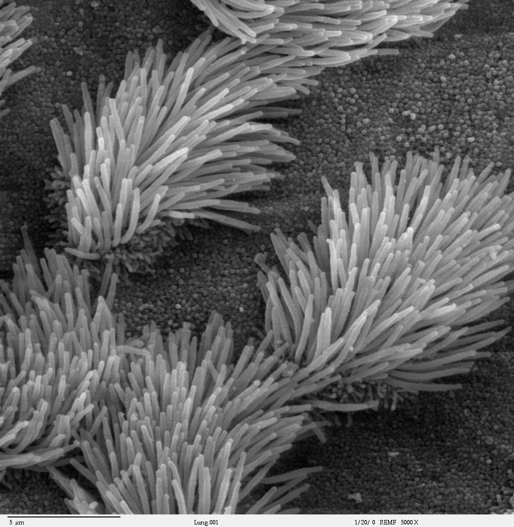
The cilium (: cilia; ; in Medieval Latin and in anatomy, ''cilium'') is a short hair-like membrane protrusion from many types of eukaryotic cell. (Cilia are absent in bacteria and archaea.) The cilium has the shape of a slender threadlike projection that extends from the surface of the much larger cell body. Eukaryotic flagella found on sperm cells and many protozoans have a similar structure to motile cilia that enables swimming through liquids; they are longer than cilia and have a different undulating motion. There are two major classes of cilia: ''motile'' and ''non-motile'' cilia, each with two subtypes, giving four types in all. A cell will typically have one primary cilium or many motile cilia. The structure of the cilium core, called the axoneme, determines the cilium class. Most motile cilia have a central pair of single microtubules surrounded by nine pairs of double microtubules called a 9+2 axoneme. Most non-motile cilia have a 9+0 axoneme that lacks the centra ... [...More Info...] [...Related Items...] OR: [Wikipedia] [Google] [Baidu] |
Respiratory Epithelium
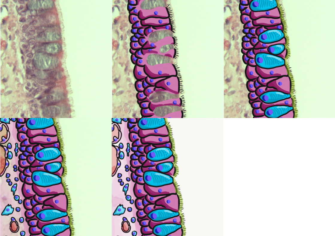
Respiratory epithelium, or airway epithelium, is ciliated pseudostratified columnar epithelium a type of columnar epithelium found lining most of the respiratory tract as respiratory mucosa, where it serves to moisten and protect the airways. It is not present in the vocal cords of the larynx, or the oropharynx and laryngopharynx, where instead the epithelium is stratified squamous. It also functions as a barrier to potential pathogens and foreign particles, preventing infection and tissue injury by the secretion of mucus and the action of mucociliary clearance. Structure The respiratory epithelium lining the upper respiratory airways is classified as ciliated pseudostratified columnar epithelium. This designation is due to the arrangement of the multiple cell types composing the respiratory epithelium. While all cells make contact with the basement membrane and are, therefore, a single layer of cells, their nuclei are not aligned in the same plane. Hence, it appear ... [...More Info...] [...Related Items...] OR: [Wikipedia] [Google] [Baidu] |
Fallopian Tube
The fallopian tubes, also known as uterine tubes, oviducts or salpinges (: salpinx), are paired tubular sex organs in the human female body that stretch from the Ovary, ovaries to the uterus. The fallopian tubes are part of the female reproductive system. In other vertebrates, they are only called oviducts. Each tube is a muscular hollow organ that is on average between in length, with an external diameter of . It has four described parts: the intramural part, isthmus, ampulla, and infundibulum with associated fimbriae. Each tube has two openings: a proximal opening nearest to the uterus, and a distal opening nearest to the ovary. The fallopian tubes are held in place by the mesosalpinx, a part of the broad ligament mesentery that wraps around the tubes. Another part of the broad ligament, the mesovarium suspends the ovaries in place. An ovum, egg cell is transported from an ovary to a fallopian tube where it may be human fertilization, fertilized in the ampulla of the tube. ... [...More Info...] [...Related Items...] OR: [Wikipedia] [Google] [Baidu] |
Ependymal Cells
The ependyma is the thin neuroepithelial ( simple columnar ciliated epithelium) lining of the ventricular system of the brain and the central canal of the spinal cord. The ependyma is one of the four types of neuroglia in the central nervous system (CNS). It is involved in the production of cerebrospinal fluid (CSF), and is shown to serve as a reservoir for neuroregeneration. Structure The ependyma is made up of ependymal cells called ependymocytes, a type of glial cell. These cells line the ventricles in the brain and the central canal of the spinal cord, which become filled with cerebrospinal fluid. These are nervous tissue cells with simple columnar shape, much like that of some mucosal epithelial cells. Early monociliated ependymal cells are differentiated to multiciliated ependymal cells for their function in circulating cerebrospinal fluid. The basal membranes of these cells are characterized by tentacle-like extensions that attach to astrocytes. The apical side is cov ... [...More Info...] [...Related Items...] OR: [Wikipedia] [Google] [Baidu] |
Trachea
The trachea (: tracheae or tracheas), also known as the windpipe, is a cartilaginous tube that connects the larynx to the bronchi of the lungs, allowing the passage of air, and so is present in almost all animals' lungs. The trachea extends from the larynx and branches into the two primary bronchi. At the top of the trachea, the cricoid cartilage attaches it to the larynx. The trachea is formed by a number of horseshoe-shaped rings, joined together vertically by overlying annular ligaments of trachea, ligaments, and by the trachealis muscle at their ends. The epiglottis closes the opening to the larynx during swallowing. The trachea begins to form in the second month of embryo development, becoming longer and more fixed in its position over time. Its epithelium is lined with columnar epithelium, column-shaped cells that have hair-like extensions called cilia, with scattered goblet cells that produce protective mucins. The trachea can be affected by inflammation or infection, usua ... [...More Info...] [...Related Items...] OR: [Wikipedia] [Google] [Baidu] |
Mucociliary Clearance
Mucociliary clearance (MCC), mucociliary transport, or the mucociliary escalator describes the self-clearing mechanism of the respiratory tract, airways in the respiratory system. It is one of the two protective processes for the lungs in removing inhaled particulates, particles including pathogens before they can reach the Parenchyma#Lung parenchyma, delicate tissue of the lungs. The other clearance mechanism is provided by the cough reflex. Mucociliary clearance has a major role in pulmonary hygiene. MCC effectiveness relies on the correct properties of the airway surface liquid produced, both of the Mucus#Respiratory system, periciliary sol layer and the overlying Mucus#Respiratory system, mucus gel layer, and of the number and quality of the cilium, cilia present in the respiratory epithelium, lining of the airways. An important factor is the rate of mucin secretion. The ion channels CFTR and ENaC work together to maintain the necessary hydration of the airway surface liquid. ... [...More Info...] [...Related Items...] OR: [Wikipedia] [Google] [Baidu] |
Flagella
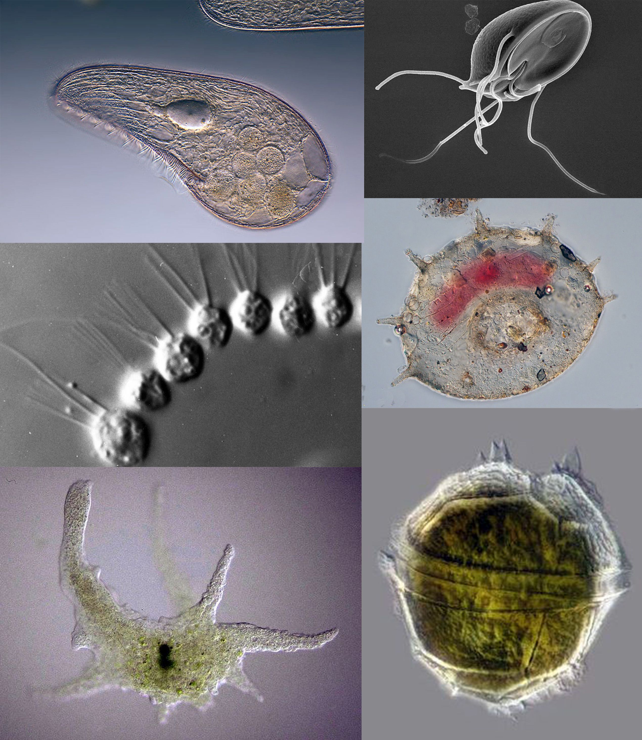
A flagellum (; : flagella) (Latin for 'whip' or 'scourge') is a hair-like appendage that protrudes from certain plant and animal sperm cells, from fungal spores ( zoospores), and from a wide range of microorganisms to provide motility. Many protists with flagella are known as flagellates. A microorganism may have from one to many flagella. A gram-negative bacterium '' Helicobacter pylori'', for example, uses its flagella to propel itself through the stomach to reach the mucous lining where it may colonise the epithelium and potentially cause gastritis, and ulcers – a risk factor for stomach cancer. In some swarming bacteria, the flagellum can also function as a sensory organelle, being sensitive to wetness outside the cell. Across the three domains of Bacteria, Archaea, and Eukaryota, the flagellum has a different structure, protein composition, and mechanism of propulsion but shares the same function of providing motility. The Latin word means " whip" to describe its ... [...More Info...] [...Related Items...] OR: [Wikipedia] [Google] [Baidu] |
Protozoan
Protozoa (: protozoan or protozoon; alternative plural: protozoans) are a polyphyletic group of single-celled eukaryotes, either free-living or parasitic, that feed on organic matter such as other microorganisms or organic debris. Historically, protozoans were regarded as "one-celled animals". When first introduced by Georg Goldfuss, in 1818, the taxon Protozoa was erected as a class within the Animalia, with the word 'protozoa' meaning "first animals", because they often possess animal-like behaviours, such as motility and predation, and lack a cell wall, as found in plants and many algae. This classification remained widespread in the 19th and early 20th century, and even became elevated to a variety of higher ranks, including phylum, subkingdom, kingdom, and then sometimes included within the paraphyletic Protoctista or Protista. By the 1970s, it became usual to require that all taxa be monophyletic (derived from a common ancestor that would also be regarded as protozo ... [...More Info...] [...Related Items...] OR: [Wikipedia] [Google] [Baidu] |
Mechanosensation
Mechanosensation is the transduction of mechanical stimuli into neural signals. Mechanosensation provides the basis for the senses of light touch, hearing, proprioception, and pain. Mechanoreceptors found in the skin, called cutaneous mechanoreceptors, are responsible for the sense of touch. Tiny cells in the inner ear, called hair cells, are responsible for hearing and balance. States of neuropathic pain, such as hyperalgesia and allodynia, are also directly related to mechanosensation. A wide array of elements are involved in the process of mechanosensation, many of which are still not fully understood. Cutaneous mechanoreceptors Cutaneous mechanoreceptors are physiologically classified with respect to conduction velocity, which is directly related to the diameter and myelination of the axon. Rapidly adapting and slowly adapting mechanoreceptors Mechanoreceptors that possess a large diameter and high myelination are called ''low-threshold mechanoreceptors''. Fibers that respond ... [...More Info...] [...Related Items...] OR: [Wikipedia] [Google] [Baidu] |
Brain
The brain is an organ (biology), organ that serves as the center of the nervous system in all vertebrate and most invertebrate animals. It consists of nervous tissue and is typically located in the head (cephalization), usually near organs for special senses such as visual perception, vision, hearing, and olfaction. Being the most specialized organ, it is responsible for receiving information from the sensory nervous system, processing that information (thought, cognition, and intelligence) and the coordination of motor control (muscle activity and endocrine system). While invertebrate brains arise from paired segmental ganglia (each of which is only responsible for the respective segmentation (biology), body segment) of the ventral nerve cord, vertebrate brains develop axially from the midline dorsal nerve cord as a brain vesicle, vesicular enlargement at the rostral (anatomical term), rostral end of the neural tube, with centralized control over all body segments. All vertebr ... [...More Info...] [...Related Items...] OR: [Wikipedia] [Google] [Baidu] |
Oviduct
The oviduct in vertebrates is the passageway from an ovary. In human females, this is more usually known as the fallopian tube. The eggs travel along the oviduct. These eggs will either be fertilized by spermatozoa to become a zygote, or will degenerate in the body. Normally, these are paired structures, but in birds and some cartilaginous fishes, one or the other side fails to develop (together with the corresponding ovary), and only one functional oviduct can be found. Except in teleosts, the oviduct is not directly in contact with the ovary. Instead, the most anterior portion ends in a funnel-shaped structure called the infundibulum, which collects eggs as they are released by the ovary into the body cavity. The only female vertebrates to lack oviducts are the jawless fishes. In these species, the single fused ovary releases eggs directly into the body cavity. The fish eventually extrudes the eggs through a small genital pore towards the rear of the body. Fish and amphibia ... [...More Info...] [...Related Items...] OR: [Wikipedia] [Google] [Baidu] |
Ventricular System
In neuroanatomy, the ventricular system is a set of four interconnected cavities known as cerebral ventricles in the brain. Within each ventricle is a region of choroid plexus which produces the circulating cerebrospinal fluid (CSF). The ventricular system is continuous with the central canal of the spinal cord from the fourth ventricle, allowing for the flow of CSF to circulate. All of the ventricular system and the central canal of the spinal cord are lined with ependyma, a specialised form of epithelium connected by tight junctions that make up the blood–cerebrospinal fluid barrier. Structure The system comprises four ventricles: * lateral ventricles right and left (one for each hemisphere) * third ventricle * fourth ventricle There are several foramina, openings acting as channels, that connect the ventricles. The interventricular foramina (also called the foramina of Monro) connect the lateral ventricles to the third ventricle through which the cerebrospinal ... [...More Info...] [...Related Items...] OR: [Wikipedia] [Google] [Baidu] |


