|
Cervical Rib
Cervical ribs are the ribs of the neck in many tetrapods. In most mammals, including humans, cervical ribs are not normally present as separate structures. They can, however, occur as a pathology. In humans, pathological cervical ribs are usually not of clinical concern, although they can cause a form of thoracic outlet syndrome. Development Like other ribs, the cervical ribs form by endochondral ossification. Variation The cervical ribs of sauropod dinosaurs were extended by ossified tendons, and could reach exceptional lengths; a cervical rib of ''Mamenchisaurus sinocanadorum'' was long. In birds, the cervical ribs are small and completely fused to the vertebrae. In therian mammals, the cervical ribs fully fuse with the cervical vertebrae to form part of the transverse processes, except in rare pathological cases. In contrast, monotremes retain the plesiomorphic condition of having separate cervical ribs. Pathological cervical ribs A cervical rib in humans is an Supernumerar ... [...More Info...] [...Related Items...] OR: [Wikipedia] [Google] [Baidu] [Amazon] |
Ribs
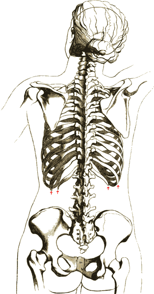
The rib cage or thoracic cage is an endoskeletal enclosure in the thorax of most vertebrates that comprises the ribs, vertebral column and sternum, which protect the vital organs of the thoracic cavity, such as the heart, lungs and great vessels and support the shoulder girdle to form the core (anatomy), core part of the axial skeleton. A typical human skeleton, human thoracic cage consists of 12 pairs of ribs and the adjoining costal cartilages, the sternum (along with the manubrium and xiphoid process), and the 12 thoracic vertebrae articulating with the ribs. The thoracic cage also provides attachments for extrinsic skeletal muscles of the neck, upper limbs, upper abdomen and back, and together with the overlying skin and associated fascia and muscles, makes up the thoracic wall. In tetrapods, the rib cage intrinsically holds the muscles of respiration (thoracic diaphragm, diaphragm, intercostal muscles, etc.) that are crucial for active inhalation and forced exhalation, and ... [...More Info...] [...Related Items...] OR: [Wikipedia] [Google] [Baidu] [Amazon] |
Congenital
A birth defect is an abnormal condition that is present at childbirth, birth, regardless of its cause. Birth defects may result in disability, disabilities that may be physical disability, physical, intellectual disability, intellectual, or developmental disability, developmental. The disabilities can range from mild to severe. Birth defects are divided into two main types: structural disorders in which problems are seen with the shape of a body part and functional disorders in which problems exist with how a body part works. Functional disorders include metabolic disorder, metabolic and degenerative disease, degenerative disorders. Some birth defects include both structural and functional disorders. Birth defects may result from genetic disorder, genetic or chromosome abnormality, chromosomal disorders, exposure to certain medications or chemicals, or certain vertically transmitted infection, infections during pregnancy. Risk factors include folate deficiency, alcohol drink, d ... [...More Info...] [...Related Items...] OR: [Wikipedia] [Google] [Baidu] [Amazon] |
Adson's Sign
Adson's sign is the loss of the radial pulse in the arm by rotating head to the ipsilateral side with extended neck following deep inspiration. It is sometimes used as a sign of thoracic outlet syndrome (TOS). It is named after Alfred Washington Adson. Limitations, and pathophysiology of thoracic outlet syndrome Adson's sign is no longer used as a positive diagnosis of TOS since many people without TOS will show a positive Adson's. There is minimal evidence of interexaminer reliability. Thoracic outlet obstruction may be caused by a number of abnormalities, including degenerative or bony disorders, trauma to the cervical spine, fibromuscular bands, vascular abnormalities, and spasm of the anterior scalene muscle. Symptoms are due to compression of the brachial plexus and subclavian vasculature, and consist of complaints ranging from diffuse arm pain to a sensation of arm fatigue, frequently aggravated by carrying anything in the ipsilateral Standard anatomical terms of lo ... [...More Info...] [...Related Items...] OR: [Wikipedia] [Google] [Baidu] [Amazon] |
Scalene Muscles
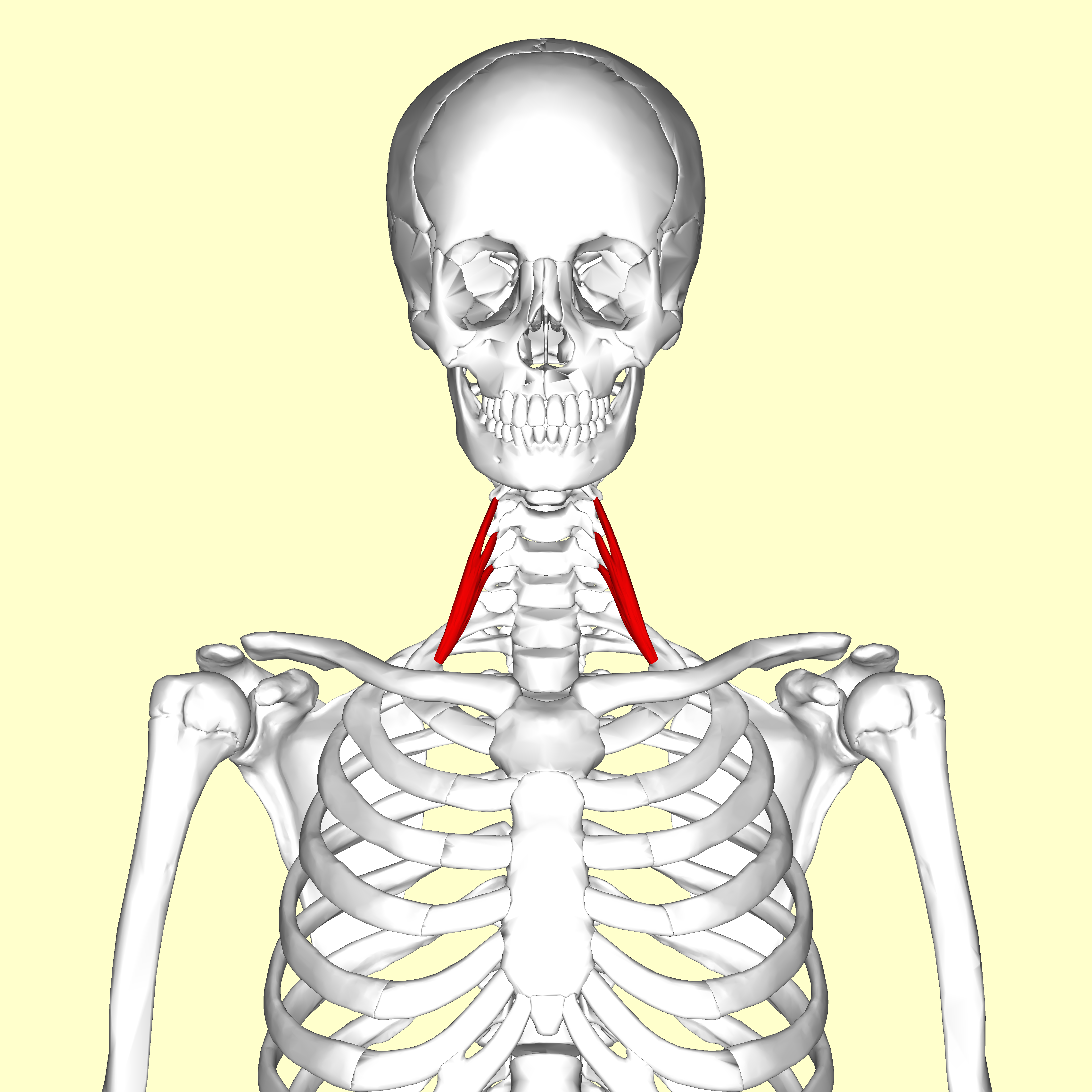
The scalene muscles are a group of three muscles on each side of the neck, identified as the anterior, the middle, and the posterior. They are innervated by the third to the eighth cervical spinal nerves (C3-C8). The anterior and middle scalene muscles lift the first rib and bend the neck to the side they are on. The posterior scalene lifts the second rib and tilts the neck to the same side. The muscles are named from the Ancient Greek (), meaning 'uneven'. Structure The scalene muscles are attached at one end to bony protrusions on vertebrae C2 to C7 and at the other end to the first and second ribs. Anterior scalene The anterior scalene muscle (), lies deeply at the side of the neck, behind the sternocleidomastoid muscle. It arises from the anterior tubercles of the transverse processes of the third, fourth, fifth, and sixth cervical vertebrae, and descending, almost vertically, is inserted by a narrow, flat tendon into the scalene tubercle on the inner border of the ... [...More Info...] [...Related Items...] OR: [Wikipedia] [Google] [Baidu] [Amazon] |
Subclavian Artery
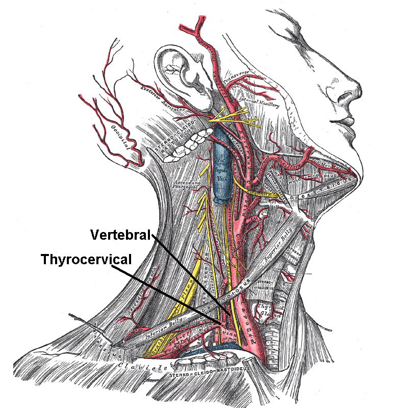
In human anatomy, the subclavian arteries are paired major arteries of the upper thorax, below the clavicle. They receive blood from the aortic arch. The left subclavian artery supplies blood to the left arm and the right subclavian artery supplies blood to the right arm, with some branches supplying the head and thorax. On the left side of the body, the subclavian comes directly off the aortic arch, while on the right side it arises from the relatively short brachiocephalic artery when it bifurcates into the subclavian and the right common carotid artery. The usual branches of the subclavian on both sides of the body are the vertebral artery, the internal thoracic artery, the thyrocervical trunk, the costocervical trunk and the dorsal scapular artery, which may branch off the transverse cervical artery, which is a branch of the thyrocervical trunk. The subclavian becomes the axillary artery at the lateral border of the first rib. Structure From its origin, the subclavian art ... [...More Info...] [...Related Items...] OR: [Wikipedia] [Google] [Baidu] [Amazon] |
Brachial Plexus
The brachial plexus is a network of nerves (nerve plexus) formed by the anterior rami of the lower four Spinal nerve#Cervical nerves, cervical nerves and first Spinal nerve#Thoracic nerves, thoracic nerve (cervical spinal nerve 5, C5, Cervical spinal nerve 6, C6, cervical spinal nerve 7, C7, cervical spinal nerve 8, C8, and thoracic spinal nerve 1, T1). This plexus extends from the spinal cord, through the cervicoaxillary canal in the neck, over the first rib, and into the axilla, armpit, it supplies Afferent nerve fiber, afferent and efferent nerve fibers to the chest, shoulder, arm, forearm, and hand. Structure The brachial plexus is divided into five ''roots'', three ''trunks'', six ''divisions'' (three anterior and three posterior), three ''cords'', and five ''branches''. There are five "terminal" branches and numerous other "pre-terminal" or "collateral" branches, such as the subscapular nerve, the thoracodorsal nerve, and the long thoracic nerve, that leave the plexus at vari ... [...More Info...] [...Related Items...] OR: [Wikipedia] [Google] [Baidu] [Amazon] |
Anatomical Terms Of Location
Standard anatomical terms of location are used to describe unambiguously the anatomy of humans and other animals. The terms, typically derived from Latin or Greek roots, describe something in its standard anatomical position. This position provides a definition of what is at the front ("anterior"), behind ("posterior") and so on. As part of defining and describing terms, the body is described through the use of anatomical planes and axes. The meaning of terms that are used can change depending on whether a vertebrate is a biped or a quadruped, due to the difference in the neuraxis, or if an invertebrate is a non-bilaterian. A non-bilaterian has no anterior or posterior surface for example but can still have a descriptor used such as proximal or distal in relation to a body part that is nearest to, or furthest from its middle. International organisations have determined vocabularies that are often used as standards for subdisciplines of anatomy. For example, '' Termi ... [...More Info...] [...Related Items...] OR: [Wikipedia] [Google] [Baidu] [Amazon] |
Journal Of Anatomy
A journal, from the Old French ''journal'' (meaning "daily"), may refer to: *Bullet journal, a method of personal organization *Diary, a record of personal secretive thoughts and as open book to personal therapy or used to feel connected to oneself. A record of what happened over the course of a day or other period *Daybook, also known as a general journal, a daily record of financial transactions *Logbook, a record of events important to the operation of a vehicle, facility, or otherwise * Transaction log, a chronological record of data processing * Travel journal, a record of the traveller's experience during the course of their journey In publishing, ''journal'' can refer to various periodicals or serials: *Academic journal, an academic or scholarly periodical **Scientific journal, an academic journal focusing on science ** Medical journal, an academic journal focusing on medicine **Law review, a professional journal focusing on legal interpretation *Magazine, non-academic or ... [...More Info...] [...Related Items...] OR: [Wikipedia] [Google] [Baidu] [Amazon] |
Ossification
Ossification (also called osteogenesis or bone mineralization) in bone remodeling is the process of laying down new bone material by cells named osteoblasts. It is synonymous with bone tissue formation. There are two processes resulting in the formation of normal, healthy bone tissue: Intramembranous ossification is the direct laying down of bone into the primitive connective tissue ( mesenchyme), while endochondral ossification involves cartilage as a precursor. In fracture healing, endochondral osteogenesis is the most commonly occurring process, for example in fractures of long bones treated by plaster of Paris, whereas fractures treated by open reduction and internal fixation with metal plates, screws, pins, rods and nails may heal by intramembranous osteogenesis. Heterotopic ossification is a process resulting in the formation of bone tissue that is often atypical, at an extraskeletal location. Calcification is often confused with ossification. Calcificatio ... [...More Info...] [...Related Items...] OR: [Wikipedia] [Google] [Baidu] [Amazon] |
Nerve
A nerve is an enclosed, cable-like bundle of nerve fibers (called axons). Nerves have historically been considered the basic units of the peripheral nervous system. A nerve provides a common pathway for the Electrochemistry, electrochemical nerve impulses called action potentials that are transmitted along each of the axons to peripheral organs or, in the case of sensory nerves, from the periphery back to the central nervous system. Each axon is an extension of an individual neuron, along with other supportive cells such as some Schwann cells that coat the axons in myelin. Each axon is surrounded by a layer of connective tissue called the endoneurium. The axons are bundled together into groups called Nerve fascicle, fascicles, and each fascicle is wrapped in a layer of connective tissue called the perineurium. The entire nerve is wrapped in a layer of connective tissue called the epineurium. Nerve cells (often called neurons) are further classified as either Sensory neuron, sens ... [...More Info...] [...Related Items...] OR: [Wikipedia] [Google] [Baidu] [Amazon] |


