|
Camptodactyly-arthropathy-coxa Vara-pericarditis Syndrome
Camptodactyly-arthropathy-coxa vara-pericarditis syndrome is a rare autosomal recessive genetic medical condition due to a mutation in the gene proteoglycan 4 (PRG4) – a mucin-type glycoprotein that acts as a lubricant for the cartilage surfaces. This gene is also known as lubricin. Presentation This condition was first described in 1986.Bulutlar G, Yazici H, Ozdogan H, Schreuder I (1986) A familial syndrome of pericarditis, arthritis, camptodactyly and coxa vara. Arthritis Rheum 29:436–438 and is a syndrome of camptodactyly, arthropathy, coxa vara, and pericarditis.Offiah AC, Woo P, Prieur AM, Hasson N, Hall CM (2005) Camptodactyly-arthropathy-coxa vara-pericarditis syndrome versus juvenile idiopathic arthropathy. AJR Am J Roentgenol 185(2):522-529 It may also include congenital cataracts.Akawi NA, Ali BR, Al-Gazali L (2012) A novel mutation in PRG4 gene underlying camptodactyly-arthropathy-coxa vara-pericarditis syndrome with the possible expansion of the phenotype to inclu ... [...More Info...] [...Related Items...] OR: [Wikipedia] [Google] [Baidu] [Amazon] |
Autosomal Recessive
In genetics, dominance is the phenomenon of one variant (allele) of a gene on a chromosome masking or overriding the Phenotype, effect of a different variant of the same gene on Homologous chromosome, the other copy of the chromosome. The first variant is termed dominant and the second is called recessive. This state of having Heterozygosity, two different variants of the same gene on each chromosome is originally caused by a mutation in one of the genes, either new (''de novo'') or Heredity, inherited. The terms autosomal dominant or autosomal recessive are used to describe gene variants on non-sex chromosomes (autosomes) and their associated traits, while those on sex chromosomes (allosomes) are termed X-linked dominant, X-linked recessive or Y-linked; these have an inheritance and presentation pattern that depends on the sex of both the parent and the child (see Sex linkage). Since there is only one Y chromosome, Y-linked traits cannot be dominant or recessive. Additionally, ... [...More Info...] [...Related Items...] OR: [Wikipedia] [Google] [Baidu] [Amazon] |
Vitronectin
Vitronectin (VTN or VN) is a glycoprotein of the hemopexin family which is synthesized and excreted by the liver, and abundantly found in serum, the extracellular matrix and bone. In humans it is encoded by the ''VTN'' gene. Vitronectin binds to integrin alpha-V beta-3 and thus promotes cell adhesion and spreading. It also inhibits the membrane-damaging effect of the terminal cytolytic complement pathway and binds to several serpins (serine protease inhibitors). It is a secreted protein and exists in either a single chain form or a clipped, two chain form held together by a disulfide bond. Vitronectin has been speculated to be involved in hemostasis and tumor malignancy. Structure Vitronectin is a 75 kDa glycoprotein, consisting of 478 amino acid residues. About one-third of the protein's molecular mass is composed of carbohydrates. On occasion, the protein is cleaved after arginine 379, to produce two-chain vitronectin, where the two parts are linked by a disulfid ... [...More Info...] [...Related Items...] OR: [Wikipedia] [Google] [Baidu] [Amazon] |
Phalanges
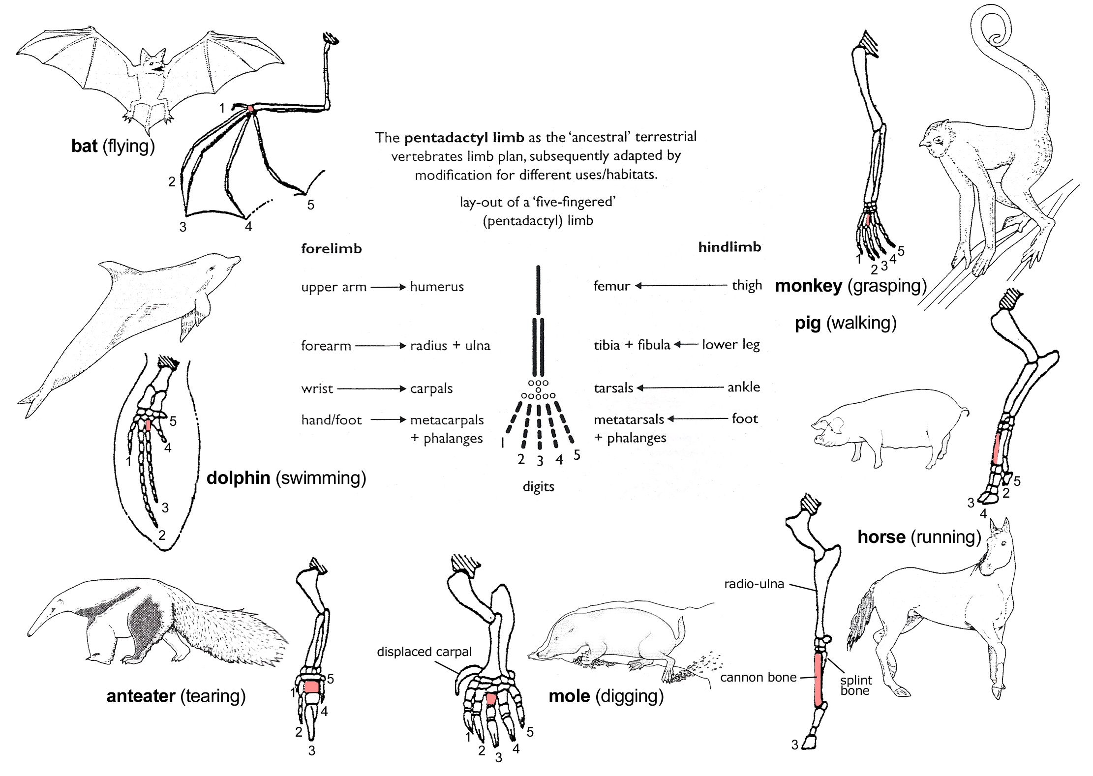
The phalanges (: phalanx ) are digit (anatomy), digital bones in the hands and foot, feet of most vertebrates. In primates, the Thumb, thumbs and Hallux, big toes have two phalanges while the other Digit (anatomy), digits have three phalanges. The phalanges are classed as long bones. Structure The phalanges are the bones that make up the fingers of the hand and the toes of the foot. There are 56 phalanges in the human body, with fourteen on each hand and foot. Three phalanges are present on each finger and toe, with the exception of the thumb and hallux, big toe, which possess only two. The middle and far phalanges of the fifth toes are often fused together (symphalangism). The phalanges of the hand are commonly known as the finger bones. The phalanges of the foot differ from the hand in that they are often shorter and more compressed, especially in the proximal phalanges, those closest to the torso. A phalanx is named according to whether it is Anatomical terms of locatio ... [...More Info...] [...Related Items...] OR: [Wikipedia] [Google] [Baidu] [Amazon] |
Metacarpal
In human anatomy, the metacarpal bones or metacarpus, also known as the "palm bones", are the appendicular bones that form the intermediate part of the hand between the phalanges (fingers) and the carpal bones ( wrist bones), which articulate with the forearm. The metacarpal bones are homologous to the metatarsal bones in the foot. Structure The metacarpals form a transverse arch to which the rigid row of distal carpal bones are fixed. The peripheral metacarpals (those of the thumb and little finger) form the sides of the cup of the palmar gutter and as they are brought together they deepen this concavity. The index metacarpal is the most firmly fixed, while the thumb metacarpal articulates with the trapezium and acts independently from the others. The middle metacarpals are tightly united to the carpus by intrinsic interlocking bone elements at their bases. The ring metacarpal is somewhat more mobile while the fifth metacarpal is semi-independent.Tubiana ''et al'' 1998, p ... [...More Info...] [...Related Items...] OR: [Wikipedia] [Google] [Baidu] [Amazon] |
Osteopenia
Osteopenia, known as "low bone mass" or "low bone density", is a condition in which bone mineral density is low. Because their bones are weaker, people with osteopenia may have a higher risk of fractures, and some people may go on to develop osteoporosis. In 2010, 43 million older adults in the US had osteopenia. Unlike osteoporosis, osteopenia does not usually cause symptoms, and losing bone density in itself does not cause pain. There is no single cause for osteopenia, although there are several risk factors, including modifiable (behavioral, including dietary and use of certain drugs) and non-modifiable (for instance, loss of bone mass with age). For people with risk factors, screening via a DXA scanner may help to detect the development and progression of low bone density. Prevention of low bone density may begin early in life and includes a healthy diet and weight-bearing exercise, as well as avoidance of tobacco and alcohol. The treatment of osteopenia is controversial ... [...More Info...] [...Related Items...] OR: [Wikipedia] [Google] [Baidu] [Amazon] |
Synovial Fluid
Synovial fluid, also called synovia, elp 1/sup> is a viscous, non-Newtonian fluid found in the cavities of synovial joints. With its egg white–like consistency, the principal role of synovial fluid is to reduce friction between the articular cartilage of synovial joints during movement. Synovial fluid is a small component of the transcellular fluid component of extracellular fluid. Structure The inner membrane of synovial joints is called the synovial membrane and secretes synovial fluid into the joints. Synovial fluid is an ultrafiltrate from blood, and contains proteins derived from the blood plasma and proteins that are produced by cells within the joint tissues. The fluid contains hyaluronan secreted by fibroblast-like cells in the synovial membrane, lubricin (proteoglycan 4; PRG4) secreted by the surface chondrocytes of the articular cartilage and interstitial fluid filtered from the blood plasma. This fluid forms a thin layer (roughly 50 μm) at the surface ... [...More Info...] [...Related Items...] OR: [Wikipedia] [Google] [Baidu] [Amazon] |
C Reactive Protein
C-reactive protein (CRP) is an annular (ring-shaped) pentameric protein found in blood plasma, whose circulating concentrations rise in response to inflammation. It is an acute-phase protein of hepatic origin that increases following interleukin-6 secretion by macrophages and T cells. Its physiological role is to bind to lysophosphatidylcholine expressed on the surface of dead or dying cells (and some types of bacteria) in order to activate the complement system via C1q. CRP is synthesized by the liver in response to factors released by macrophages, T cells and fat cells ( adipocytes). It is a member of the pentraxin family of proteins. It is not related to C-peptide (insulin) or protein C (blood coagulation). C-reactive protein was the first pattern recognition receptor (PRR) to be identified. History and etymology Discovered by Tillett and Francis in 1930, it was initially thought that CRP might be a pathogenic secretion since it was elevated in a variety of illnes ... [...More Info...] [...Related Items...] OR: [Wikipedia] [Google] [Baidu] [Amazon] |
Erythrocyte Sedimentation Rate
The erythrocyte sedimentation rate (ESR or sed rate) is the rate at which red blood cells in anticoagulated whole blood descend in a standardized tube over a period of one hour. It is a common hematology test, and is a non-specific measure of inflammation. To perform the test, anticoagulated blood is traditionally placed in an upright tube, known as a Westergren tube, and the distance which the red blood cells fall is measured and reported in millimetres at the end of one hour. Since the introduction of automated analyzers into the clinical laboratory, the ESR test has been automatically performed. The ESR is influenced by the aggregation of red blood cells: blood plasma proteins, mainly fibrinogen, promote the formation of red cell clusters called '' rouleaux'' or larger structures (interconnected rouleaux, irregular clusters). As according to Stokes' law the sedimentation velocity varies like the square of the object's diameter, larger aggregates settle faster. While aggrega ... [...More Info...] [...Related Items...] OR: [Wikipedia] [Google] [Baidu] [Amazon] |
Hemopexin
Hemopexin (or haemopexin; Hpx; Hx), also known as beta-1B-glycoprotein, is a glycoprotein that in humans is encoded by the ''HPX'' gene and belongs to the hemopexin family of proteins. Hemopexin is the plasma protein with the highest binding affinity for heme. Hemoglobin ''itself'' circulating ''alone'' in the blood plasma (called ''free hemoglobin'', as opposed to the hemoglobin situated in and circulating with the red blood cell.) will soon be oxidized into met-hemoglobin which then further disassociates into ''free'' heme along with globin chain. The free heme will then be oxidized into free met-heme and sooner or later the hemopexin will come to bind free met-heme together, forming a complex of met-heme and hemopexin, continuing their journey in the circulation until reaching a receptor, such as LRP1, on hepatocytes or macrophages within the spleen, liver and bone marrow. Hemopexin's arrival and subsequent binding to the free heme not only prevent heme's pro-oxidant and ... [...More Info...] [...Related Items...] OR: [Wikipedia] [Google] [Baidu] [Amazon] |
Heparin
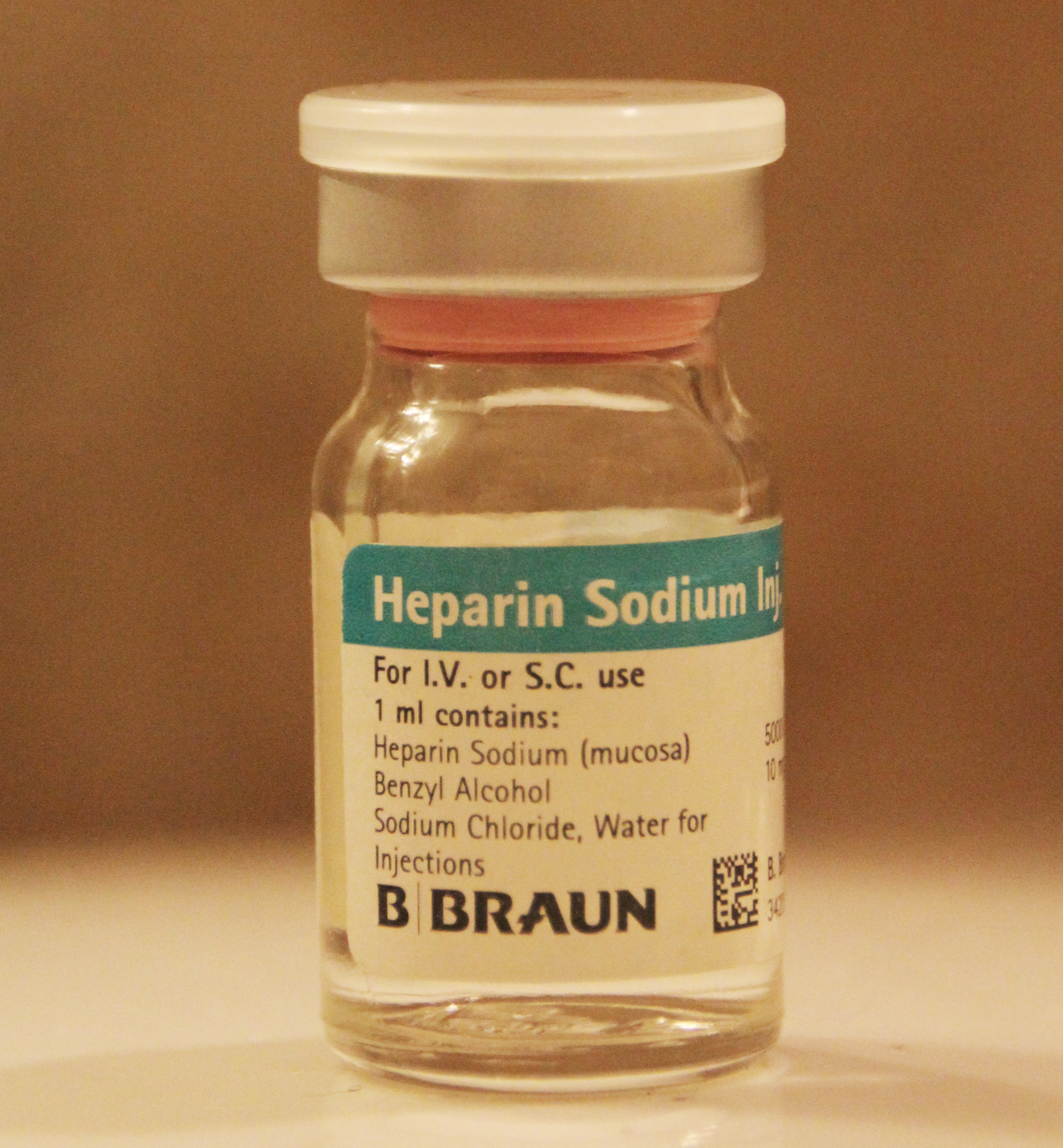
Heparin, also known as unfractionated heparin (UFH), is a medication and naturally occurring glycosaminoglycan. Heparin is a blood anticoagulant that increases the activity of antithrombin. It is used in the treatment of myocardial infarction, heart attacks and unstable angina. It can be given intravenously or by subcutaneous injection, injection under the skin. Its anticoagulant properties make it useful to prevent blood clotting in blood specimen test tubes and kidney dialysis machines. Common side effects include bleeding, pain at the injection site, and thrombocytopenia, low blood platelets. Serious side effects include heparin-induced thrombocytopenia. Greater care is needed in those with poor kidney function. Heparin is contraindicated for suspected cases of Post-vaccination embolic and thrombotic events, vaccine-induced pro-thrombotic immune thrombocytopenia (VIPIT) secondary to SARS-CoV-2 vaccination, as heparin may further increase the risk of bleeding in an anti-PF4 ... [...More Info...] [...Related Items...] OR: [Wikipedia] [Google] [Baidu] [Amazon] |
Articular Cartilage
Hyaline cartilage is the glass-like (hyaline) and translucent cartilage found on many joint surfaces. It is also most commonly found in the ribs, nose, larynx, and trachea. Hyaline cartilage is pearl-gray in color, with a firm consistency and has a considerable amount of collagen. It contains no nerves or blood vessels, and its structure is relatively simple. Structure Hyaline cartilage is the most common kind of cartilage in the human body. It is primarily composed of type II collagen and proteoglycans. Hyaline cartilage is located in the trachea, nose, epiphyseal plate, sternum, and ribs. Hyaline cartilage is covered externally by a fibrous membrane known as the perichondrium. The primary cells of cartilage are chondrocytes, which are in a Matrix (biology), matrix of fibrous tissue, proteoglycans and glycosaminoglycans. As cartilage does not have lymph glands or blood vessels, the movements of solutes, including nutrients, occur via diffusion within the fluid compartments con ... [...More Info...] [...Related Items...] OR: [Wikipedia] [Google] [Baidu] [Amazon] |


