|
Calcium Induced Calcium Release
Ryanodine receptors (RyR) make up a class of high-conductance, intracellular calcium channels present in various forms, such as animal muscles and neurons. There are three major isoforms of the ryanodine receptor, which are found in different tissues and participate in various signaling pathways involving calcium release from intracellular organelles. Structure Ryanodine receptors are multidomain homotetramers which regulate intracellular calcium ion release from the sarcoplasmic and endoplasmic reticula. They are the largest known ion channels, with weights exceeding 2 megadaltons, and their structural complexity enables a wide variety of allosteric regulation mechanisms. RyR1 cryo-EM structure revealed a large cytosolic assembly built on an extended Alpha solenoid, α-solenoid scaffold connecting key regulatory domains to the pore. The RyR1 pore architecture shares the general structure of the six-transmembrane ion channel superfamily. A unique domain inserted between the secon ... [...More Info...] [...Related Items...] OR: [Wikipedia] [Google] [Baidu] |
Ryanodine Receptor 2
Ryanodine receptor 2 (RYR2) is one of a class of ryanodine receptors and a protein found primarily in cardiac muscle. In humans, it is encoded by the ''RYR2'' gene. In the process of cardiac calcium-induced calcium release, RYR2 is the major mediator for sarcoplasmic release of stored calcium ions. Structure The channel is composed of RYR2 homotetramers and FK506-binding proteins found in a 1:4 stoichiometric ratio. Calcium channel function is affected by the specific type of FK506 isomer interacting with the RYR2 protein, due to binding differences and other factors. Function The RYR2 protein functions as the major component of a calcium channel located in the sarcoplasmic reticulum that supplies ions to the cardiac muscle during systole. To enable cardiac muscle contraction, calcium influx through voltage-gated L-type calcium channels in the plasma membrane allows calcium ions to bind to RYR2 located on the sarcoplasmic reticulum. This bindi ... [...More Info...] [...Related Items...] OR: [Wikipedia] [Google] [Baidu] |
Drosophila Melanogaster
''Drosophila melanogaster'' is a species of fly (an insect of the Order (biology), order Diptera) in the family Drosophilidae. The species is often referred to as the fruit fly or lesser fruit fly, or less commonly the "vinegar fly", "pomace fly", or "banana fly". In the wild, ''D. melanogaster'' are attracted to rotting fruit and fermenting beverages, and are often found in orchards, kitchens and pubs. Starting with Charles W. Woodworth's 1901 proposal of the use of this species as a model organism, ''D. melanogaster'' continues to be widely used for biological research in genetics, physiology, microbial pathogenesis, and Life history theory, life history evolution. ''D. melanogaster'' was the first animal to be Fruit flies in space, launched into space in 1947. As of 2017, six Nobel Prizes have been awarded to drosophilists for their work using the insect. ''Drosophila melanogaster'' is typically used in research owing to its rapid life cycle, relatively simple genetics with on ... [...More Info...] [...Related Items...] OR: [Wikipedia] [Google] [Baidu] |
Muscle Contraction
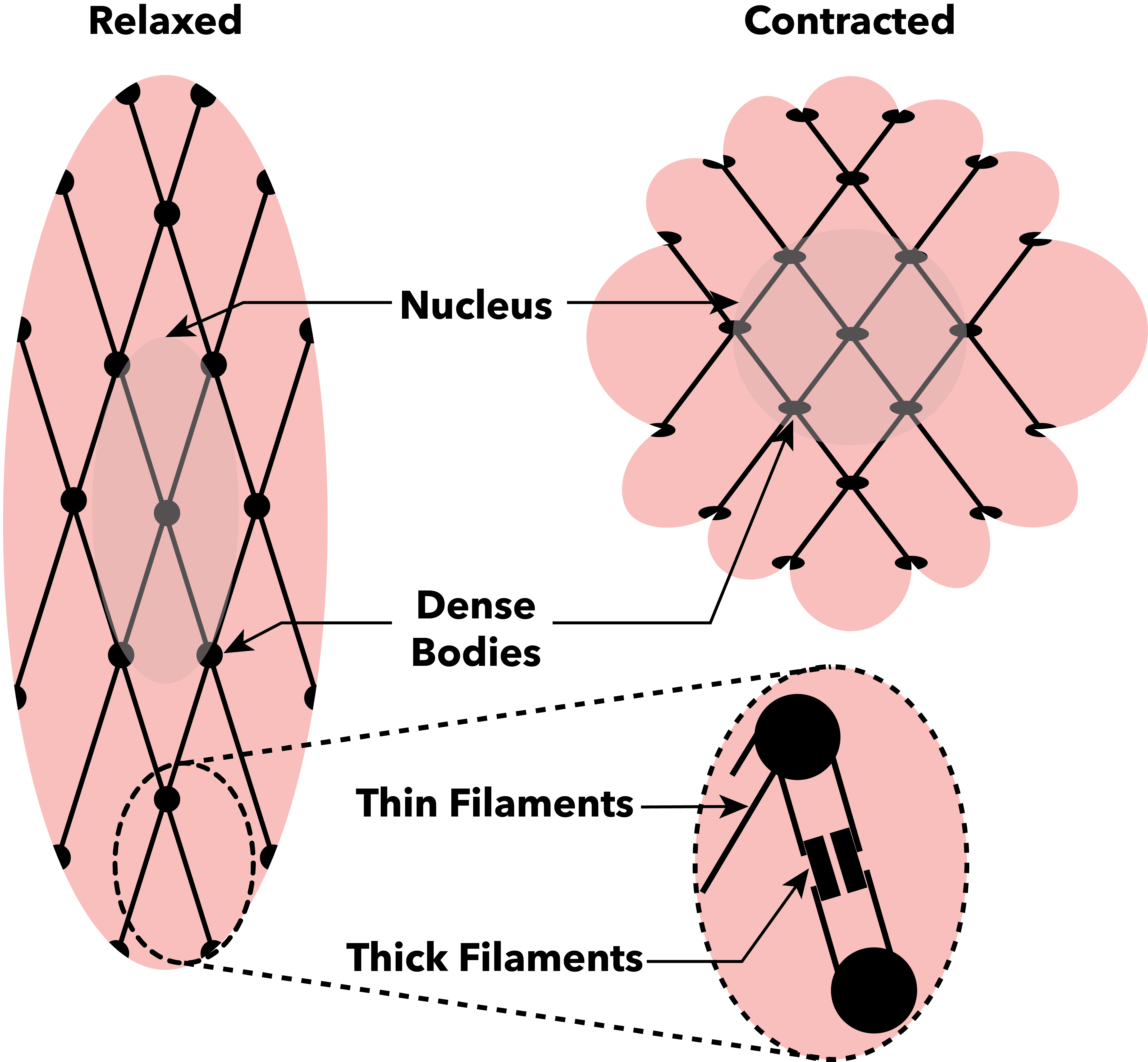
Muscle contraction is the activation of Tension (physics), tension-generating sites within muscle cells. In physiology, muscle contraction does not necessarily mean muscle shortening because muscle tension can be produced without changes in muscle length, such as when holding something heavy in the same position. The termination of muscle contraction is followed by muscle relaxation, which is a return of the muscle fibers to their low tension-generating state. For the contractions to happen, the muscle cells must rely on the change in action of two types of Myofilament, filaments: thin and thick filaments. The major constituent of thin filaments is a chain formed by helical coiling of two strands of actin, and thick filaments dominantly consist of chains of the Motor protein, motor-protein myosin. Together, these two filaments form myofibrils - the basic functional organelles in the skeletal muscle system. In vertebrates, Muscle cell#Muscle contraction in striated muscle, skele ... [...More Info...] [...Related Items...] OR: [Wikipedia] [Google] [Baidu] |
Endoplasmic Reticulum
The endoplasmic reticulum (ER) is a part of a transportation system of the eukaryote, eukaryotic cell, and has many other important functions such as protein folding. The word endoplasmic means "within the cytoplasm", and reticulum is Latin for "little net". It is a type of organelle made up of two subunits – rough endoplasmic reticulum (RER), and smooth endoplasmic reticulum (SER). The endoplasmic reticulum is found in most eukaryotic cells and forms an interconnected network of flattened, membrane-enclosed sacs known as cisternae (in the RER), and tubular structures in the SER. The membranes of the ER are continuous with the outer nuclear membrane. The endoplasmic reticulum is not found in red blood cells, or spermatozoa. There are two types of ER that share many of the same proteins and engage in certain common activities such as the synthesis of certain lipids and cholesterol. Different types of Cell (biology), cells contain different ratios of the two types of ER dependin ... [...More Info...] [...Related Items...] OR: [Wikipedia] [Google] [Baidu] |
Sarcoplasmic Reticulum
The sarcoplasmic reticulum (SR) is a membrane-bound structure found within muscle cells that is similar to the smooth endoplasmic reticulum in other cells. The main function of the SR is to store calcium ions (Ca2+). Calcium ion levels are kept relatively constant, with the concentration of calcium ions within a cell being 10,000 times smaller than the concentration of calcium ions outside the cell. This means that small increases in calcium ions within the cell are easily detected and can bring about important cellular changes (the calcium is said to be a second messenger). Calcium is used to make calcium carbonate (found in chalk) and calcium phosphate, two compounds that the body uses to make teeth and bones. This means that too much calcium within the cells can lead to hardening (calcification) of certain intracellular structures, including the mitochondria, leading to cell death. Therefore, it is vital that calcium ion levels are controlled tightly, and can be released int ... [...More Info...] [...Related Items...] OR: [Wikipedia] [Google] [Baidu] |
Calcium In Biology
Calcium ions (Ca2+) contribute to the physiology and biochemistry of organisms' cells. They play an important role in signal transduction pathways, where they act as a second messenger, in neurotransmitter release from neurons, in contraction of all muscle cell types, and in fertilization. Many enzymes require calcium ions as a cofactor, including several of the coagulation factors. Extracellular calcium is also important for maintaining the potential difference across excitable cell membranes, as well as proper bone formation. Plasma calcium levels in mammals are tightly regulated, electronic-book electronic- with bone acting as the major mineral storage site. Calcium ions, Ca2+, are released from bone into the bloodstream under controlled conditions. Calcium is transported through the bloodstream as dissolved ions or bound to proteins such as serum albumin. Parathyroid hormone secreted by the parathyroid gland regulates the resorption of Ca2+ from bone, reabsorptio ... [...More Info...] [...Related Items...] OR: [Wikipedia] [Google] [Baidu] |
Coupling Of The Muscle Action Potential To Ca++ Release From The Sarcoplasmic Reticulum - The Dihydropyridine Receptor And Ryanodine Receptor
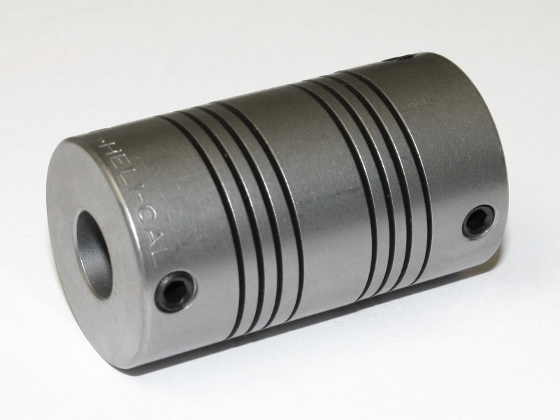
A coupling is a device used to connect two shafts together at their ends for the purpose of transmitting power. The primary purpose of couplings is to join two pieces of rotating equipment while permitting some degree of misalignment or end movement or both. In a more general context, a coupling can also be a mechanical device that serves to connect the ends of adjacent parts or objects. Couplings do not normally allow disconnection of shafts during operation, however there are torque-limiting couplings which can slip or disconnect when some torque limit is exceeded. Selection, installation and maintenance of couplings can lead to reduced maintenance time and maintenance cost. Uses Shaft couplings are used in machinery for several purposes. A primary function is to transfer power from one end to another end (ex: motor transfer power to pump through coupling). Other common uses: * To alter the vibration characteristics of rotating units * To connect the driving and the driven ... [...More Info...] [...Related Items...] OR: [Wikipedia] [Google] [Baidu] |
RYR3
Ryanodine receptor 3 is one of a class of ryanodine receptors and a protein that in humans is encoded by the ''RYR3'' gene. The protein encoded by this gene is both a calcium channel and a receptor for the plant alkaloid ryanodine. RYR3 and RYR1 control the resting calcium ion concentration in skeletal muscle Skeletal muscle (commonly referred to as muscle) is one of the three types of vertebrate muscle tissue, the others being cardiac muscle and smooth muscle. They are part of the somatic nervous system, voluntary muscular system and typically are a .... See also * Ryanodine receptor References Further reading * * * * * * * * * * * * * * * * * * External links * Ion channels EF-hand-containing proteins {{gene-15-stub ... [...More Info...] [...Related Items...] OR: [Wikipedia] [Google] [Baidu] |
Ryanodine Receptor 3
Ryanodine receptor 3 is one of a class of ryanodine receptors and a protein that in humans is encoded by the ''RYR3'' gene. The protein encoded by this gene is both a calcium channel and a receptor for the plant alkaloid ryanodine. RYR3 and RYR1 control the resting calcium ion concentration in skeletal muscle. See also *Ryanodine receptor Ryanodine receptors (RyR) make up a class of high-conductance, intracellular calcium channels present in various forms, such as animal muscles and neurons. There are three major isoforms of the ryanodine receptor, which are found in different tissu ... References Further reading * * * * * * * * * * * * * * * * * * External links * Ion channels EF-hand-containing proteins {{gene-15-stub ... [...More Info...] [...Related Items...] OR: [Wikipedia] [Google] [Baidu] |
RYR2
Ryanodine receptor 2 (RYR2) is one of a class of ryanodine receptors and a protein found primarily in cardiac muscle. In humans, it is encoded by the ''RYR2'' gene. In the process of cardiac calcium-induced calcium release, RYR2 is the major mediator for sarcoplasmic release of stored calcium ions. Structure The channel is composed of RYR2 homotetramers and FK506-binding proteins found in a 1:4 stoichiometric ratio. Calcium channel function is affected by the specific type of FK506 isomer interacting with the RYR2 protein, due to binding differences and other factors. Function The RYR2 protein functions as the major component of a calcium channel located in the sarcoplasmic reticulum that supplies ions to the cardiac muscle during systole. To enable cardiac muscle contraction, calcium influx through voltage-gated L-type calcium channels in the plasma membrane allows calcium ions to bind to RYR2 located on the sarcoplasmic reticulum. This b ... [...More Info...] [...Related Items...] OR: [Wikipedia] [Google] [Baidu] |
Ryanodine Receptor 2
Ryanodine receptor 2 (RYR2) is one of a class of ryanodine receptors and a protein found primarily in cardiac muscle. In humans, it is encoded by the ''RYR2'' gene. In the process of cardiac calcium-induced calcium release, RYR2 is the major mediator for sarcoplasmic release of stored calcium ions. Structure The channel is composed of RYR2 homotetramers and FK506-binding proteins found in a 1:4 stoichiometric ratio. Calcium channel function is affected by the specific type of FK506 isomer interacting with the RYR2 protein, due to binding differences and other factors. Function The RYR2 protein functions as the major component of a calcium channel located in the sarcoplasmic reticulum that supplies ions to the cardiac muscle during systole. To enable cardiac muscle contraction, calcium influx through voltage-gated L-type calcium channels in the plasma membrane allows calcium ions to bind to RYR2 located on the sarcoplasmic reticulum. This bindi ... [...More Info...] [...Related Items...] OR: [Wikipedia] [Google] [Baidu] |
RYR1
Ryanodine receptor 1 (RYR-1) also known as skeletal muscle calcium release channel or skeletal muscle-type ryanodine receptor is one of a class of ryanodine receptors and a protein found primarily in skeletal muscle. In humans, it is encoded by the ''RYR1'' gene. Function RYR1 functions as a calcium release channel in the sarcoplasmic reticulum, as well as a connection between the sarcoplasmic reticulum and the transverse tubule. RYR1 is associated with the dihydropyridine receptor (L-type calcium channels) within the sarcolemma of the T-tubule, which opens in response to depolarization, and thus effectively means that the RYR1 channel opens in response to depolarization of the cell. RYR1 plays a signaling role during embryonic skeletal myogenesis. A correlation exists between RYR1-mediated Ca2+ signaling and the expression of multiple molecules involved in key myogenic signaling pathways. Of these, more than 10 differentially expressed genes belong to the Wnt family which a ... [...More Info...] [...Related Items...] OR: [Wikipedia] [Google] [Baidu] |


