|
Beta-LH
Luteinizing hormone subunit beta also known as lutropin subunit beta or LHβ is a polypeptide that in association with an alpha subunit common to all gonadotropin hormones forms the reproductive signaling molecule luteinizing hormone. In humans it is encoded by the ''LHB'' gene. Gene The luteinizing hormone beta subunit is encoded by a single gene in all mammals. In primates, this gene is located within a cluster that arose through gene duplication, and also includes multiple redundant genes encoding the beta subunit of chorionic gonadotropin as well as several nonfunctional pseudogenes. In humans these are contiguous on chromosome 19q13.3. In equids the beta subunit polypeptides of luteinizing hormone and chorionic gonadotropin are identical in sequence, differing only in their carbohydrate side-chains, and are the product of a single gene. Function This gene is a member of the glycoprotein hormone beta chain family and encodes the beta subunit of luteinizing hormone ( ... [...More Info...] [...Related Items...] OR: [Wikipedia] [Google] [Baidu] |
Human Chorionic Gonadotropin
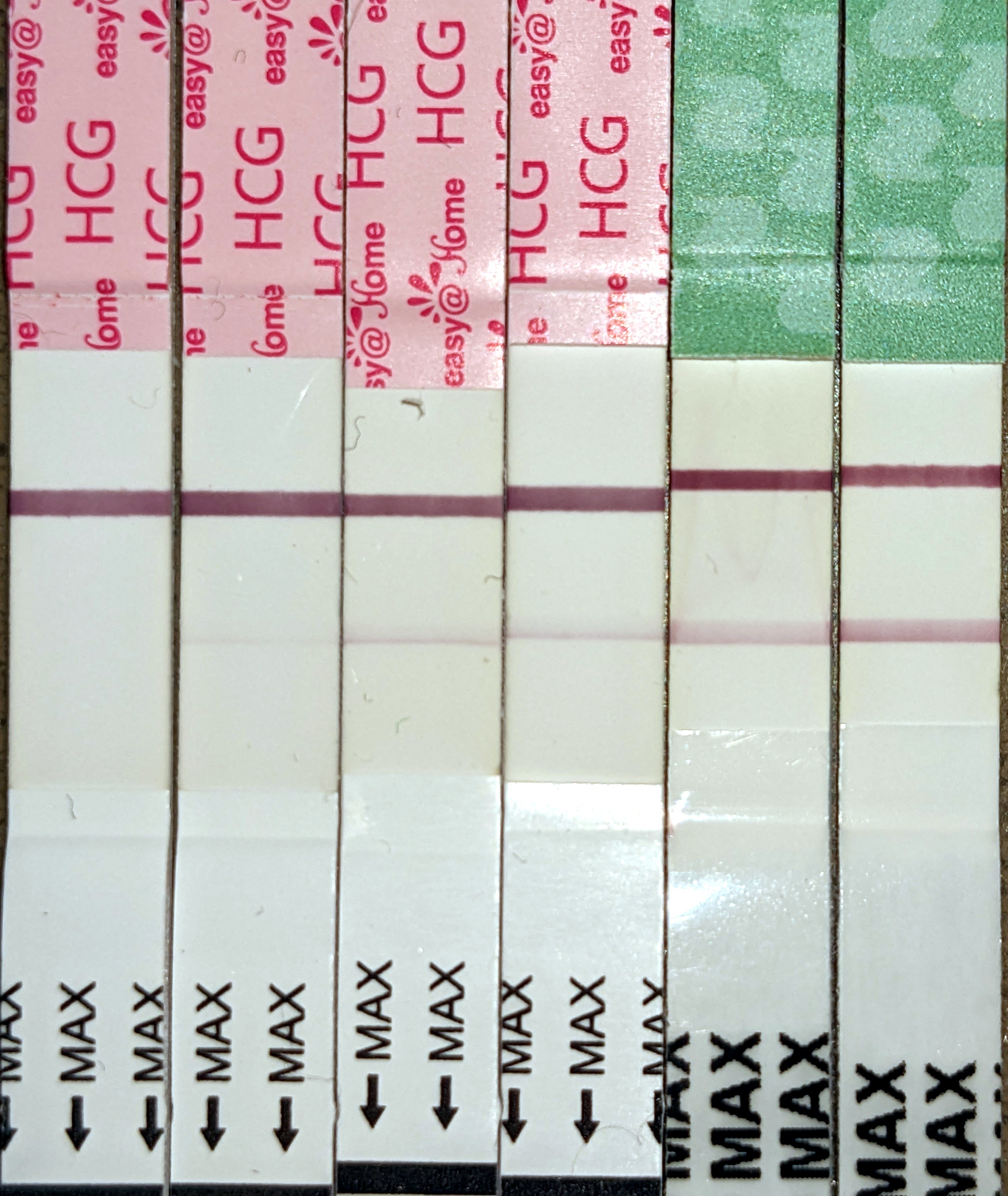
Human chorionic gonadotropin (hCG) is a hormone for the maternal recognition of pregnancy produced by trophoblast cells that are surrounding a growing embryo (syncytiotrophoblast initially), which eventually forms the placenta after implantation. The presence of hCG is detected in some pregnancy tests (HCG pregnancy strip tests). Some cancerous tumors produce this hormone; therefore, elevated levels measured when the patient is not pregnant may lead to a cancer diagnosis and, if high enough, paraneoplastic syndromes, however, it is unknown whether this production is a contributing cause or an effect of carcinogenesis. The pituitary analog of hCG, known as luteinizing hormone (LH), is produced in the pituitary gland of males and females of all ages. Beta-hCG is initially secreted by the syncytiotrophoblast. Structure Human chorionic gonadotropin is a glycoprotein composed of 237 amino acids with a molecular mass of 36.7 kDa, approximately 14.5kDa αhCG and 22.2kDa βhCG ... [...More Info...] [...Related Items...] OR: [Wikipedia] [Google] [Baidu] |
Peptide
Peptides are short chains of amino acids linked by peptide bonds. A polypeptide is a longer, continuous, unbranched peptide chain. Polypeptides that have a molecular mass of 10,000 Da or more are called proteins. Chains of fewer than twenty amino acids are called oligopeptides, and include dipeptides, tripeptides, and tetrapeptides. Peptides fall under the broad chemical classes of biological polymers and oligomers, alongside nucleic acids, oligosaccharides, polysaccharides, and others. Proteins consist of one or more polypeptides arranged in a biologically functional way, often bound to ligands such as coenzymes and cofactors, to another protein or other macromolecule such as DNA or RNA, or to complex macromolecular assemblies. Amino acids that have been incorporated into peptides are termed residues. A water molecule is released during formation of each amide bond.. All peptides except cyclic peptides have an N-terminal (amine group) and C-terminal (carboxyl g ... [...More Info...] [...Related Items...] OR: [Wikipedia] [Google] [Baidu] |
Heterodimer
In biochemistry, a protein dimer is a macromolecular complex or multimer formed by two protein monomers, or single proteins, which are usually non-covalently bound. Many macromolecules, such as proteins or nucleic acids, form dimers. The word ''dimer'' has roots meaning "two parts", '' di-'' + '' -mer''. A protein dimer is a type of protein quaternary structure. A protein homodimer is formed by two identical proteins while a protein heterodimer is formed by two different proteins. Most protein dimers in biochemistry are not connected by covalent bonds. An example of a non-covalent heterodimer is the enzyme reverse transcriptase, which is composed of two different amino acid chains. An exception is dimers that are linked by disulfide bridges such as the homodimeric protein NEMO. Some proteins contain specialized domains to ensure dimerization (dimerization domains) and specificity. The G protein-coupled cannabinoid receptors have the ability to form both homo- and hetero ... [...More Info...] [...Related Items...] OR: [Wikipedia] [Google] [Baidu] |
Ovaries
The ovary () is a gonad in the female reproductive system that produces ova; when released, an ovum travels through the fallopian tube/oviduct into the uterus. There is an ovary on the left and the right side of the body. The ovaries are endocrine glands, secreting various hormones that play a role in the Menstruation (mammal), menstrual cycle and Fecundity, fertility. The ovary progresses through many stages beginning in the prenatal development, prenatal period through menopause. Structure Each ovary is whitish in color and located alongside the lateral wall of the uterus in a region called the ovarian fossa. The ovarian fossa is the region that is bounded by the external iliac artery and in front of the ureter and the internal iliac artery. This area is about 4 cm x 3 cm x 2 cm in size.Daftary, Shirish; Chakravarti, Sudip (2011). Manual of Obstetrics, 3rd Edition. Elsevier. pp. 1-16. . The ovaries are surrounded by a capsule, and have an outer cortex and an in ... [...More Info...] [...Related Items...] OR: [Wikipedia] [Google] [Baidu] |
Testes
A testicle or testis ( testes) is the gonad in all male bilaterians, including humans, and is homologous to the ovary in females. Its primary functions are the production of sperm and the secretion of androgens, primarily testosterone. The release of testosterone is regulated by luteinizing hormone (LH) from the anterior pituitary gland. Sperm production is controlled by follicle-stimulating hormone (FSH) from the anterior pituitary gland and by testosterone produced within the gonads. Structure Appearance Males have two testicles of similar size contained within the scrotum, which is an extension of the abdominal wall. Scrotal asymmetry, in which one testicle extends farther down into the scrotum than the other, is common. This is because of the differences in the vasculature's anatomy. For 85% of men, the right testis hangs lower than the left one. Measurement and volume The volume of the testicle can be estimated by palpating it and comparing it to ellipsoids (an ... [...More Info...] [...Related Items...] OR: [Wikipedia] [Google] [Baidu] |
Ovulation
Ovulation is an important part of the menstrual cycle in female vertebrates where the egg cells are released from the ovaries as part of the ovarian cycle. In female humans ovulation typically occurs near the midpoint in the menstrual cycle and after the follicular phase. Ovulation is stimulated by an increase in luteinizing hormone (LH). The ovarian follicles rupture and release the secondary oocyte ovarian cells. After ovulation, during the luteal phase, the egg will be available to be fertilized by sperm. If it is not, it will break down in less than a day. Meanwhile, the uterine lining ( endometrium) continues to thicken to be able to receive a fertilized egg. If no conception occurs, the uterine lining will eventually break down and be shed from the body via the vagina during menstruation. Some people choose to track ovulation in order to improve or aid becoming pregnant by timing intercourse with their ovulation. The signs of ovulation may include cervical muc ... [...More Info...] [...Related Items...] OR: [Wikipedia] [Google] [Baidu] |
Spermatogenesis
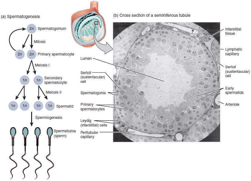
Spermatogenesis is the process by which haploid spermatozoa develop from germ cells in the seminiferous tubules of the testicle. This process starts with the Mitosis, mitotic division of the stem cells located close to the basement membrane of the tubules. These cells are called Spermatogonial Stem Cells, spermatogonial stem cells. The mitotic division of these produces two types of cells. Type A cells replenish the stem cells, and type B cells differentiate into primary spermatocytes. The primary spermatocyte divides meiotically (Meiosis I) into two secondary spermatocytes; each secondary spermatocyte divides into two equal haploid spermatids by Meiosis II. The spermatids are transformed into spermatozoa (sperm) by the process of spermiogenesis. These develop into mature spermatozoa, also known as sperm, sperm cells. Thus, the primary spermatocyte gives rise to two cells, the secondary spermatocytes, and the two secondary spermatocytes by their subdivision produce four spermatoz ... [...More Info...] [...Related Items...] OR: [Wikipedia] [Google] [Baidu] |
Pituitary Gland
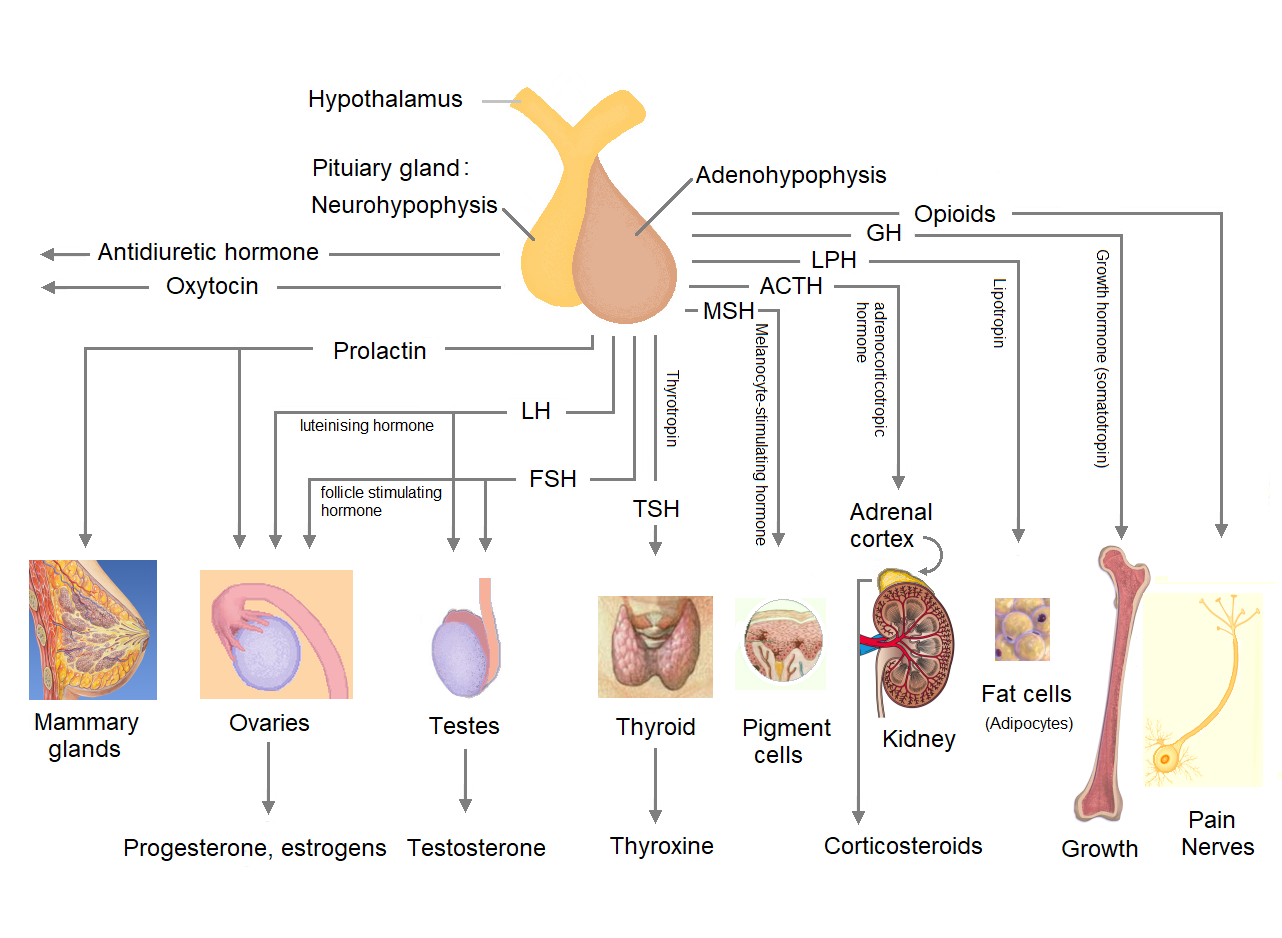
The pituitary gland or hypophysis is an endocrine gland in vertebrates. In humans, the pituitary gland is located at the base of the human brain, brain, protruding off the bottom of the hypothalamus. The pituitary gland and the hypothalamus control much of the body's endocrine system. It is seated in part of the sella turcica a fossa (anatomy), depression in the sphenoid bone, known as the hypophyseal fossa. The human pituitary gland is ovoid, oval shaped, about 1 cm in diameter, in weight on average, and about the size of a kidney bean. Digital version. There are two main lobes of the pituitary, an anterior pituitary, anterior lobe, and a posterior pituitary, posterior lobe joined and separated by a small intermediate lobe. The anterior lobe (adenohypophysis) is the glandular part that produces and secretes several hormones. The posterior lobe (neurohypophysis) secretes neurohypophysial hormones produced in the hypothalamus. Both lobes have different origins and they are both co ... [...More Info...] [...Related Items...] OR: [Wikipedia] [Google] [Baidu] |
Chorionic Gonadotropin Alpha
The chorion is the outermost fetal membrane around the embryo in mammals, birds and reptiles (amniotes). It is also present around the embryo of other animals, like insects and molluscs. Structure In humans and other therian mammals, the chorion is one of the fetal membranes that exist during pregnancy between the developing fetus and mother. The chorion and the amnion together form the amniotic sac. In humans it is formed by extraembryonic mesoderm and the two layers of trophoblast that surround the embryo and other membranes; the chorionic villi emerge from the chorion, invade the endometrium, and allow the transfer of nutrients from maternal blood to fetal blood. Layers The chorion consists of two layers: an outer formed by the trophoblast, and an inner formed by the extra-embryonic mesoderm. The trophoblast is made up of an internal layer of cubical or prismatic cells, the cytotrophoblast or layer of Langhans, and an external multinucleated layer, the syncytiotrophob ... [...More Info...] [...Related Items...] OR: [Wikipedia] [Google] [Baidu] |
Glycoprotein
Glycoproteins are proteins which contain oligosaccharide (sugar) chains covalently attached to amino acid side-chains. The carbohydrate is attached to the protein in a cotranslational or posttranslational modification. This process is known as glycosylation. Secreted extracellular proteins are often glycosylated. In proteins that have segments extending extracellularly, the extracellular segments are also often glycosylated. Glycoproteins are also often important integral membrane proteins, where they play a role in cell–cell interactions. It is important to distinguish endoplasmic reticulum-based glycosylation of the secretory system from reversible cytosolic-nuclear glycosylation. Glycoproteins of the cytosol and nucleus can be modified through the reversible addition of a single GlcNAc residue that is considered reciprocal to phosphorylation and the functions of these are likely to be an additional regulatory mechanism that controls phosphorylation-based signalling. In ... [...More Info...] [...Related Items...] OR: [Wikipedia] [Google] [Baidu] |
Equine Chorionic Gonadotropin
Equinae is a subfamily of the family Equidae, known from the Hemingfordian stage of the Early Miocene (16 million years ago) onwards. They originated in North America, before dispersing to every continent except Australia and Antarctica. They are thought to be a monophyletic grouping. Members of the subfamily are referred to as equines; the only extant equines are the horse The horse (''Equus ferus caballus'') is a domesticated, one-toed, hoofed mammal. It belongs to the taxonomic family Equidae and is one of two extant subspecies of ''Equus ferus''. The horse has evolved over the past 45 to 55 mi ...s, asses, and zebras of the genus ''Equus'', with two other genera '' Haringtonhippus'' and '' Hippidion'' becoming extinct at the beginning of the Holocene, around 11–12,000 years ago. The subfamily contains two tribes, the Equini and the Hipparionini, as well as two unplaced genera, '' Merychippus'' and '' Scaphohippus''. Members of the family ancestr ... [...More Info...] [...Related Items...] OR: [Wikipedia] [Google] [Baidu] |


