|
Back (horse)
The back is the area of Equine anatomy, horse anatomy where the saddle goes, and in popular usage extends to include the loin or lumbar region behind the thoracic vertebrae that also is crucial to a horse's weight-carrying ability. These two sections of the vertebral column beginning at the withers, the start of the thoracic vertebrae, and extend to the last Lumbar vertebrae, lumbar vertebra. Because horses are equestrianism, ridden by humans, the strength and structure of the horse's back is critical to the animal's usefulness. The thoracic vertebrae are the true "back" vertebral structures of the skeleton, providing the underlying support of the saddle, and the lumbar vertebrae of the loin provide the ''coupling'' that joins the back to the horse anatomy, hindquarters. Integral to the back structure is the rib cage, which also provides support to the horse and rider. A complex design of bone, muscle, tendons and ligaments all work together to allow a horse to support the weight ... [...More Info...] [...Related Items...] OR: [Wikipedia] [Google] [Baidu] [Amazon] |
Ambling
An ambling gait or amble is any of several four-beat intermediate horse gaits, all of which are faster than a walk but usually slower than a canter and always slower than a gallop. Horses that amble are sometimes referred to as " gaited", particularly in the United States. Ambling gaits are smoother for a rider than either the two-beat trot or pace and most can be sustained for relatively long periods, making them particularly desirable for trail riding and other tasks where a rider must spend long periods in the saddle. Historically, horses able to amble were highly desired for riding long distances on poor roads. Once roads improved and carriage travel became popular, their use declined in Europe but continued in popularity in the Americas, particularly in areas where plantation agriculture was practiced and the inspection of fields and crops necessitated long daily rides. The ability to perform an ambling gait is usually an inherited trait. In 2012, a DNA study found th ... [...More Info...] [...Related Items...] OR: [Wikipedia] [Google] [Baidu] [Amazon] |
Poll (livestock)
The poll is a name of the part of an animal's head, alternatively referencing a point immediately behind or right between the ears. This area of the anatomy is of particular significance for the horse The horse (''Equus ferus caballus'') is a domesticated, one-toed, hoofed mammal. It belongs to the taxonomic family Equidae and is one of two extant subspecies of ''Equus ferus''. The horse has evolved over the past 45 to 55 mi .... Specifically, the "poll" refers to the occipital protrusion at the back of the skull. However, in common usage, many horsemen refer to the poll joint, between the atlas (C1) and skull as the poll. The area at the joint has a slight depression, and is a sensitive location. Thus, because the crownpiece of a bridle passes over the poll joint, a rider can indirectly exert pressure on the horse's poll by means of the reins, bit, and bridle. See also * Polled livestock, for information on naturally or mechanically dehorned animals ... [...More Info...] [...Related Items...] OR: [Wikipedia] [Google] [Baidu] [Amazon] |
Nuchal Ligament
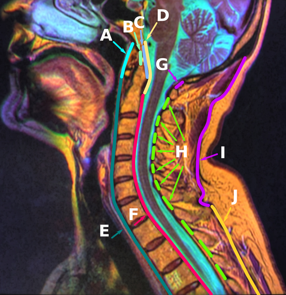
The nuchal ligament is a ligament at the back of the neck that is continuous with the supraspinous ligament. Structure The nuchal ligament extends from the external occipital protuberance on the skull and median nuchal line to the spinous process of the seventh cervical vertebra in the lower part of the neck. From the anterior border of the nuchal ligament, a fibrous lamina is given off. This is attached to the posterior tubercle of the atlas, and to the spinous processes of the cervical vertebrae, and forms a septum between the muscles on either side of the neck. The trapezius and splenius capitis muscle attach to the nuchal ligament. Function It is a tendon-like structure that has developed independently in humans and other animals well adapted for running. In some four-legged animals, particularly ungulates and canids, the nuchal ligament serves to sustain the weight of the head. Clinical significance In Chiari malformation treatment, decompression and duraplas ... [...More Info...] [...Related Items...] OR: [Wikipedia] [Google] [Baidu] [Amazon] |
Supraspinous Ligament
The supraspinous ligament (also known as the supraspinal ligament) is a ligament extending across the tips of the spinous processes of the vertebra of the vertebral column. Anatomy The supraspinous ligament connects the tips of the spinous processes from the seventh cervical vertebra to the sacrum. Superior to the 7th cervical vertebra, the supraspinous ligament is continuous with the nuchal ligament. It is thicker and broader in the lumbar region than in the thoracic region, and intimately blended with the neighboring fascia in both these regions. Inferior to L4, the supraspinous ligament becomes indistinct, lost amid the prominent lumbar fascia. Between the spinous processes, the supraspinous ligament is continuous with the interspinous ligaments. Structure The most superficial fibers of this ligament extend across 3-4 vertebrae, deeper fibres extend across 2-3 vertebrae, while the deepest connect the spinous processes of adjacent vertebrae. Function The supraspinous ligam ... [...More Info...] [...Related Items...] OR: [Wikipedia] [Google] [Baidu] [Amazon] |
Pelvis
The pelvis (: pelves or pelvises) is the lower part of an Anatomy, anatomical Trunk (anatomy), trunk, between the human abdomen, abdomen and the thighs (sometimes also called pelvic region), together with its embedded skeleton (sometimes also called bony pelvis or pelvic skeleton). The pelvic region of the trunk includes the bony pelvis, the pelvic cavity (the space enclosed by the bony pelvis), the pelvic floor, below the pelvic cavity, and the perineum, below the pelvic floor. The pelvic skeleton is formed in the area of the back, by the sacrum and the coccyx and anteriorly and to the left and right sides, by a pair of hip bones. The two hip bones connect the spine with the lower limbs. They are attached to the sacrum posteriorly, connected to each other anteriorly, and joined with the two femurs at the hip joints. The gap enclosed by the bony pelvis, called the pelvic cavity, is the section of the body underneath the abdomen and mainly consists of the reproductive organs and ... [...More Info...] [...Related Items...] OR: [Wikipedia] [Google] [Baidu] [Amazon] |
Abdominal External Oblique Muscle
The abdominal external oblique muscle (also external oblique muscle or exterior oblique) is the largest and outermost of the three flat Abdomen#Muscles, abdominal muscles of the lateral anterior abdomen. Structure The external oblique is situated on the lateral and anterior parts of the abdomen. It is broad, thin, and irregularly quadrilateral, its muscular portion occupying the side, its aponeurosis the anterior wall of the abdomen. In most humans, the oblique is not visible, due to subcutaneous adipose, fat deposits and the small size of the muscle. It arises from eight fleshy digitations, each from the external surfaces and inferior borders of the fifth to twelfth ribs (lower eight ribs). These digitations are arranged in an oblique line which runs inferiorly and anteriorly, with the upper digitations being attached close to the cartilages of the corresponding ribs, the lowest to the apex of the cartilage of the last rib, the intermediate ones to the ribs at some distance fr ... [...More Info...] [...Related Items...] OR: [Wikipedia] [Google] [Baidu] [Amazon] |
Intercostal Muscle
The intercostal muscles comprise many different groups of muscle Muscle is a soft tissue, one of the four basic types of animal tissue. There are three types of muscle tissue in vertebrates: skeletal muscle, cardiac muscle, and smooth muscle. Muscle tissue gives skeletal muscles the ability to muscle contra ...s that run between the ribs, and help form and move the chest wall. The intercostal muscles are mainly involved in the mechanical aspect of breathing by helping expand and shrink the size of the chest cavity. Structure There are three principal layers: # External intercostal muscles also known as intercostalis externus aid in quiet and forced inhalation. They originate on ribs 1–11 and have their insertion on ribs 2–12. The external intercostals are responsible for the elevation of the ribs and bending them more open, thus expanding the transverse dimensions of the thoracic cavity. The muscle fibers are directed downwards, forwards and medially in the anteri ... [...More Info...] [...Related Items...] OR: [Wikipedia] [Google] [Baidu] [Amazon] |
Longissimus Dorsi
The longissimus () is the muscle lateral to the semispinalis muscles. It is the longest subdivision of the erector spinae muscles that extends forward into the transverse processes of the posterior cervical vertebrae. Structure Longissimus thoracis et lumborum The longissimus thoracis et lumborum is the intermediate and largest of the continuations of the erector spinae. In the lumbar region (longissimus lumborum), where it is as yet blended with the iliocostalis, some of its fibers are attached to the whole length of the posterior surfaces of the transverse processes and the accessory processes of the lumbar vertebrae, and to the anterior layer of the lumbodorsal fascia. In the thoracic region (longissimus thoracis), it is inserted, by rounded tendons, into the tips of the transverse processes of all the thoracic vertebrae, and by fleshy processes into the lower nine or ten ribs between their tubercles and angles. Longissimus cervicis The longissimus cervicis (transver ... [...More Info...] [...Related Items...] OR: [Wikipedia] [Google] [Baidu] [Amazon] |
Trapezius
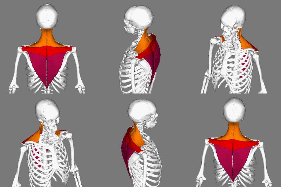
The trapezius is a large paired trapezoid-shaped surface muscle that extends longitudinally from the occipital bone to the lower thoracic vertebrae of the human spine, spine and laterally to the spine of the scapula. It moves the scapula and supports the arm. The trapezius has three functional parts: * an upper (descending) part which supports the weight of the arm; * a middle region (transverse), which retracts the scapula; and * a lower (ascending) part which medially rotates and depresses the scapula. Name and history The trapezius muscle resembles a trapezoid, trapezium, also known as a trapezoid, or diamond-shaped quadrilateral. The word "spinotrapezius" refers to the human trapezius, although it is not commonly used in modern texts. In other mammals, it refers to a portion of the analogous muscle. Structure The ''superior'' or ''upper'' (or descending) fibers of the trapezius originate from the spinous process of C7, the external occipital protuberance, the me ... [...More Info...] [...Related Items...] OR: [Wikipedia] [Google] [Baidu] [Amazon] |


