|
B-scan
Medical ultrasound includes diagnostic techniques (mainly imaging) using ultrasound, as well as therapeutic applications of ultrasound. In diagnosis, it is used to create an image of internal body structures such as tendons, muscles, joints, blood vessels, and internal organs, to measure some characteristics (e.g., distances and velocities) or to generate an informative audible sound. The usage of ultrasound to produce visual images for medicine is called medical ultrasonography or simply sonography, or echography. The practice of examining pregnant women using ultrasound is called obstetric ultrasonography, and was an early development of clinical ultrasonography. The machine used is called an ultrasound machine, a sonograph or an echograph. The visual image formed using this technique is called an ultrasonogram, a sonogram or an echogram. Ultrasound is composed of sound waves with frequencies greater than 20,000 Hz, which is the approximate upper threshold of human ... [...More Info...] [...Related Items...] OR: [Wikipedia] [Google] [Baidu] |
B-flow Ultrasonography Of Venous Reflux
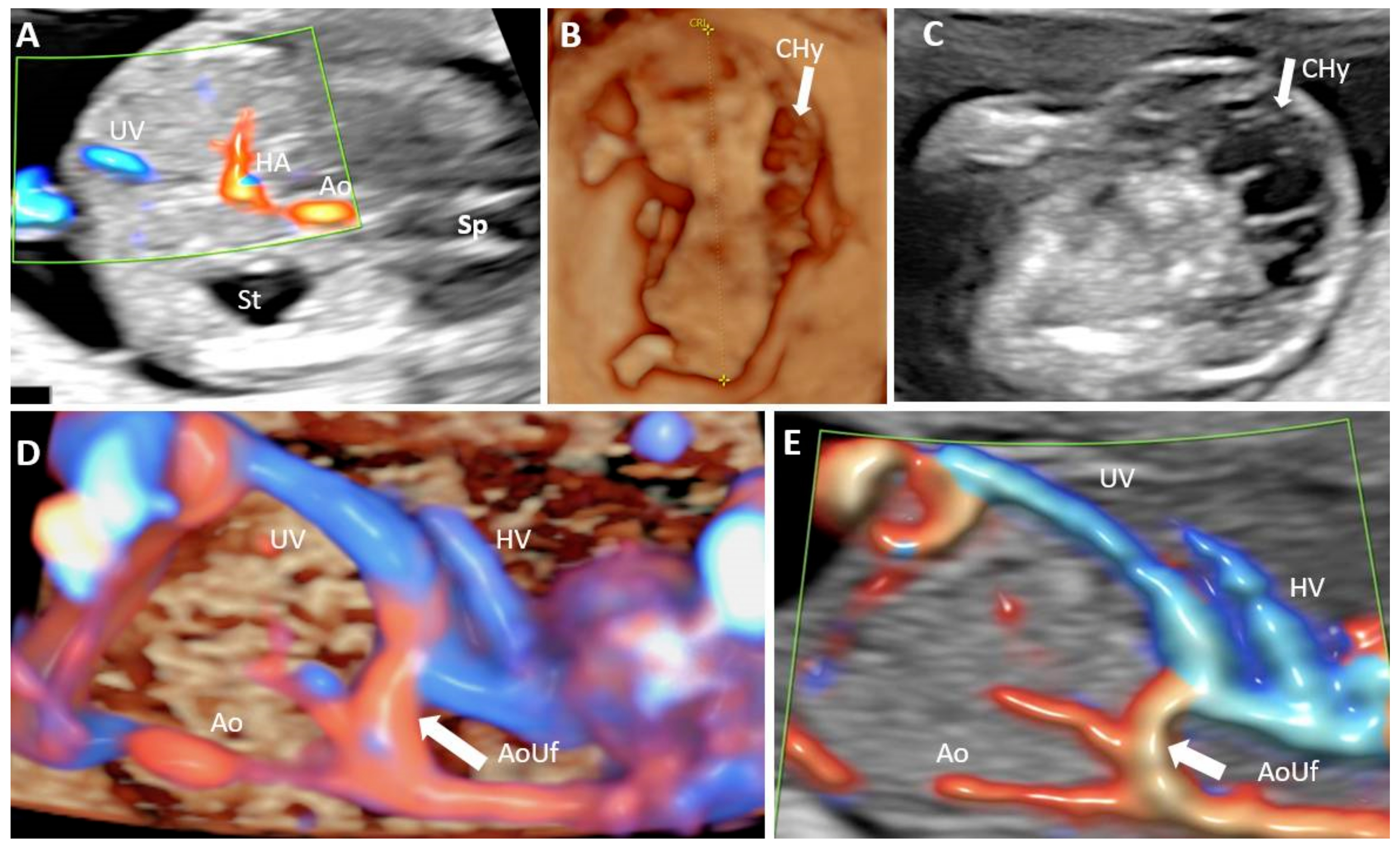
Medical ultrasound includes Medical diagnosis, diagnostic techniques (mainly medical imaging, imaging) using ultrasound, as well as therapeutic ultrasound, therapeutic applications of ultrasound. In diagnosis, it is used to create an image of internal body structures such as tendons, muscles, joints, blood vessels, and internal organs, to measure some characteristics (e.g., distances and velocities) or to generate an informative audible sound. The usage of ultrasound to produce visual images for medicine is called medical ultrasonography or simply sonography, or echography. The practice of examining pregnant women using ultrasound is called obstetric ultrasonography, and was an early development of clinical ultrasonography. The machine used is called an ultrasound machine, a sonograph or an echograph. The visual image formed using this technique is called an ultrasonogram, a sonogram or an echogram. Ultrasound is composed of sound waves with frequency, frequencies greater than ... [...More Info...] [...Related Items...] OR: [Wikipedia] [Google] [Baidu] |
Medical Imaging
Medical imaging is the technique and process of imaging the interior of a body for clinical analysis and medical intervention, as well as visual representation of the function of some organs or tissues (physiology). Medical imaging seeks to reveal internal structures hidden by the skin and bones, as well as to diagnose and treat disease. Medical imaging also establishes a database of normal anatomy and physiology to make it possible to identify abnormalities. Although imaging of removed organ (anatomy), organs and Tissue (biology), tissues can be performed for medical reasons, such procedures are usually considered part of pathology instead of medical imaging. Measurement and recording techniques that are not primarily designed to produce images, such as electroencephalography (EEG), magnetoencephalography (MEG), electrocardiography (ECG), and others, represent other technologies that produce data susceptible to representation as a parameter graph versus time or maps that contain ... [...More Info...] [...Related Items...] OR: [Wikipedia] [Google] [Baidu] |
Echo Sounding
Echo sounding or depth sounding is the use of sonar for ranging, normally to determine the depth (coordinate), depth of water (bathymetry). It involves transmitting acoustic waves into water and recording the time interval between emission and return of a pulse; the resulting time of flight, along with knowledge of the speed of sound in water, allows determining the distance between sonar and target. This information is then typically used for navigation purposes or in order to obtain depths for Nautical chart, charting purposes. Echo sounding can also be used for ranging to other targets, such as fish schools. Hydroacoustic assessments have traditionally employed mobile surveys from boats to evaluate fish biomass and spatial distributions. Conversely, fixed-location techniques use stationary transducers to monitor passing fish. The word ''Depth sounding, sounding'' is used for all types of depth measurements, including those that don't use sound, and is unrelated in origin to ... [...More Info...] [...Related Items...] OR: [Wikipedia] [Google] [Baidu] |
Ultrasonic Transducer
Ultrasonic transducers and ultrasonic sensors are devices that generate or sense ultrasound energy. They can be divided into three broad categories: transmitters, receivers and transceivers. Transmitters convert signal (electrical engineering), electrical signals into ultrasound, receivers convert ultrasound into electrical signals, and transceivers can both transmit and receive ultrasound. Applications and performance Ultrasound can be used for measuring wind speed and direction (anemometer), tank or channel fluid level, and speed through air or water. For measuring speed or direction, a device uses multiple detectors and calculates the speed from the relative distances to particulates in the air or water. To measure tank or channel liquid level, and also sea level (tide gauge), the sensor measures the distance (ranging) to the surface of the fluid. Further applications include: humidifiers, sonar, medical ultrasonography, burglar alarms and non-destructive testing. Systems typ ... [...More Info...] [...Related Items...] OR: [Wikipedia] [Google] [Baidu] |
Bone
A bone is a rigid organ that constitutes part of the skeleton in most vertebrate animals. Bones protect the various other organs of the body, produce red and white blood cells, store minerals, provide structure and support for the body, and enable mobility. Bones come in a variety of shapes and sizes and have complex internal and external structures. They are lightweight yet strong and hard and serve multiple functions. Bone tissue (osseous tissue), which is also called bone in the uncountable sense of that word, is hard tissue, a type of specialised connective tissue. It has a honeycomb-like matrix internally, which helps to give the bone rigidity. Bone tissue is made up of different types of bone cells. Osteoblasts and osteocytes are involved in the formation and mineralisation of bone; osteoclasts are involved in the resorption of bone tissue. Modified (flattened) osteoblasts become the lining cells that form a protective layer on the bone surface. The mine ... [...More Info...] [...Related Items...] OR: [Wikipedia] [Google] [Baidu] |
Portable Ultrasound
Portable ultrasound is a modality of medical ultrasonography that utilizes small and light devices, compared to the console-style ultrasound machines that preceded them. In most cases these mobile ultrasound systems could be carried by hand and in some cases even operated for a time on battery power alone. The first portable ultrasound machines arrived in the early 1980s but battery powered systems that could be easily carried did not arrive until the late 1990s. History The ADR 2130, designed by Marty Wilcox was the first portable ultrasound unit commercially available in the United States, being released in 1975. This unit weighed about 25 lbs, had 3 linear probes to choose from and used an oscilloscope for a display instead of a TV monitor. ADR was later purchased by ATL which later spun off its portable technology into a company that became Sonosite. ATL was later purchased by Philips while Sonosite came out with its first battery powered portable offering, the Sonosite 180 ... [...More Info...] [...Related Items...] OR: [Wikipedia] [Google] [Baidu] |
Therapeutic Ultrasound
Therapeutic ultrasound refers generally to the use of ultrasound for the treatment of a medical condition or for therapeutic benefit. Physiotherapeutic ultrasound was introduced into clinical practice in the 1950s, with lithotripsy introduced in the 1980s. Other uses of ultrasound for therapeutic benefit are at various stages in transitioning from research to clinical use and include: Focused ultrasound, high-intensity focused ultrasound (HIFU), targeted ultrasound drug delivery, trans-dermal ultrasound drug delivery, ultrasound hemostasis, cancer therapy, and ultrasound assisted thrombolysis Ultrasound used for therapeutic benefit often use focused ultrasound waves, however, unfocused ultrasound waves may also be used. In the above applications, the ultrasound passes through human tissue where it is the main source of the observed biological effect (the oscillation of abrasive dental tools at ultrasonic frequencies therefore do not belong to this class). The ultrasound within ti ... [...More Info...] [...Related Items...] OR: [Wikipedia] [Google] [Baidu] |
Red Blood Cell
Red blood cells (RBCs), referred to as erythrocytes (, with -''cyte'' translated as 'cell' in modern usage) in academia and medical publishing, also known as red cells, erythroid cells, and rarely haematids, are the most common type of blood cell and the vertebrate's principal means of delivering oxygen () to the body tissue (biology), tissues—via blood flow through the circulatory system. Erythrocytes take up oxygen in the lungs, or in fish the gills, and release it into tissues while squeezing through the body's capillary, capillaries. The cytoplasm of a red blood cell is rich in hemoglobin (Hb), an iron-containing biomolecule that can bind oxygen and is responsible for the red color of the cells and the blood. Each human red blood cell contains approximately 270 million hemoglobin molecules. The cell membrane is composed of proteins and lipids, and this structure provides properties essential for physiological Cell (biology), cell function such as erythrocyte deformabil ... [...More Info...] [...Related Items...] OR: [Wikipedia] [Google] [Baidu] |
Fourier Analysis
In mathematics, Fourier analysis () is the study of the way general functions may be represented or approximated by sums of simpler trigonometric functions. Fourier analysis grew from the study of Fourier series, and is named after Joseph Fourier, who showed that representing a function as a sum of trigonometric functions greatly simplifies the study of heat transfer. The subject of Fourier analysis encompasses a vast spectrum of mathematics. In the sciences and engineering, the process of decomposing a function into oscillatory components is often called Fourier analysis, while the operation of rebuilding the function from these pieces is known as Fourier synthesis. For example, determining what component frequencies are present in a musical note would involve computing the Fourier transform of a sampled musical note. One could then re-synthesize the same sound by including the frequency components as revealed in the Fourier analysis. In mathematics, the term ''Fourier an ... [...More Info...] [...Related Items...] OR: [Wikipedia] [Google] [Baidu] |
Contrast Agents
A contrast agent (or contrast medium) is a substance used to increase the contrast of structures or fluids within the body in medical imaging. Contrast agents absorb or alter external electromagnetism or ultrasound, which is different from radiopharmaceuticals, which emit radiation themselves. In X-ray imaging, contrast agents enhance the radiodensity in a target tissue or structure. In magnetic resonance imaging (MRI), contrast agents shorten (or in some instances increase) the relaxation times of nuclei within body tissues in order to alter the contrast in the image. Contrast agents are commonly used to improve the visibility of blood vessels and the gastrointestinal tract. The types of contrast agent are classified according to their intended imaging modalities. Radiocontrast media For radiography, which is based on X-rays, iodine and barium are the most common types of contrast agent. Various sorts of iodinated contrast agents exist, with variations occurring between the ... [...More Info...] [...Related Items...] OR: [Wikipedia] [Google] [Baidu] |
Doppler Effect
The Doppler effect (also Doppler shift) is the change in the frequency of a wave in relation to an observer who is moving relative to the source of the wave. The ''Doppler effect'' is named after the physicist Christian Doppler, who described the phenomenon in 1842. A common example of Doppler shift is the change of pitch heard when a vehicle sounding a horn approaches and recedes from an observer. Compared to the emitted frequency, the received frequency is higher during the approach, identical at the instant of passing by, and lower during the recession. When the source of the sound wave is moving towards the observer, each successive cycle of the wave is emitted from a position closer to the observer than the previous cycle. Hence, from the observer's perspective, the time between cycles is reduced, meaning the frequency is increased. Conversely, if the source of the sound wave is moving away from the observer, each cycle of the wave is emitted from a position farther from ... [...More Info...] [...Related Items...] OR: [Wikipedia] [Google] [Baidu] |
Doppler Ultrasonography
Doppler ultrasonography is medical ultrasonography that employs the Doppler effect to perform imaging of the movement of tissues and body fluids (usually blood), and their relative velocity to the probe. By calculating the frequency shift of a particular sample volume, for example, flow in an artery or a jet of blood flow over a heart valve, its speed and direction can be determined and visualized. Duplex ultrasonography sometimes refers to Doppler ultrasonography or spectral Doppler ultrasonography. Doppler ultrasonography consists of two components: brightness mode (B-mode) showing anatomy of the organs, and Doppler mode (showing blood flow) superimposed on the B-mode. Meanwhile, spectral Doppler ultrasonography consists of three components: B-mode, Doppler mode, and spectral waveform displayed at the lower half of the image. Therefore, "duplex ultrasonography" is a misnomer for spectral Doppler ultrasonography, and more exact name should be "triplex ultrasonography". This is ... [...More Info...] [...Related Items...] OR: [Wikipedia] [Google] [Baidu] |


