|
Articularis Genus Muscle
The articularis genus (also known as the subcrureus muscle) is a small skeletal muscle located anteriorly on the thigh just above the knee. Structure It arises from the anterior surface of the lower part of the body of the femur, deep to the vastus intermedius, close to the knee and from the deep fibers of the vastus intermedius. Its insertion is on the synovial membrane of the knee-joint. Blood supply It is supplied by the lateral femoral circumflex artery. Innervation It is innervated by branches of the femoral nerve (L2-L4). Variation Flat, wispy and highly variable, sometimes consisting of several separate muscular bundles, this muscle is without a distinct investing fascia and ranges 1.5–3 cm in width. It is usually distinct from the vastus intermedius, but occasionally blended with it. Function Articularis genus pulls the suprapatellar bursa superiorly during extension of the knee, and prevents impingement of the synovial membrane between the patel ... [...More Info...] [...Related Items...] OR: [Wikipedia] [Google] [Baidu] |
Femur
The femur (; : femurs or femora ), or thigh bone is the only long bone, bone in the thigh — the region of the lower limb between the hip and the knee. In many quadrupeds, four-legged animals the femur is the upper bone of the hindleg. The Femoral head, top of the femur fits into a socket in the pelvis called the hip joint, and the bottom of the femur connects to the shinbone (tibia) and kneecap (patella) to form the knee. In humans the femur is the largest and thickest bone in the body. Structure The femur is the only bone in the upper Human leg, leg. The two femurs converge Anatomical terms of location, medially toward the knees, where they articulate with the Anatomical terms of location, proximal ends of the tibiae. The angle at which the femora converge is an important factor in determining the femoral-tibial angle. In females, thicker pelvic bones cause the femora to converge more than in males. In the condition genu valgum, ''genu valgum'' (knock knee), the femurs conve ... [...More Info...] [...Related Items...] OR: [Wikipedia] [Google] [Baidu] |
Bursae Of The Knee Joint
A synovial bursa, usually simply bursa (: bursae or bursas), is a small fluid-filled sac lined by synovial membrane with an inner capillary layer of viscous synovial fluid (similar in consistency to that of a raw egg white). It provides a cushion between bones and tendons and/or muscles around a joint. This helps to reduce friction between the bones and allows free movement. Bursae are found around most major joints of the body. Structure Based on location, there are three types of bursa: subcutaneous, submuscular and subtendinous. A subcutaneous bursa is located between the skin and an underlying bone. It allows skin to move smoothly over the bone. Examples include the prepatellar bursa located over the kneecap and the olecranon bursa at the tip of the elbow. A submuscular bursa is found between a muscle and an underlying bone, or between adjacent muscles. These prevent rubbing of the muscle during movements. A large submuscular bursa, the trochanteric bursa, is found at the la ... [...More Info...] [...Related Items...] OR: [Wikipedia] [Google] [Baidu] |
Femoral Artery
The femoral artery is a large artery in the thigh and the main arterial supply to the thigh and leg. The femoral artery gives off the deep femoral artery and descends along the anteromedial part of the thigh in the femoral triangle. It enters and passes through the adductor canal, and becomes the popliteal artery as it passes through the adductor hiatus in the adductor magnus near the junction of the middle and distal thirds of the thigh. The femoral artery proximal to the origin of the deep femoral artery is referred to as the ''common femoral artery'', whereas the femoral artery distal to this origin is referred to as the ''superficial femoral artery''. Structure The femoral artery represents the continuation of the external iliac artery beyond the inguinal ligament underneath which the vessel passes to enter the thigh. The vessel passes under the inguinal ligament just medial of the midpoint of this ligament, midway between the anterior superior iliac spine and ... [...More Info...] [...Related Items...] OR: [Wikipedia] [Google] [Baidu] |
Femoral Nerve
The femoral nerve is a nerve in the thigh that supplies skin on the upper thigh and inner leg, and the muscles that extend the knee. It is the largest branch of the lumbar plexus. Structure The femoral nerve is the major nerve supplying the anterior compartment of the thigh. It is the largest branch of the lumbar plexus, and arises from the dorsal divisions of the ventral rami of the second, third, and fourth lumbar nerves (L2, L3, and L4). The nerve enters Scarpa's triangle by passing beneath the inguinal ligament, just lateral to the femoral artery. In the thigh, the nerve lies in a groove between iliacus muscle and psoas major muscle, outside the femoral sheath, and lateral to the femoral artery. After a short course of about 4 cm in the thigh, the nerve is divided into anterior and posterior divisions, separated by lateral femoral circumflex artery. The branches are shown below: Muscular branches * The nerve to the pectineus muscle arises immediately above the ... [...More Info...] [...Related Items...] OR: [Wikipedia] [Google] [Baidu] |
Skeletal Muscle
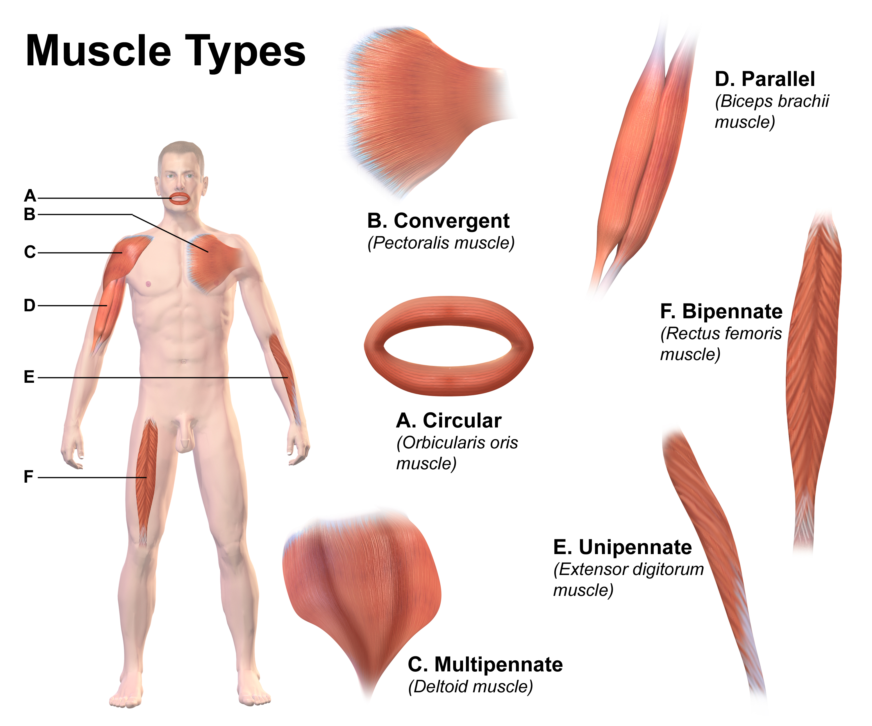
Skeletal muscle (commonly referred to as muscle) is one of the three types of vertebrate muscle tissue, the others being cardiac muscle and smooth muscle. They are part of the somatic nervous system, voluntary muscular system and typically are attached by tendons to bones of a skeleton. The skeletal muscle cells are much longer than in the other types of muscle tissue, and are also known as ''muscle fibers''. The tissue of a skeletal muscle is striated muscle tissue, striated – having a striped appearance due to the arrangement of the sarcomeres. A skeletal muscle contains multiple muscle fascicle, fascicles – bundles of muscle fibers. Each individual fiber and each muscle is surrounded by a type of connective tissue layer of fascia. Muscle fibers are formed from the cell fusion, fusion of developmental myoblasts in a process known as myogenesis resulting in long multinucleated cells. In these cells, the cell nucleus, nuclei, termed ''myonuclei'', are located along the inside ... [...More Info...] [...Related Items...] OR: [Wikipedia] [Google] [Baidu] |
Thigh
In anatomy, the thigh is the area between the hip (pelvis) and the knee. Anatomically, it is part of the lower limb. The single bone in the thigh is called the femur. This bone is very thick and strong (due to the high proportion of bone tissue), and forms a ball and socket joint at the hip, and a modified hinge joint at the knee. Structure Bones The femur is the only bone in the thigh and serves as an attachment site for all thigh muscles. The head of the femur articulates with the acetabulum in the pelvic bone forming the hip joint, while the distal part of the femur articulates with the tibia and patella forming the knee. By most measures, the femur is the strongest and longest bone in the body. The femur is categorised as a long bone and comprises a diaphysis, the shaft (or body) and two epiphyses, the lower extremity and the upper extremity of femur, that articulate with adjacent bones in the hip and knee. Muscular compartments In cross-section, the thigh is d ... [...More Info...] [...Related Items...] OR: [Wikipedia] [Google] [Baidu] |
Knee
In humans and other primates, the knee joins the thigh with the leg and consists of two joints: one between the femur and tibia (tibiofemoral joint), and one between the femur and patella (patellofemoral joint). It is the largest joint in the human body. The knee is a modified hinge joint, which permits flexion and extension (kinesiology), extension as well as slight internal and external rotation. The knee is vulnerable to injury and to the development of osteoarthritis. It is often termed a ''compound joint'' having tibiofemoral and patellofemoral components. (The fibular collateral ligament is often considered with tibiofemoral components.) Structure The knee is a modified hinge joint, a type of synovial joint, which is composed of three functional compartments: the patellofemoral articulation, consisting of the patella, or "kneecap", and the patellar groove on the front of the femur through which it slides; and the medial and lateral tibiofemoral articulations linking the ... [...More Info...] [...Related Items...] OR: [Wikipedia] [Google] [Baidu] |
Vastus Intermedius Muscle
The vastus intermedius () (Cruraeus) arises from the front and lateral surfaces of the body of the femur in its upper two-thirds, sitting under the rectus femoris muscle and from the lower part of the lateral intermuscular septum. Its fibers end in a superficial aponeurosis, which forms the deep part of the quadriceps femoris tendon. The vastus medialis and vastus intermedius appear to be inseparably united, but when the rectus femoris has been reflected during dissection a narrow interval will be observed extending upward from the medial border of the patella between the two muscles, and the separation may be continued as far as the lower part of the intertrochanteric line, where, however, the two muscles are frequently continuous. Due to being the deeper middle-most of the quadriceps muscle group, the intermedius is the most difficult to stretch once maximum knee flexion is attained. It cannot be further stretched by hip extension as the rectus femoris can, nor is it accessi ... [...More Info...] [...Related Items...] OR: [Wikipedia] [Google] [Baidu] |
Synovial Membrane
Synovial () may refer to: * Synovial fluid * Synovial joint A synovial joint, also known as diarthrosis, joins bones or cartilage with a fibrous joint capsule that is continuous with the periosteum of the joined bones, constitutes the outer boundary of a synovial cavity, and surrounds the bones' articulati ... * Synovial membrane * Synovial bursa {{disambiguation ... [...More Info...] [...Related Items...] OR: [Wikipedia] [Google] [Baidu] |
Lateral Circumflex Femoral Artery
The lateral circumflex femoral artery (also known as the lateral femoral circumflex artery or the external circumflex artery) is an artery in the upper thigh. It is usually a branch of the profunda femoris artery, and produces three branches. It is mostly distributed to the muscles of the lateral thigh, supplying arterial blood to muscles of the knee extensor group. Structure Origin The lateral femoral circumflex artery usually arises from the lateral side of the profunda femoris artery, but may occasionally arise directly from the femoral artery. It is the largest branch of the profunda femoris artery. Course and relations The lateral circumflex femoral artery usually courses anterior to the femoral neck. It passes horizontally between the divisions of the femoral nerve. It passes posterior to the sartorius muscle and rectus femoris muscle. It passes laterally across the hip joint capsule. It divides into ascending, transverse, and descending branches. Branches The lat ... [...More Info...] [...Related Items...] OR: [Wikipedia] [Google] [Baidu] |
Femoral Nerve
The femoral nerve is a nerve in the thigh that supplies skin on the upper thigh and inner leg, and the muscles that extend the knee. It is the largest branch of the lumbar plexus. Structure The femoral nerve is the major nerve supplying the anterior compartment of the thigh. It is the largest branch of the lumbar plexus, and arises from the dorsal divisions of the ventral rami of the second, third, and fourth lumbar nerves (L2, L3, and L4). The nerve enters Scarpa's triangle by passing beneath the inguinal ligament, just lateral to the femoral artery. In the thigh, the nerve lies in a groove between iliacus muscle and psoas major muscle, outside the femoral sheath, and lateral to the femoral artery. After a short course of about 4 cm in the thigh, the nerve is divided into anterior and posterior divisions, separated by lateral femoral circumflex artery. The branches are shown below: Muscular branches * The nerve to the pectineus muscle arises immediately above the ... [...More Info...] [...Related Items...] OR: [Wikipedia] [Google] [Baidu] |
Fascia
A fascia (; : fasciae or fascias; adjective fascial; ) is a generic term for macroscopic membranous bodily structures. Fasciae are classified as superficial, visceral or deep, and further designated according to their anatomical location. The knowledge of fascial structures is essential in surgery, as they create borders for infectious processes (for example Psoas abscess) and haematoma. An increase in pressure may result in a compartment syndrome, where a prompt fasciotomy may be necessary. For this reason, profound descriptions of fascial structures are available in anatomical literature from the 19th century. Function Fasciae were traditionally thought of as passive structures that transmit mechanical tension generated by muscular activities or external forces throughout the body. An important function of muscle fasciae is to reduce friction of muscular force. In doing so, fasciae provide a supportive and movable wrapping for nerves and blood vessels as they pass thro ... [...More Info...] [...Related Items...] OR: [Wikipedia] [Google] [Baidu] |


