|
Appendectomies
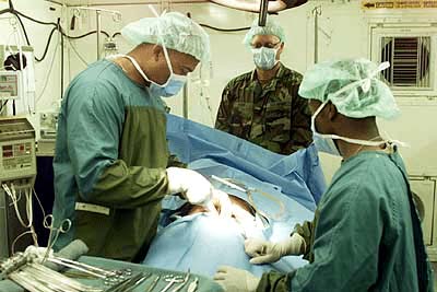
An appendectomy (American English) or appendicectomy (British English) is a surgical operation in which the vermiform appendix (a portion of the intestine) is removed. Appendectomy is normally performed as an urgent or emergency procedure to treat complicated acute appendicitis. Appendectomy may be performed laparoscopically (as minimally invasive surgery) or as an open operation. Over the 2010s, surgical practice has increasingly moved towards routinely offering laparoscopic appendicectomy; for example in the United Kingdom over 95% of adult appendicectomies are planned as laparoscopic procedures. Laparoscopy is often used if the diagnosis is in doubt, or in order to leave a less visible surgical scar. Recovery may be slightly faster after laparoscopic surgery, although the laparoscopic procedure itself is more expensive and resource-intensive than open surgery and generally takes longer. Advanced pelvic sepsis occasionally requires a lower midline laparotomy. Complicated (perfo ... [...More Info...] [...Related Items...] OR: [Wikipedia] [Google] [Baidu] |
Appendectomy Incision Locations
An appendectomy (American English) or appendicectomy (British English) is a surgical operation in which the vermiform appendix (a portion of the intestine) is removed. Appendectomy is normally performed as an urgent or emergency procedure to treat complicated acute appendicitis. Appendectomy may be performed laparoscopically (as minimally invasive surgery) or as an open operation. Over the 2010s, surgical practice has increasingly moved towards routinely offering laparoscopic appendicectomy; for example in the United Kingdom over 95% of adult appendicectomies are planned as laparoscopic procedures. Laparoscopy is often used if the diagnosis is in doubt, or in order to leave a less visible surgical scar. Recovery may be slightly faster after laparoscopic surgery, although the laparoscopic procedure itself is more expensive and resource-intensive than open surgery and generally takes longer. Advanced pelvic sepsis occasionally requires a lower midline laparotomy. Complicated ( ... [...More Info...] [...Related Items...] OR: [Wikipedia] [Google] [Baidu] |
Appendicitis
Appendicitis is inflammation of the Appendix (anatomy), appendix. Symptoms commonly include right lower abdominal pain, nausea, vomiting, fever and anorexia (symptom), decreased appetite. However, approximately 40% of people do not have these typical symptoms. Severe complications of a ruptured appendix include widespread, agonising and awful peritonitis, inflammation of the inner lining of the abdominal wall and sepsis. Appendicitis is primarily caused by a blockage of the Lumen (anatomy), hollow portion in the appendix. This blockage typically results from a Fecalith, faecolith, a calcified "stone" made of feces. Some studies show a correlation between appendicoliths and disease severity. Other factors such as inflamed Mucosa-associated lymphoid tissue, lymphoid tissue from a viral infection, Human parasite, intestinal parasites, gallstone, or Neoplasm, tumors may also lead to this blockage. When the appendix becomes blocked, it experiences increased pressure, reduced blood f ... [...More Info...] [...Related Items...] OR: [Wikipedia] [Google] [Baidu] |
McBurney's Point
McBurney's point is the point over the right side of the abdomen that is one-third of the distance from the anterior superior iliac spine to the Navel, umbilicus (navel). This is near the most common location of the Vermiform appendix, appendix. Location McBurney's point is located one third of the distance from the right anterior superior iliac spine to the Navel, umbilicus (navel). This point roughly corresponds to the most common location of the base of the Vermiform appendix, appendix, where it is attached to the cecum. Appendicitis Deep Pain, tenderness at McBurney's point, known as McBurney's sign, is a sign of acute appendicitis. The clinical sign of referred pain in the epigastrium when pressure is applied is also known as Aaron's sign. Specific localization of tenderness to McBurney's point indicates that inflammation is no longer limited to the lumen of the bowel (which localizes pain poorly), and is irritating the lining of the peritoneum at the place where the pe ... [...More Info...] [...Related Items...] OR: [Wikipedia] [Google] [Baidu] |
Anterior Superior Iliac Spine
The anterior superior iliac spine (ASIS) is a bony projection of the iliac bone, and an important landmark of surface anatomy. It refers to the anterior extremity of the iliac crest of the pelvis. It provides attachment for the inguinal ligament, and the sartorius muscle. The tensor fasciae latae muscle attaches to the lateral aspect of the superior anterior iliac spine, and also about 5 cm away at the iliac tubercle. Structure The anterior superior iliac spine refers to the anterior extremity of the iliac crest of the pelvis. This is a key surface landmark, and easily palpated. It provides attachment for the inguinal ligament, the sartorius muscle, and the tensor fasciae latae muscle. A variety of structures lie close to the anterior superior iliac spine, including the subcostal nerve, the femoral artery (which passes between it and the pubic symphysis), and the iliohypogastric nerve. Clinical significance The anterior superior iliac spine provides a clue i ... [...More Info...] [...Related Items...] OR: [Wikipedia] [Google] [Baidu] |
Muscle Relaxant
A muscle relaxant is a drug that affects skeletal muscle function and decreases the muscle tone. It may be used to alleviate symptoms such as muscle spasms, pain, and hyperreflexia. The term "muscle relaxant" is used to refer to two major therapeutic groups: Neuromuscular-blocking drugs, neuromuscular blockers and Antispasmodic, spasmolytics. Neuromuscular blockers act by interfering with transmission at the neuromuscular end plate and have no central nervous system (CNS) activity. They are often used during surgical procedures and in intensive care and emergency medicine to cause temporary paralysis. Spasmolytics, also known as "centrally acting" muscle relaxant, are used to alleviate musculoskeletal pain and spasms and to reduce spasticity in a variety of neurological conditions. While both neuromuscular blockers and spasmolytics are often grouped together as muscle relaxant, [...More Info...] [...Related Items...] OR: [Wikipedia] [Google] [Baidu] |
Supine Position
The supine position () means lying horizontally, with the face and torso facing up, as opposed to the prone position, which is face down. When used in surgical procedures, it grants access to the peritoneal, thoracic, and pericardium, pericardial regions; as well as the head, neck, and extremities. Using anatomical terms of location, the dorsal side is down, and the ventral side is up, when supine. Semi-supine In scientific literature "semi-supine" commonly refers to positions where the upper body is tilted (at 45° or variations) and not completely horizontal. Relation to sudden infant death syndrome The decline in death due to sudden infant death syndrome (SIDS) is said to be attributable to having babies sleep in the supine position. The realization that infants sleeping face down, or in a prone position, had an increased mortality rate re-emerged into medical awareness at the end of the 1980s when two researchers, Susan Beal in Australia and Gus De Jonge in the Nether ... [...More Info...] [...Related Items...] OR: [Wikipedia] [Google] [Baidu] |
Abdomen
The abdomen (colloquially called the gut, belly, tummy, midriff, tucky, or stomach) is the front part of the torso between the thorax (chest) and pelvis in humans and in other vertebrates. The area occupied by the abdomen is called the abdominal cavity. In arthropods, it is the posterior (anatomy), posterior tagma (biology), tagma of the body; it follows the thorax or cephalothorax. In humans, the abdomen stretches from the thorax at the thoracic diaphragm to the pelvis at the pelvic brim. The pelvic brim stretches from the lumbosacral joint (the intervertebral disc between Lumbar vertebrae, L5 and Vertebra#Sacrum, S1) to the pubic symphysis and is the edge of the pelvic inlet. The space above this inlet and under the thoracic diaphragm is termed the abdominal cavity. The boundary of the abdominal cavity is the abdominal wall in the front and the peritoneal surface at the rear. In vertebrates, the abdomen is a large body cavity enclosed by the abdominal muscles, at the front an ... [...More Info...] [...Related Items...] OR: [Wikipedia] [Google] [Baidu] |
General Surgery
General surgery is a Surgical specialties, surgical specialty that focuses on alimentary canal and Abdomen, abdominal contents including the esophagus, stomach, small intestine, large intestine, liver, pancreas, gallbladder, Appendix (anatomy), appendix and bile ducts, and often the thyroid gland. General surgeons also deal with diseases involving the human skin, skin, breast, soft tissue, trauma (medicine), trauma, peripheral artery disease and hernias and perform endoscopic as such as gastroscopy, colonoscopy and laparoscopic procedures. Scope General surgeons may sub-specialise into one or more of the following disciplines: Trauma surgery In many parts of the world including North America, Australia and the UK, United Kingdom, the overall responsibility for major trauma, trauma care falls under the auspices of general surgery. Some general surgeons obtain advanced training in this field (most commonly surgical critical care) and specialty certification surgical critical ... [...More Info...] [...Related Items...] OR: [Wikipedia] [Google] [Baidu] |
Navel
The navel (clinically known as the umbilicus; : umbilici or umbilicuses; also known as the belly button or tummy button) is a protruding, flat, or hollowed area on the abdomen at the attachment site of the umbilical cord. Structure The umbilicus is used to visually separate the abdomen into quadrants. The umbilicus is a prominent Scar#Umbilical, scar on the abdomen, with its position being relatively consistent among humans. The skin around the waist at the level of the umbilicus is supplied by the tenth thoracic spinal nerve (T10 dermatome (anatomy), dermatome). The umbilicus itself typically lies at a vertical level corresponding to the junction between the L3 and L4 vertebrae, with a normal variation among people between the L3 and L5 vertebrae. Parts of the adult navel include the "umbilical cord remnant" or "umbilical tip", which is the often protruding scar left by the detachment of the umbilical cord. This is located in the center of the navel, sometimes described ... [...More Info...] [...Related Items...] OR: [Wikipedia] [Google] [Baidu] |
Endotracheal Tube
A tracheal tube is a catheter that is inserted into the trachea for the primary purpose of establishing and maintaining a patent airway and to ensure the adequate exchange of oxygen and carbon dioxide. Many different types of tracheal tubes are available, suited for different specific applications: * An endotracheal tube is a specific type of tracheal tube that is nearly always inserted through the mouth (orotracheal) or nose (nasotracheal). * A tracheostomy tube is another type of tracheal tube; this curved metal or plastic tube may be inserted into a tracheostomy stoma (following a tracheotomy) to maintain a patent lumen. * A tracheal button is a rigid plastic cannula about in length that can be placed into the tracheostomy after removal of a tracheostomy tube to maintain patency of the lumen. History Portex Medical (England and France) produced the first cuff-less plastic 'Ivory' endotracheal tubes. Ivan Magill later added a cuff (these were glued on by hand to make the famo ... [...More Info...] [...Related Items...] OR: [Wikipedia] [Google] [Baidu] |
External Oblique
The abdominal external oblique muscle (also external oblique muscle or exterior oblique) is the largest and outermost of the three flat abdominal muscles of the lateral anterior abdomen. Structure The external oblique is situated on the lateral and anterior parts of the abdomen. It is broad, thin, and irregularly quadrilateral, its muscular portion occupying the side, its aponeurosis the anterior wall of the abdomen. In most humans, the oblique is not visible, due to subcutaneous fat deposits and the small size of the muscle. It arises from eight fleshy digitations, each from the external surfaces and inferior borders of the fifth to twelfth ribs (lower eight ribs). These digitations are arranged in an oblique line which runs inferiorly and anteriorly, with the upper digitations being attached close to the cartilages of the corresponding ribs, the lowest to the apex of the cartilage of the last rib, the intermediate ones to the ribs at some distance from their cartilages. The ... [...More Info...] [...Related Items...] OR: [Wikipedia] [Google] [Baidu] |


