|
Aortic Coarctation
Coarctation of the aorta (CoA) is a congenital condition whereby the aorta is narrow, usually in the area where the ductus arteriosus (ligamentum arteriosum after regression) inserts. The word ''coarctation'' means "pressing or drawing together; narrowing". Coarctations are most common in the aortic arch. The arch may be small in babies with coarctations. Other heart defects may also occur when coarctation is present, typically occurring on the left side of the heart. When a patient has a coarctation, the left ventricle has to work harder. Since the aorta is narrowed, the left ventricle must generate a much higher pressure than normal in order to force enough blood through the aorta to deliver blood to the lower part of the body. If the narrowing is severe enough, the left ventricle may not be strong enough to push blood through the coarctation, thus resulting in a lack of blood to the lower half of the body. Physiologically its complete form is manifested as interrupted aortic ... [...More Info...] [...Related Items...] OR: [Wikipedia] [Google] [Baidu] |
Congenital Condition
A birth defect is an abnormal condition that is present at birth, regardless of its cause. Birth defects may result in disabilities that may be physical, intellectual, or developmental. The disabilities can range from mild to severe. Birth defects are divided into two main types: structural disorders in which problems are seen with the shape of a body part and functional disorders in which problems exist with how a body part works. Functional disorders include metabolic and degenerative disorders. Some birth defects include both structural and functional disorders. Birth defects may result from genetic or chromosomal disorders, exposure to certain medications or chemicals, or certain infections during pregnancy. Risk factors include folate deficiency, drinking alcohol or smoking during pregnancy, poorly controlled diabetes, and a mother over the age of 35 years old. Many birth defects are believed to involve multiple factors. Birth defects may be visible at birth or diag ... [...More Info...] [...Related Items...] OR: [Wikipedia] [Google] [Baidu] |
Subclavian Artery
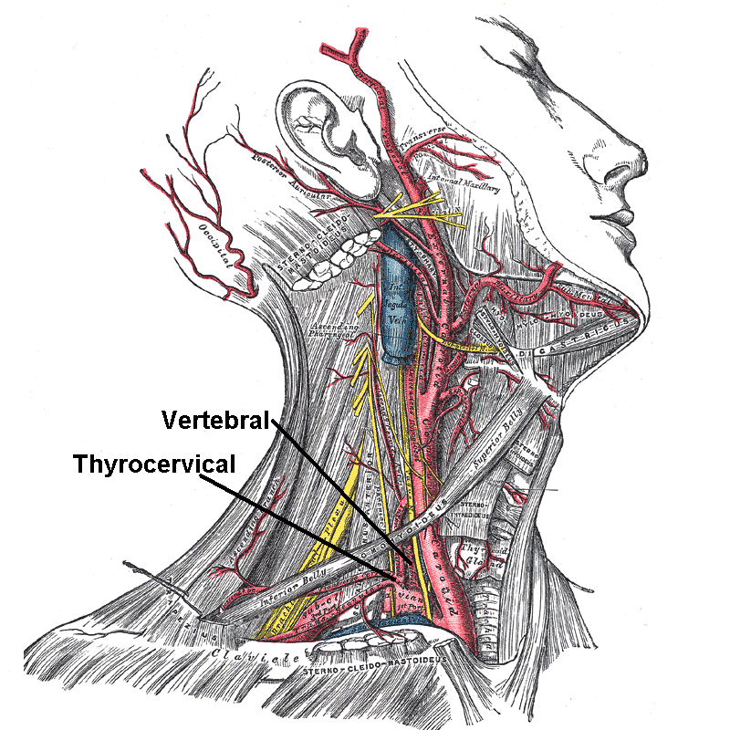
In human anatomy, the subclavian arteries are paired major arteries of the upper thorax, below the clavicle. They receive blood from the aortic arch. The left subclavian artery supplies blood to the left arm and the right subclavian artery supplies blood to the right arm, with some branches supplying the head and thorax. On the left side of the body, the subclavian comes directly off the aortic arch, while on the right side it arises from the relatively short brachiocephalic artery when it bifurcates into the subclavian and the right common carotid artery. The usual branches of the subclavian on both sides of the body are the vertebral artery, the internal thoracic artery, the thyrocervical trunk, the costocervical trunk and the dorsal scapular artery, which may branch off the transverse cervical artery, which is a branch of the thyrocervical trunk. The subclavian becomes the axillary artery at the lateral border of the first rib. Structure From its origin, the subclavian art ... [...More Info...] [...Related Items...] OR: [Wikipedia] [Google] [Baidu] |
Clarence Crafoord
Clarence Crafoord (28 May 1899 – 25 February 1984) was a Swedish cardiovascular system, cardiovascular surgery, surgeon, best known for performing the first successful repair of aortic coarctation on 19 October 1944, one year before Robert E. Gross (surgeon), Robert E. Gross. Crafoord also introduced heparin as thrombosis prophylaxis in the 1930s and he pioneered mechanical positive-pressure ventilation during thoracic operations in the 1940s. Crafoord was professor of thoracic surgery at Karolinska Institute from 1948 to 1966. References * * * * * * * 1899 births 1983 deaths Swedish cardiac surgeons Academic staff of the Karolinska Institute 20th-century Swedish physicians History of surgery 20th-century surgeons People from Hudiksvall {{Sweden-med-bio-stub ... [...More Info...] [...Related Items...] OR: [Wikipedia] [Google] [Baidu] |
Arterial Hypertension
Hypertension, also known as high blood pressure, is a long-term medical condition in which the blood pressure in the arteries is persistently elevated. High blood pressure usually does not cause symptoms itself. It is, however, a major risk factor for stroke, coronary artery disease, heart failure, atrial fibrillation, peripheral arterial disease, vision loss, chronic kidney disease, and dementia. Hypertension is a major cause of premature death worldwide. High blood pressure is classified as primary (essential) hypertension or secondary hypertension. About 90–95% of cases are primary, defined as high blood pressure due to non-specific lifestyle and genetic factors. Lifestyle factors that increase the risk include excess salt in the diet, excess body weight, smoking, physical inactivity and alcohol use. The remaining 5–10% of cases are categorized as secondary hypertension, defined as high blood pressure due to a clearly identifiable cause, such as chronic kidney di ... [...More Info...] [...Related Items...] OR: [Wikipedia] [Google] [Baidu] |
Open Aortic Surgery
Open aortic surgery (OAS), also known as open aortic repair (OAR), describes a technique whereby an abdominal, thoracic or retroperitoneal surgical incision is used to visualize and control the aorta for purposes of treatment, usually by the replacement of the affected segment with a prosthetic Vascular grafting, graft. OAS is used to treat Abdominal aortic aneurysm, aneurysms of the abdominal and thoracic aorta, aortic dissection, acute aortic syndrome, and aortic ruptures. Aortobifemoral bypass is also used to treat atherosclerotic disease of the abdominal aorta below the level of the renal arteries. In 2003, OAS was surpassed by endovascular aneurysm repair (EVAR) as the most common technique for repairing abdominal aortic aneurysms in the United States. Depending on the extent of the aorta repaired, an open aortic operation may be called an Infrarenal aortic repair, a Thoracic aortic repair, or a Thoracoabdominal aortic repair. A thoracoabdominal aortic repair is a more extensi ... [...More Info...] [...Related Items...] OR: [Wikipedia] [Google] [Baidu] |
Bicuspid Aortic Valve
Bicuspid aortic valve (BAV) is a form of heart disease in which two of the leaflets of the aortic valve fuse during development in the womb resulting in a two-leaflet (bicuspid) valve instead of the normal three-leaflet (tricuspid) valve. BAV is the most common cause of heart disease present at birth and affects approximately 1.3% of adults. Normally, the mitral valve is the only bicuspid valve and this is situated between the heart's left atrium and left ventricle. Heart valves play a crucial role in ensuring the unidirectional flow of blood from the atria to the ventricles, or from the ventricle to the aorta or pulmonary trunk. BAV is normally inherited. Signs and symptoms In many cases, a bicuspid aortic valve will cause no problems. People with BAV may become tired more easily than those with normal valvular function and have difficulty maintaining stamina for cardio-intensive activities due to poor heart performance caused by stress on the aortic wall.Maredia A.K., G ... [...More Info...] [...Related Items...] OR: [Wikipedia] [Google] [Baidu] |
Phase Contrast Magnetic Resonance Imaging
Phase contrast magnetic resonance imaging (PC-MRI) is a specific type of magnetic resonance imaging used primarily to determine flow velocities. PC-MRI can be considered a method of Magnetic Resonance Velocimetry. It also provides a method of magnetic resonance angiography. Since modern PC-MRI is typically time-resolved, it provides a means of 4D imaging (three spatial dimensions plus time). How it Works Atoms with an odd number of protons or neutrons have a randomly aligned angular spin momentum. When placed in a strong magnetic field, some of these spins align with the axis of the external field, which causes a net 'longitudinal' magnetization. These spins precess about the axis of the external field at a frequency proportional to the strength of that field. Then, energy is added to the system through a Radio frequency (RF) pulse to 'excite' the spins, changing the axis that the spins precess about. These spins can then be observed by receiver coils (Radiofrequency coils) usin ... [...More Info...] [...Related Items...] OR: [Wikipedia] [Google] [Baidu] |
Descending Aorta
In human anatomy, the descending aorta is part of the aorta, the largest artery in the body. The descending aorta begins at the aortic arch and runs down through the chest and abdomen. The descending aorta anatomically consists of two portions or segments, the thoracic and the abdominal aorta, in correspondence with the two great cavities of the trunk in which it is situated. Within the abdomen, the descending aorta branches into the two common iliac arteries which serve the pelvis and eventually legs. The ductus arteriosus connects to the junction between the pulmonary artery and the descending aorta in foetal life. This artery later regresses as the ligamentum arteriosum. The descending aorta has important functions within the body. The descending aorta transports oxygenated blood from the heart to the rest of the body. See also * Abbott artery References External links * – "Left side of the mediastinum The mediastinum (from ;: mediastina) is the central c ... [...More Info...] [...Related Items...] OR: [Wikipedia] [Google] [Baidu] |
MRI Contrast Agent
MRI contrast agents are contrast agents used to improve the visibility of internal body structures in magnetic resonance imaging (MRI). The most commonly used compounds for contrast enhancement are gadolinium-based contrast agents (GBCAs). Such MRI contrast agents shorten the Relaxation (NMR), relaxation times of nuclei within body tissues following oral or intravenous administration. Due to safety concerns, these products carry a Boxed warning, Black Box Warning in the USA. Theory of operation In MRI scanners, sections of the body are exposed to a strong magnetic field causing primarily the hydrogen nuclei ("spins") of water in tissues to be polarized in the direction of the magnetic field. An intense radiofrequency pulse is applied that tips the magnetization generated by the hydrogen nuclei in the direction of the receiver coil where the spin polarization can be detected. Random molecular rotational oscillations matching the resonance frequency of the nuclear spins provide ... [...More Info...] [...Related Items...] OR: [Wikipedia] [Google] [Baidu] |
Body Surface Area
In physiology and medicine, the body surface area (BSA) is the measured or calculated surface area of a human body. For many clinical purposes, BSA is a better indicator of metabolic mass than body weight because it is less affected by abnormal adipose mass. Nevertheless, there have been several important critiques of the use of BSA in determining the dosage of medications with a narrow therapeutic index, such as chemotherapy. Typically there is a 4–10 fold variation in drug clearance between individuals due to differing the activity of drug elimination processes related to genetic and environmental factors. This can lead to significant overdosing and underdosing (and increased risk of disease recurrence). It is also thought to be a distorting factor in Phase I and II trials that may result in potentially helpful medications being prematurely rejected. The trend to personalized medicine is one approach to counter this weakness. Uses Examples of uses of the BSA: * Renal clea ... [...More Info...] [...Related Items...] OR: [Wikipedia] [Google] [Baidu] |
Echocardiogram
Echocardiography, also known as cardiac ultrasound, is the use of ultrasound to examine the heart. It is a type of medical imaging, using standard ultrasound or Doppler ultrasound. The visual image formed using this technique is called an echocardiogram, a cardiac echo, or simply an echo. Echocardiography is routinely used in the diagnosis, management, and follow-up of patients with any suspected or known heart diseases. It is one of the most widely used diagnostic imaging modalities in cardiology. It can provide a wealth of helpful information, including the size and shape of the heart (internal chamber size quantification), pumping capacity, location and extent of any tissue damage, and assessment of valves. An echocardiogram can also give physicians other estimates of heart function, such as a calculation of the cardiac output, ejection fraction, and diastolic function (how well the heart relaxes). Echocardiography is an important tool in assessing wall motion abnormality ... [...More Info...] [...Related Items...] OR: [Wikipedia] [Google] [Baidu] |
Magnetic Resonance Angiography
Magnetic resonance angiography (MRA) is a group of techniques based on magnetic resonance imaging (MRI) to image blood vessels. Magnetic resonance angiography is used to generate images of arteries (and less commonly veins) in order to evaluate them for stenosis (abnormal narrowing), Vascular occlusion, occlusions, aneurysms (vessel wall dilatations, at risk of rupture) or other abnormalities. MRA is often used to evaluate the arteries of the neck and brain, the thoracic and abdominal aorta, the renal arteries, and the legs (the latter exam is often referred to as a "run-off"). Acquisition A variety of techniques can be used to generate the pictures of blood vessels, both artery, arteries and veins, based on flow effects or on contrast (inherent or pharmacologically generated). The most frequently applied MRA methods involve the use intravenous MRI contrast agent, contrast agents, particularly those containing gadolinium to shorten the Spin–lattice relaxation, ''T''1 of bloo ... [...More Info...] [...Related Items...] OR: [Wikipedia] [Google] [Baidu] |


