|
Anatomical Pathology
Anatomical pathology (''Commonwealth'') or anatomic pathology (''U.S.'') is a medical specialty that is concerned with the diagnosis of disease based on the macroscopic, microscopic, biochemical, immunologic and molecular examination of organs and tissues. Over the 20th century, surgical pathology has evolved tremendously: from historical examination of whole bodies (autopsy) to a more modernized practice, centered on the diagnosis and prognosis of cancer to guide treatment decision-making in oncology. Its modern founder was the Italian scientist Giovanni Battista Morgagni from Forlì. Anatomical pathology is one of two branches of pathology, the other being clinical pathology, the diagnosis of disease through the laboratory analysis of bodily fluids or tissues. Often, pathologists practice both anatomical and clinical pathology, a combination known as general pathology. Similar specialties exist in veterinary pathology. Differences with clinical pathology Anatom ... [...More Info...] [...Related Items...] OR: [Wikipedia] [Google] [Baidu] |
Breast Invasive Scirrhous Carcinoma Histopathology (1)
The breasts are two prominences located on the upper ventral region of the torso among humans and other primates. Both sexes develop breasts from the same embryological tissues. The relative size and development of the breasts is a major secondary sex distinction between females and males. There is also considerable variation in size between individuals. Permanent breast growth during puberty is caused by estrogens in conjunction with the growth hormone. Female humans are the only mammals that permanently develop breasts at puberty; all other mammals develop their mammary tissue during the latter period of pregnancy. In females, the breast serves as the mammary gland, which produces and secretes milk to feed infants. Subcutaneous fat covers and envelops a network of ducts that converge on the nipple, and these tissues give the breast its distinct size and globular shape. At the ends of the ducts are lobules, or clusters of alveoli, where milk is produced and stored i ... [...More Info...] [...Related Items...] OR: [Wikipedia] [Google] [Baidu] |
Medical Laboratory
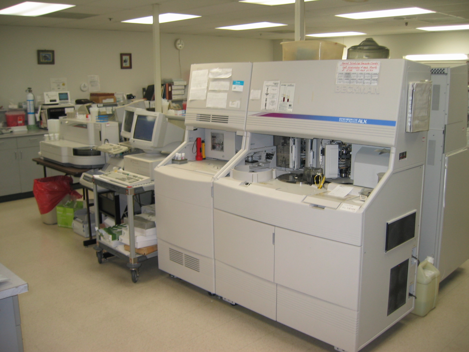
A medical laboratory or clinical laboratory is a laboratory where tests are conducted out on clinical specimens to obtain information about the health of a patient to aid in diagnosis, treatment, and prevention of disease. Clinical medical laboratories are an example of applied science, as opposed to research laboratory, research laboratories that focus on basic science, such as found in some academia, academic institutions. Medical laboratories vary in size and complexity and so offer a variety of testing services. More comprehensive services can be found in acute-care hospitals and medical centers, where 70% of clinical decisions are based on laboratory testing. Doctors offices and clinics, as well as skilled nursing and Nursing home, long-term care facilities, may have laboratories that provide more basic testing services. Commercial medical laboratories operate as independent businesses and provide testing that is otherwise not provided in other settings due to low test vol ... [...More Info...] [...Related Items...] OR: [Wikipedia] [Google] [Baidu] |
Immunohistochemistry
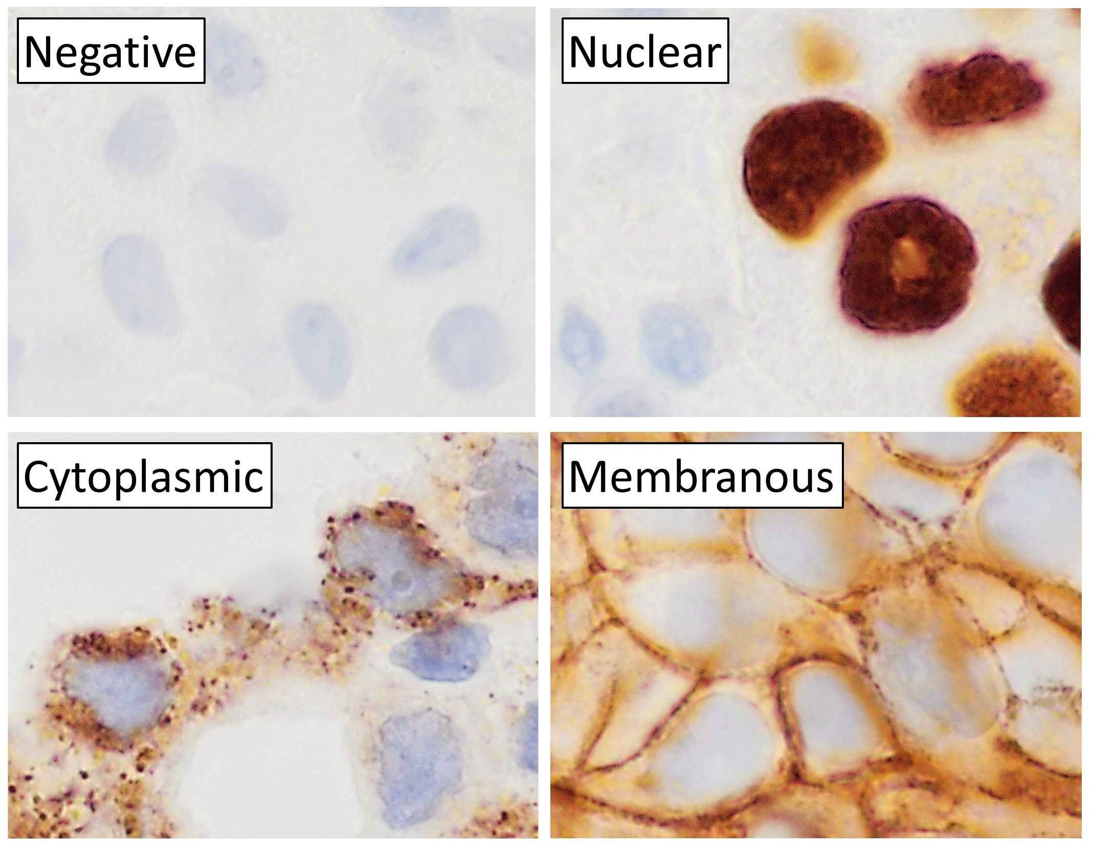
Immunohistochemistry is a form of immunostaining. It involves the process of selectively identifying antigens in cells and tissue, by exploiting the principle of Antibody, antibodies binding specifically to antigens in biological tissues. Albert Coons, Albert Hewett Coons, Ernst Berliner, Ernest Berliner, Norman Jones and Hugh J Creech was the first to develop immunofluorescence in 1941. This led to the later development of immunohistochemistry. Immunohistochemical staining is widely used in the diagnosis of abnormal cells such as those found in cancerous tumors. In some cancer cells certain tumor antigens are expressed which make it possible to detect. Immunohistochemistry is also widely used in basic research, to understand the distribution and localization of biomarkers and differentially expressed proteins in different parts of a biological tissue. Sample preparation Immunohistochemistry can be performed on tissue that has been fixed and embedded in Paraffin wax, paraffin, ... [...More Info...] [...Related Items...] OR: [Wikipedia] [Google] [Baidu] |
Histochemistry
Immunohistochemistry is a form of immunostaining. It involves the process of selectively identifying antigens in cells and tissue, by exploiting the principle of antibodies binding specifically to antigens in biological tissues. Albert Hewett Coons, Ernest Berliner, Norman Jones and Hugh J Creech was the first to develop immunofluorescence in 1941. This led to the later development of immunohistochemistry. Immunohistochemical staining is widely used in the diagnosis of abnormal cells such as those found in cancerous tumors. In some cancer cells certain tumor antigens are expressed which make it possible to detect. Immunohistochemistry is also widely used in basic research, to understand the distribution and localization of biomarkers and differentially expressed proteins in different parts of a biological tissue. Sample preparation Immunohistochemistry can be performed on tissue that has been fixed and embedded in paraffin, but also cryopreservated (frozen) tissue. Based on ... [...More Info...] [...Related Items...] OR: [Wikipedia] [Google] [Baidu] |
Eosin
Eosin is the name of several fluorescent acidic compounds which bind to and from salts with basic, or eosinophilic, compounds like proteins containing basic amino acid residues such as histidine, arginine and lysine, and stains them dark red or pink as a result of the actions of bromine on eosin. In addition to staining proteins in the cytoplasm, it can be used to stain collagen and muscle fibers for examination under the microscope. Structures that stain readily with eosin are termed eosinophilic. In the field of histology, Eosin Y is the form of eosin used most often as a histologic stain. History and etymology Eosin was named by its inventor Heinrich Caro after the nickname (Eos) of a childhood friend, Anna Peters. It was commercialized (mainly for the textile industry) in 1874, in the same year when it was invented. Variants There are actually two very closely related compounds commonly referred to as ''eosin''. Most often used is in histology is eosin Y, which is a t ... [...More Info...] [...Related Items...] OR: [Wikipedia] [Google] [Baidu] |
Haematoxylin
Haematoxylin American and British English spelling differences#ae and oe, or hematoxylin (), also called natural black 1 or Colour Index International, C.I. 75290, is a chemical compound, compound extracted from wood#Heartwood and sapwood, heartwood of the logwood tree (''Haematoxylum campechianum'') with a chemical formula of . This Natural dye, naturally derived dye has been used as a staining, histologic stain, as an ink and as a dye in the textile and leather industry. As a dye, haematoxylin has been called palo de Campeche, logwood extract, bluewood and blackwood. In histology, haematoxylin staining is commonly followed by counterstain, counterstaining with eosin. When paired, this staining procedure is known as H&E staining and is one of the most commonly used combinations in histology. In addition to its use in the H&E stain, haematoxylin is also a component of the Papanicolaou stain (or Pap stain) which is widely used in the study of cytology specimens. Although the stain ... [...More Info...] [...Related Items...] OR: [Wikipedia] [Google] [Baidu] |
Histology
Histology, also known as microscopic anatomy or microanatomy, is the branch of biology that studies the microscopic anatomy of biological tissue (biology), tissues. Histology is the microscopic counterpart to gross anatomy, which looks at larger structures visible without a microscope. Although one may divide microscopic anatomy into ''organology'', the study of organs, ''histology'', the study of tissues, and ''cytology'', the study of cell (biology), cells, modern usage places all of these topics under the field of histology. In medicine, histopathology is the branch of histology that includes the microscopic identification and study of diseased tissue. In the field of paleontology, the term paleohistology refers to the histology of fossil organisms. Biological tissues Animal tissue classification There are four basic types of animal tissues: muscle tissue, nervous tissue, connective tissue, and epithelial tissue. All animal tissues are considered to be subtypes of these ... [...More Info...] [...Related Items...] OR: [Wikipedia] [Google] [Baidu] |
Microscope
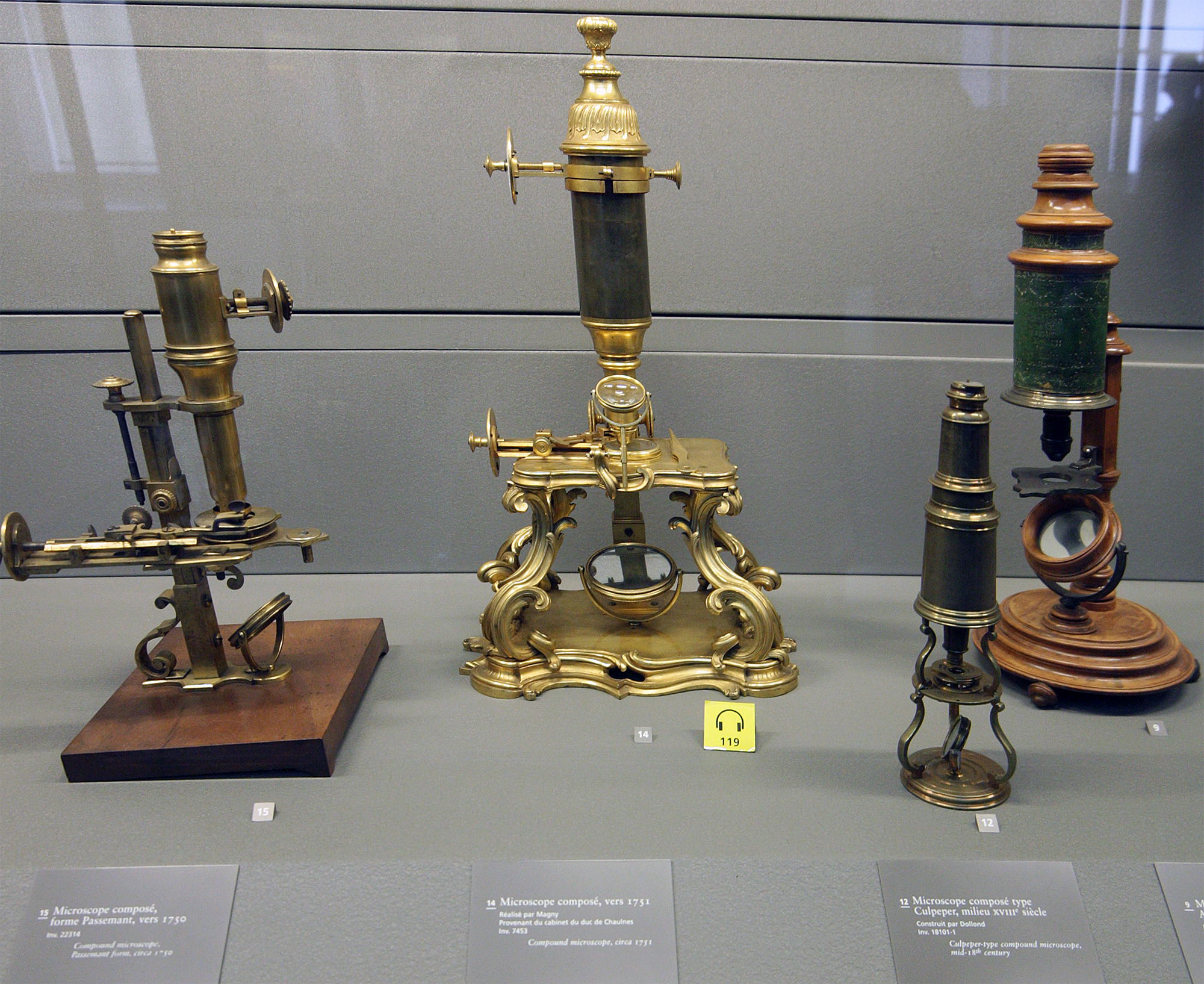
A microscope () is a laboratory equipment, laboratory instrument used to examine objects that are too small to be seen by the naked eye. Microscopy is the science of investigating small objects and structures using a microscope. Microscopic means being invisible to the eye unless aided by a microscope. There are many types of microscopes, and they may be grouped in different ways. One way is to describe the method an instrument uses to interact with a sample and produce images, either by sending a beam of light or electrons through a sample in its optical path, by detecting fluorescence, photon emissions from a sample, or by scanning across and a short distance from the surface of a sample using a probe. The most common microscope (and the first to be invented) is the optical microscope, which uses lenses to refract visible light that passed through a microtome, thinly sectioned sample to produce an observable image. Other major types of microscopes are the fluorescence micro ... [...More Info...] [...Related Items...] OR: [Wikipedia] [Google] [Baidu] |
Optical Microscope
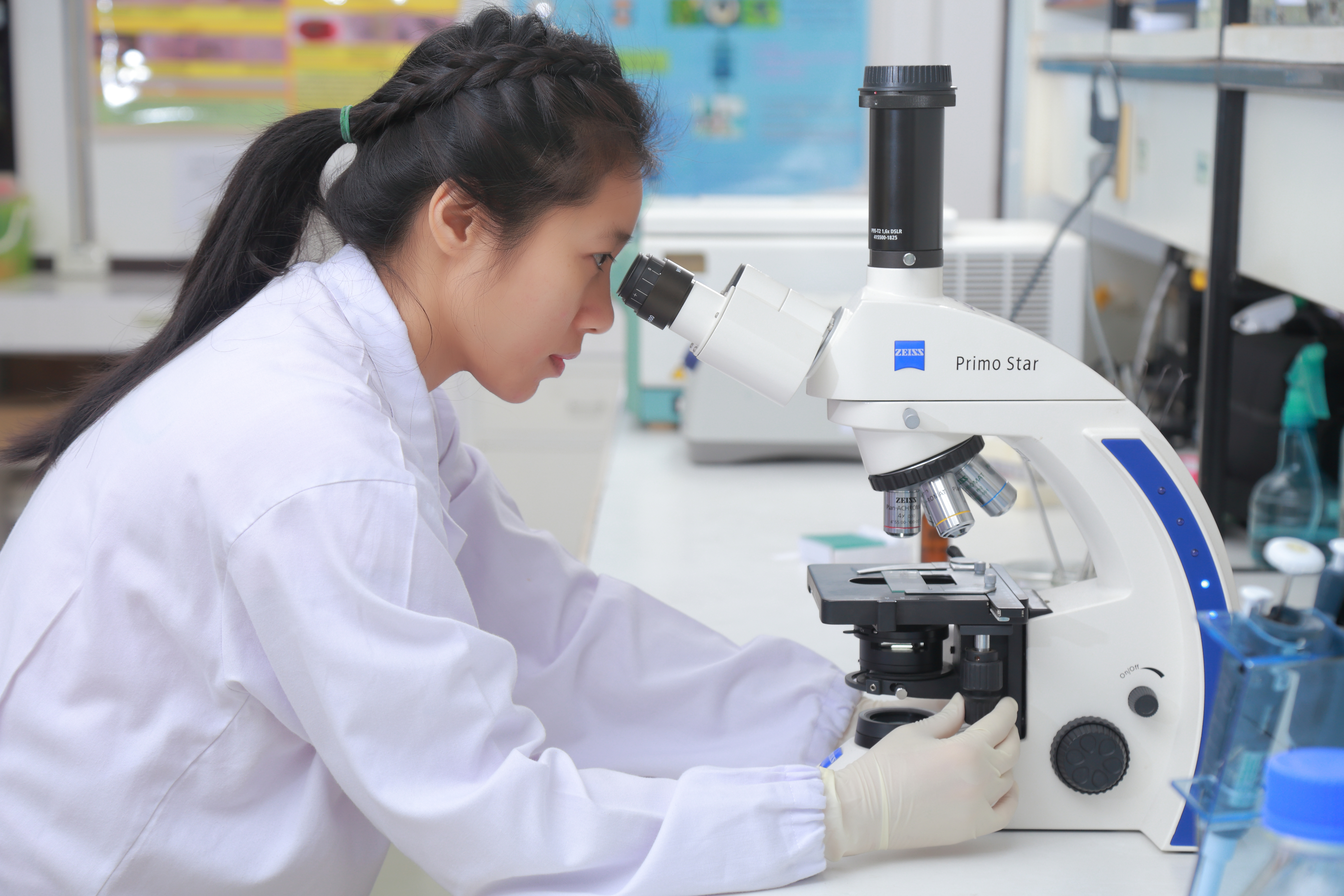
The optical microscope, also referred to as a light microscope, is a type of microscope that commonly uses visible light and a system of lenses to generate magnified images of small objects. Optical microscopes are the oldest design of microscope and were possibly invented in their present compound form in the 17th century. Basic optical microscopes can be very simple, although many complex designs aim to improve resolution and sample contrast. The object is placed on a stage and may be directly viewed through one or two eyepieces on the microscope. In high-power microscopes, both eyepieces typically show the same image, but with a stereo microscope, slightly different images are used to create a 3-D effect. A camera is typically used to capture the image (micrograph). The sample can be lit in a variety of ways. Transparent objects can be lit from below and solid objects can be lit with light coming through ( bright field) or around ( dark field) the objective lens. Polar ... [...More Info...] [...Related Items...] OR: [Wikipedia] [Google] [Baidu] |
Magnifying Glass
A magnifying glass is a convex lens—usually mounted in a frame with a handle—that is used to produce a magnified image of an object. A magnifying glass can also be used to focus light, such as to concentrate the Sun's radiation to create a hot spot at the focus for fire starting. Evidence of magnifying glasses exists from antiquity. The magnifying glass is an icon of detective fiction, particularly that of Sherlock Holmes. An alternative to a magnifying glass is a sheet magnifier, which comprises many very narrow concentric ring-shaped lenses, such that the combination acts as a single lens but is much thinner. Use The convex lens of a magnifying glass can be used to produce a magnified image An image or picture is a visual representation. An image can be Two-dimensional space, two-dimensional, such as a drawing, painting, or photograph, or Three-dimensional space, three-dimensional, such as a carving or sculpture. Images may be di ... of an object. A magnify ... [...More Info...] [...Related Items...] OR: [Wikipedia] [Google] [Baidu] |
Gross Examination
Gross processing, "grossing" or "gross pathology" is the process by which pathology specimens undergo examination with the bare eye to obtain diagnosis, diagnostic information, as well as cutting and tissue sampling in order to prepare material for subsequent histopathology, microscopic ''examination.'' Responsibility Gross examination of surgical pathology, surgical specimens is typically performed by a pathology, pathologist, or by a pathologists' assistant working within a pathology practice. Individuals trained in these fields are often able to gather diagnostically critical information in this stage of processing, including the stage and margin status of surgically removed tumors. Steps The initial step in any examination of a clinical specimen is confirmation of the identity of the patient and the anatomy, anatomical site from which the specimen was obtained. Sufficient clinical data should be communicated by the clinical team to the pathology team in order to guide the app ... [...More Info...] [...Related Items...] OR: [Wikipedia] [Google] [Baidu] |
Urinalysis
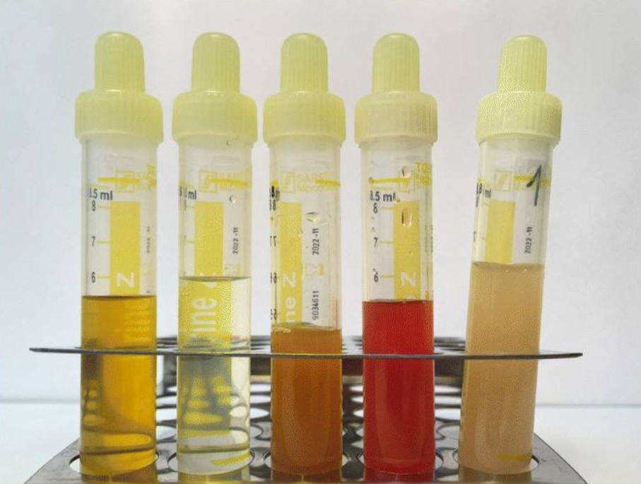
Urinalysis, a portmanteau of the words ''urine'' and ''analysis'', is a Test panel, panel of medical tests that includes physical (macroscopic) examination of the urine, chemical evaluation using urine test strips, and #Microscopic examination, microscopic examination. Macroscopic examination targets parameters such as color, clarity, odor, and specific gravity; urine test strips measure chemical properties such as pH, glucose concentration, and protein levels; and microscopy is performed to identify elements such as Cell (biology), cells, urinary casts, Crystalluria, crystals, and organisms. Background Urine is produced by the filtration of blood in the kidneys. The formation of urine takes place in microscopic structures called nephrons, about one million of which are found in a normal human kidney. Blood enters the kidney though the renal artery and flows through the kidney's vasculature into the Glomerulus (kidney), glomerulus, a tangled knot of capillaries surrounded by Bow ... [...More Info...] [...Related Items...] OR: [Wikipedia] [Google] [Baidu] |

