|
Air Bronchogram
An air bronchogram is defined as a pattern of air-filled bronchi on a background of airless lung. Material was copied from this source, which is available under Creative Commons Attribution 4.0 International License __TOC__ Consolidations In pulmonary consolidations and infiltrates, air bronchograms are most commonly caused by pneumonia or pulmonary edema (especially with alveolar edema). Other potential causes of consolidations or infiltrates with air bronchograms are: * Pulmonary edema * Non-obstructive atelectasis * Severe interstitial lung disease * Pulmonary infarct * Pulmonary hemorrhage * Normal expiration Lung nodules For lung nodules, air bronchograms used to be associated with infectious causes of consolidation and, therefore to be benign. However, in the setting of a lung nodule, an air bronchogram is actually more frequent in malignant than in benign nodules. studied the tumour-bronchus relationship and described five types: * In “Type 1” the bronchial lumen is ... [...More Info...] [...Related Items...] OR: [Wikipedia] [Google] [Baidu] |
CT With Consolidations With Air Bronchograms In Legionnaires' Disease
CT or ct may refer to: In arts and media * ''c't'' (''Computer Technik''), a German computer magazine * '' Carrick Times'', Northern Irish newspaper * Freelancer Agent Connecticut (C.T.), a fictional character in the web series ''Red vs. Blue'' * Christianity Today, an American evangelical Christian magazine Businesses and organizations * CT Corp, an Indonesian conglomerate * CT Corporation, an umbrella brand for two businesses: CT Corporation and CT Liena * C/T Group, formerly Crosby Textor Group, social research and political polling company * Canadian Tire, a Canadian company engaged in retailing, financial services and petroleum * Calgary Transit, the public transit service in Calgary, Alberta, Canada * Central Trains (National Rail abbreviation), a former train operating company in the United Kingdom * Czech Television, the public television broadcaster in the Czech Republic * Community Transit, the public transit service in Snohomish County, Washington, U.S. * Comunión T ... [...More Info...] [...Related Items...] OR: [Wikipedia] [Google] [Baidu] |
CC-BY Icon
A Creative Commons (CC) license is one of several public copyright licenses that enable the free distribution of an otherwise copyrighted "work". A CC license is used when an author wants to give other people the right to share, use, and build upon a work that the author has created. CC provides an author flexibility (for example, they might choose to allow only non-commercial uses of a given work) and protects the people who use or redistribute an author's work from concerns of copyright infringement as long as they abide by the conditions that are specified in the license by which the author distributes the work. There are several types of Creative Commons licenses. Each license differs by several combinations that condition the terms of distribution. They were initially released on December 16, 2002, by Creative Commons, a U.S. non-profit corporation founded in 2001. There have also been five versions of the suite of licenses, numbered 1.0 through 4.0. Released in November ... [...More Info...] [...Related Items...] OR: [Wikipedia] [Google] [Baidu] |
Pulmonary Consolidation
A pulmonary consolidation is a region of normally compressible lung tissue that has filled with liquid instead of air. The condition is marked by induration (swelling or hardening of normally soft tissue) of a normally aerated lung. It is considered a radiologic sign. Consolidation occurs through accumulation of inflammatory cellular exudate in the alveoli and adjoining ducts. The liquid can be pulmonary edema, inflammatory exudate, pus, inhaled water, or blood (from bronchial tree or hemorrhage from a pulmonary artery). Consolidation must be present to diagnose pneumonia: the signs of lobar pneumonia are characteristic and clinically referred to as consolidation. Signs Signs that consolidation may have occurred include: *Expansion of the thorax on inspiration is reduced on the affected side *Vocal fremitus is increased on the affected side *Percussion note is impaired in the affected area *Breath sounds are bronchial *Possible medium, late, or pan-inspiratory crackles *Vocal re ... [...More Info...] [...Related Items...] OR: [Wikipedia] [Google] [Baidu] |
Pulmonary Infiltrate
A pulmonary infiltrate is a substance denser than air, such as pus, blood, or protein, which lingers within the parenchyma of the lungs. Pulmonary infiltrates are associated with pneumonia, tuberculosis, and sarcoidosis. Pulmonary infiltrates can be observed on a chest radiograph. See also * Ground-glass opacity * Pulmonary consolidation References Pulmonology {{Pulmonology-stub} ... [...More Info...] [...Related Items...] OR: [Wikipedia] [Google] [Baidu] |
Pneumonia
Pneumonia is an Inflammation, inflammatory condition of the lung primarily affecting the small air sacs known as Pulmonary alveolus, alveoli. Symptoms typically include some combination of Cough#Classification, productive or dry cough, chest pain, fever, and Shortness of breath, difficulty breathing. The severity of the condition is variable. Pneumonia is usually caused by infection with viruses or bacteria, and less commonly by other microorganisms. Identifying the responsible pathogen can be difficult. Diagnosis is often based on symptoms and physical examination. Chest X-rays, blood tests, and Microbiological culture, culture of the sputum may help confirm the diagnosis. The disease may be classified by where it was acquired, such as community- or hospital-acquired or healthcare-associated pneumonia. Risk factors for pneumonia include cystic fibrosis, chronic obstructive pulmonary disease (COPD), sickle cell disease, asthma, diabetes, heart failure, a history of smoking, ... [...More Info...] [...Related Items...] OR: [Wikipedia] [Google] [Baidu] |
Pulmonary Edema
Pulmonary edema (British English: oedema), also known as pulmonary congestion, is excessive fluid accumulation in the tissue or air spaces (usually alveoli) of the lungs. This leads to impaired gas exchange, most often leading to shortness of breath ( dyspnea) which can progress to hypoxemia and respiratory failure. Pulmonary edema has multiple causes and is traditionally classified as cardiogenic (caused by the heart) or noncardiogenic (all other types not caused by the heart). Various laboratory tests ( CBC, troponin, BNP, etc.) and imaging studies (chest x-ray, CT scan, ultrasound) are often used to diagnose and classify the cause of pulmonary edema. Treatment is focused on three aspects: * improving respiratory function, * treating the underlying cause, and * preventing further damage and allow full recovery to the lung. Pulmonary edema can cause permanent organ damage, and when sudden (acute), can lead to respiratory failure or cardiac arrest due to hypoxia ... [...More Info...] [...Related Items...] OR: [Wikipedia] [Google] [Baidu] |
Radiopaedia
Radiopaedia is a wiki-based international collaborative educational web resource containing a radiology encyclopedia and imaging case repository. It is currently the largest freely available radiology related resource in the world with more than 50,000 patient cases and over 16,000 reference articles on radiology-related topics. The open edit nature of articles allows radiologists, radiology trainees, radiographers, sonographers, and other healthcare professionals interested in medical imaging to refine most content through time. An editorial board peer reviews all contributions. Background Radiopaedia was started as a past-time project to store radiology notes and cases online by the Australian neuroradiologist Associate Professor Frank Gaillard in December 2005, while he was a radiology resident. Frank previously had tried to print out films but they were bulky to carry. He also experimented with saving digital images on thumb drive or hard drives but found that it was diffi ... [...More Info...] [...Related Items...] OR: [Wikipedia] [Google] [Baidu] |
Pulmonary Edema
Pulmonary edema (British English: oedema), also known as pulmonary congestion, is excessive fluid accumulation in the tissue or air spaces (usually alveoli) of the lungs. This leads to impaired gas exchange, most often leading to shortness of breath ( dyspnea) which can progress to hypoxemia and respiratory failure. Pulmonary edema has multiple causes and is traditionally classified as cardiogenic (caused by the heart) or noncardiogenic (all other types not caused by the heart). Various laboratory tests ( CBC, troponin, BNP, etc.) and imaging studies (chest x-ray, CT scan, ultrasound) are often used to diagnose and classify the cause of pulmonary edema. Treatment is focused on three aspects: * improving respiratory function, * treating the underlying cause, and * preventing further damage and allow full recovery to the lung. Pulmonary edema can cause permanent organ damage, and when sudden (acute), can lead to respiratory failure or cardiac arrest due to hypoxia ... [...More Info...] [...Related Items...] OR: [Wikipedia] [Google] [Baidu] |
Atelectasis
Atelectasis is the partial collapse or closure of a lung resulting in reduced or absence in gas exchange. It is usually unilateral, affecting part or all of one lung. It is a condition where the Pulmonary alveolus, alveoli are deflated down to little or no volume, as distinct from pulmonary consolidation, in which they are filled with liquid. It is often referred to wikt:Appendix:Glossary#informal, informally as a collapsed lung, although more accurately it usually involves only a partial collapse, and that ambiguous term is also informally used for a fully collapsed lung caused by a pneumothorax. It is a very common finding in chest X-rays and other radiological studies, and may be caused by normal exhalation or by various medical conditions. Although frequently described as a ''collapse of lung tissue'', atelectasis is not synonymous with a pneumothorax, which is a more specific condition that can cause atelectasis. Acute atelectasis may occur as a post-operative complication ... [...More Info...] [...Related Items...] OR: [Wikipedia] [Google] [Baidu] |
Interstitial Lung Disease
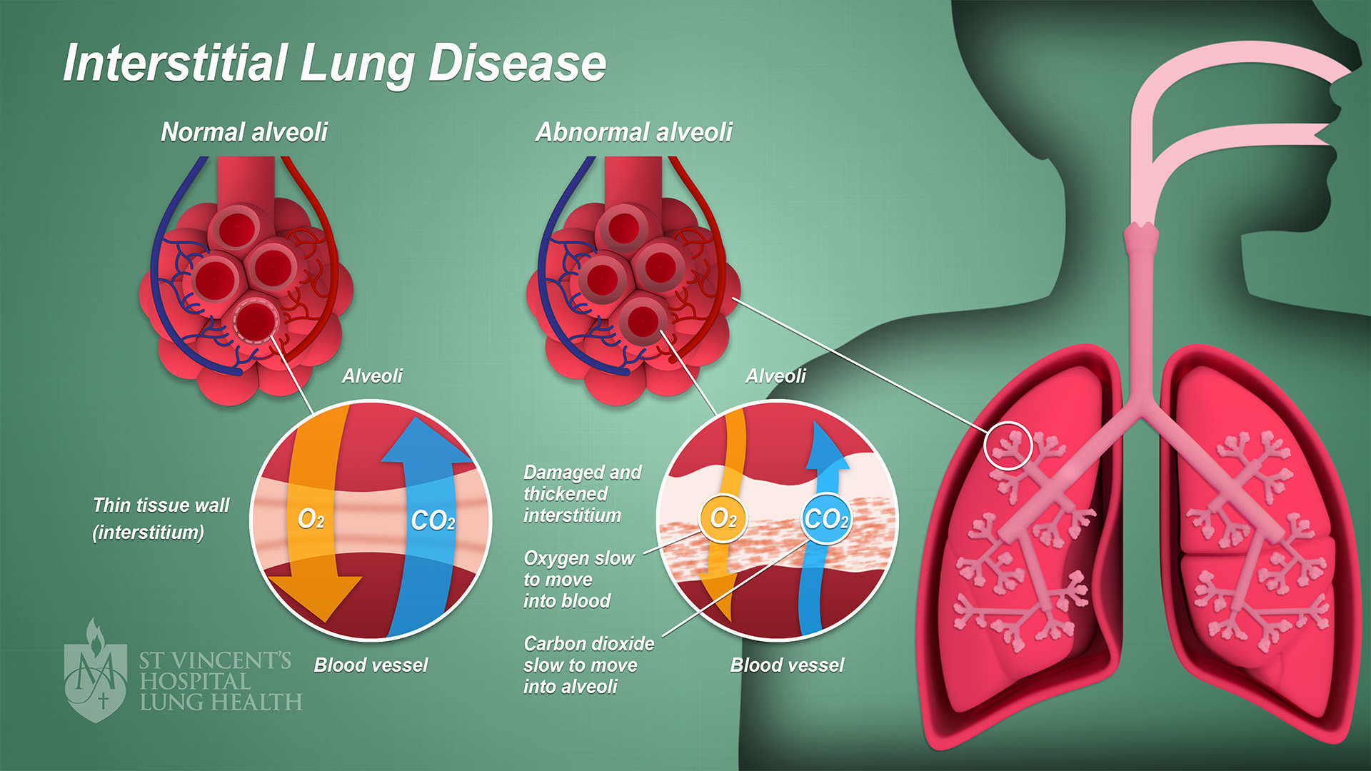
Interstitial lung disease (ILD), or diffuse parenchymal lung disease (DPLD), is a group of respiratory diseases affecting the interstitium (the tissue) and space around the alveoli (air sacs) of the lungs. It concerns alveolar epithelium, pulmonary capillary endothelium, basement membrane, and perivascular and perilymphatic tissues. It may occur when an injury to the lungs triggers an abnormal healing response. Ordinarily, the body generates just the right amount of tissue to repair damage, but in interstitial lung disease, the repair process is disrupted, and the tissue around the air sacs (alveoli) becomes scarred and thickened. This makes it more difficult for oxygen to pass into the bloodstream. The disease presents itself with the following symptoms: shortness of breath, nonproductive coughing, fatigue, and weight loss, which tend to develop slowly, over several months. The average rate of survival for someone with this disease is between three and five years. The term IL ... [...More Info...] [...Related Items...] OR: [Wikipedia] [Google] [Baidu] |
Pulmonary Infarct
Lung infarction or pulmonary infarction occurs when an artery to the lung becomes blocked and part of the lung dies. It is most often caused by a pulmonary embolism. Because of the dual blood supply to the lungs from both the bronchial circulation and the pulmonary circulation, this tissue is more resistant to infarction. An occlusion of the bronchial circulation does not cause infarction, but it can still occur in pulmonary embolism when the pulmonary circulation is blocked and the bronchial circulation cannot fully compensate for it. File:CT of lung infarction with reverse halo sign, annotated.png, CT scan of a lung infarction because of chronic pulmonary embolism Pulmonary embolism (PE) is a blockage of an pulmonary artery, artery in the lungs by a substance that has moved from elsewhere in the body through the bloodstream (embolism). Symptoms of a PE may include dyspnea, shortness of breath, chest pain ... (white arrow). The infarcted area (black arrow) has a reverse h ... [...More Info...] [...Related Items...] OR: [Wikipedia] [Google] [Baidu] |
Pulmonary Hemorrhage
Pulmonary hemorrhage (or pulmonary haemorrhage) is an acute bleeding from the lung, from the upper respiratory tract and the trachea, and the pulmonary alveoli. When evident clinically, the condition is usually massive.Pulmonary Hemorrhage Intensive Care Nursery House Staff Manual. UCSF Children's Hospital at UCSF Medical Center. 2004:The Regents of the . Retrieved 2008-10-28. The onset of pulmonary hemorrhage is characterized by a cough productive of ( |


