|
Adductor Magnus
The adductor magnus is a large triangular muscle, situated on the medial side of the thigh. It consists of two parts. The portion which arises from the ischiopubic ramus (a small part of the inferior ramus of the pubis, and the inferior ramus of the ischium) is called the pubofemoral portion, adductor portion, or adductor minimus, and the portion arising from the tuberosity of the ischium is called the ischiocondylar portion, extensor portion, or "hamstring portion". Due to its common embryonic origin, innervation, and action the ischiocondylar portion (or hamstring portion) is often considered part of the hamstring group of muscles. The ischiocondylar portion of the adductor magnus is considered a muscle of the posterior compartment of the thigh while the pubofemoral portion of the adductor magnus is considered a muscle of the medial compartment. Structure Pubofemoral (adductor) portion Those fibers which arise from the ramus of the pubis are short, horizontal in direc ... [...More Info...] [...Related Items...] OR: [Wikipedia] [Google] [Baidu] [Amazon] |
Hip-joint
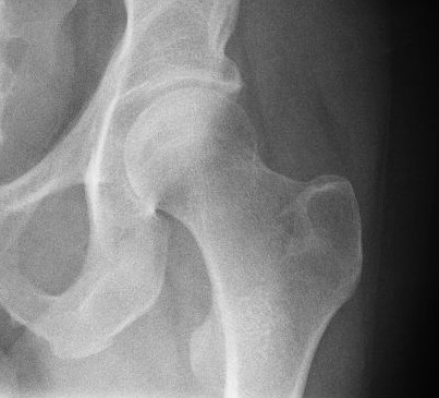
In vertebrate anatomy, the hip, or coxaLatin ''coxa'' was used by Celsus in the sense "hip", but by Pliny the Elder in the sense "hip bone" (Diab, p 77) (: ''coxae'') in medical terminology, refers to either an list of human anatomical regions, anatomical region or a joint on the outer (lateral) side of the pelvis. The hip region is located lateral (anatomy), lateral and anterior (anatomy), anterior to the Buttocks, gluteal region, inferior (anatomy), inferior to the iliac crest, and lateral to the obturator foramen, with muscle tendons and soft tissues overlying the greater trochanter of the femur. In adults, the three pelvic bones (ilium (bone), ilium, ischium and pubis (bone), pubis) have fused into one hip bone, which forms the superomedial/deep wall of the hip region. The hip joint, scientifically referred to as the acetabulofemoral joint (''art. coxae''), is the ball-and-socket joint between the pelvic acetabulum and the femoral head. Its primary function is to weight-bear ... [...More Info...] [...Related Items...] OR: [Wikipedia] [Google] [Baidu] [Amazon] |
Inferior Ramus Of The Ischium
The ischium (; : ischia) is a paired bone forming the lower and back part of the . Situated below the ilium and behind the pubis, it is one of three regions whose fusion creates the . The superior portion of this region forms approximately one-third of the |
Femoral Vein
In the human body, the femoral vein is the vein that accompanies the femoral artery in the femoral sheath. It is a deep vein that begins at the adductor hiatus (an opening in the adductor magnus muscle) as the continuation of the popliteal vein. The great saphenous vein (a superficial vein), and the deep femoral vein drain into the femoral vein in the femoral triangle when it becomes known as the common femoral vein. It ends at the inferior margin of the inguinal ligament where it becomes the external iliac vein. Its major tributaries are the deep femoral vein, and the great saphenous vein. The femoral vein contains valves. Structure The femoral vein bears valves which are mostly bicuspid and whose number is variable between individuals and often between left and right leg. Course The femoral vein continues into the thigh as the continuation from the popliteal vein at the back of the knee. It drains blood from the deep thigh muscles and thigh bone. Proximal to th ... [...More Info...] [...Related Items...] OR: [Wikipedia] [Google] [Baidu] [Amazon] |
Femoral Artery
The femoral artery is a large artery in the thigh and the main arterial supply to the thigh and leg. The femoral artery gives off the deep femoral artery and descends along the anteromedial part of the thigh in the femoral triangle. It enters and passes through the adductor canal, and becomes the popliteal artery as it passes through the adductor hiatus in the adductor magnus near the junction of the middle and distal thirds of the thigh. The femoral artery proximal to the origin of the deep femoral artery is referred to as the ''common femoral artery'', whereas the femoral artery distal to this origin is referred to as the ''superficial femoral artery''. Structure The femoral artery represents the continuation of the external iliac artery beyond the inguinal ligament underneath which the vessel passes to enter the thigh. The vessel passes under the inguinal ligament just medial of the midpoint of this ligament, midway between the anterior superior iliac spine and ... [...More Info...] [...Related Items...] OR: [Wikipedia] [Google] [Baidu] [Amazon] |
Adductor Longus Muscle
In the human body, the adductor longus is a skeletal muscle located in the thigh. One of the adductor muscles of the hip, its main function is to adduct the thigh and it is innervated by the obturator nerve. It forms the medial wall of the femoral triangle. Structure The adductor longus arises from the body of pubis inferior to pubic crest and lateral to pubic symphysis. It lies ventrally on the adductor magnus, and near the femur, the adductor brevis is interposed between these two muscles. Distally, the fibers of the adductor longus extend into the adductor canal. It is inserted into the middle third of the medial lip of the '' linea aspera''. Innervation As part of the medial compartment of the thigh, the adductor longus is innervated by the anterior division (sometimes the posterior division) of the obturator nerve. The obturator nerve exits via the anterior rami of the spinal cord The spinal cord is a long, thin, tubular structure made up of nervous tissue ... [...More Info...] [...Related Items...] OR: [Wikipedia] [Google] [Baidu] [Amazon] |
Adductor Brevis Muscle
The adductor brevis is a muscle in the thigh situated immediately deep to the pectineus and adductor longus. It belongs to the adductor muscle group. The main function of the adductor brevis is to pull the thigh medially. The adductor brevis and the rest of the adductor muscle group is also used to stabilize left to right movements of the trunk, when standing on both feet, or to balance when standing on a moving surface. The adductor muscle group is used pressing the thighs together to ride a horse, and kicking with the inside of the foot in soccer or swimming. Last, they contribute to flexion Motion, the process of movement, is described using specific anatomical terminology, anatomical terms. Motion includes movement of Organ (anatomy), organs, joints, Limb (anatomy), limbs, and specific sections of the body. The terminology used de ... of the thigh when running or against resistance (squats, jumping, etc.). Structure It is somewhat triangular in form, and arises b ... [...More Info...] [...Related Items...] OR: [Wikipedia] [Google] [Baidu] [Amazon] |
Pectineus Muscle
The pectineus muscle (, from the Latin word ''pecten'', meaning comb) is a flat, quadrangular muscle, situated at the anterior (front) part of the upper and medial (inner) aspect of the thigh. The pectineus muscle is the most anterior adductor of the hip. The muscle's primary action is hip flexion; it also produces adduction and external rotation of the hip. It can be classified in the medial compartment of thigh (when the function is emphasized) or the anterior compartment of thigh (when the nerve is emphasized). Structure The pectineus muscle arises from the pectineal line of the pubis and to a slight extent from the surface of bone in front of it, between the iliopectineal eminence and pubic tubercle, and from the fascia covering the anterior surface of the muscle; the fibers pass downward, backward, and lateral, to be inserted into the pectineal line of the femur which leads from the lesser trochanter to the linea aspera. Relations The pectineus is in relation by its ... [...More Info...] [...Related Items...] OR: [Wikipedia] [Google] [Baidu] [Amazon] |
Medial Condyle Of Femur
The medial condyle is one of the two projections on the lower extremity of femur, the other being the lateral condyle. The medial condyle is larger than the lateral (outer) condyle due to more weight bearing caused by the centre of mass being medial to the knee. On the posterior surface of the condyle the linea aspera The linea aspera () is a ridge of roughened surface on the posterior surface of the shaft of the femur. It is the site of attachments of muscles and the intermuscular septum. Its margins diverge above and below. The linea aspera is a prominent ... (a ridge with two lips: medial and lateral; running down the posterior shaft of the femur) turns into the medial and lateral supracondylar ridges, respectively. The outermost protrusion on the medial surface of the medial condyle is referred to as the "medial epicondyle" and can be palpated by running fingers medially from the patella with the knee in flexion. It is important to take into consideration the diff ... [...More Info...] [...Related Items...] OR: [Wikipedia] [Google] [Baidu] [Amazon] |
Aponeurosis
An aponeurosis (; : aponeuroses) is a flattened tendon by which muscle attaches to bone or fascia. Aponeuroses exhibit an ordered arrangement of collagen fibres, thus attaining high tensile strength in a particular direction while being vulnerable to tensional or shear forces in other directions. They have a shiny, whitish-silvery color, are histologically similar to tendons, and are very sparingly supplied with blood vessels and nerves. When dissected, aponeuroses are papery and peel off by sections. The primary regions with thick aponeuroses are in the ventral abdominal region, the dorsal lumbar region, the ventriculus in birds, and the palmar (palms) and plantar (soles) regions. Anatomy Anterior abdominal aponeuroses The anterior abdominal aponeuroses are located just superficial to the rectus abdominis muscle. It has for its borders the external oblique, pectoralis muscles, and the latissimus dorsi. Posterior lumbar aponeuroses The posterior lumbar aponeuroses are sit ... [...More Info...] [...Related Items...] OR: [Wikipedia] [Google] [Baidu] [Amazon] |
Ischium
The ischium (; : ischia) is a paired bone forming the lower and back part of the hip bone. Situated below the ilium (bone), ilium and behind the pubis (bone), pubis, it is one of three regions whose fusion creates the coxal bone. The superior portion of this region forms approximately one-third of the acetabulum. Structure The ischium is made up of three parts–the body, the superior ramus and the inferior ramus. The body contains a prominent ischial spine, spine, which serves as the origin for the superior gemellus muscle. The indentation inferior to the spine is the lesser sciatic notch. Continuing down the posterior side, the ischial tuberosity is a thick, rough-surfaced prominence below the lesser sciatic notch. This is the portion ...[...More Info...] [...Related Items...] OR: [Wikipedia] [Google] [Baidu] [Amazon] |
Gluteus Maximus Muscle
The gluteus maximus is the main extensor muscle of the hip in humans. It is the largest and outermost of the three gluteal muscles and makes up a large part of the shape and appearance of each side of the hips. It is the single largest muscle in the human body. Its thick fleshy mass, in a quadrilateral shape, forms the prominence of the buttocks. The other gluteal muscles are the medius and minimus, and sometimes informally these are collectively referred to as the glutes. Its large size is one of the most characteristic features of the muscular system in humans,Norman Eizenberg et al., ''General Anatomy: Principles and Applications'' (2008), p. 17. connected as it is with the power of maintaining the trunk in the erect posture. Other primates have much flatter hips and cannot sustain standing erectly. The muscle is made up of muscle fascicles lying parallel with one another, and are collected together into larger bundles separated by fibrous septa. Structure The gluteus ma ... [...More Info...] [...Related Items...] OR: [Wikipedia] [Google] [Baidu] [Amazon] |
Linea Aspera
The linea aspera () is a ridge of roughened surface on the posterior surface of the shaft of the femur. It is the site of attachments of muscles and the intermuscular septum. Its margins diverge above and below. The linea aspera is a prominent longitudinal ridge or crest, on the middle third of the bone, presenting a medial and a lateral lip, and a narrow rough, intermediate line. It is an important insertion point for the adductors and the lateral and medial intermuscular septa that divides the thigh into three compartments. The tension generated by muscle attached to the bones is responsible for the formation of the ridges. Structure Above Above, the linea aspera is prolonged by three ridges. * The lateral ridge is very rough, and runs almost vertically upward to the base of the greater trochanter. It is termed the gluteal tuberosity, and gives attachment to part of the gluteus maximus: its upper part is often elongated into a roughened crest, on which a more or less wel ... [...More Info...] [...Related Items...] OR: [Wikipedia] [Google] [Baidu] [Amazon] |


