|
White Adipose Tissue
White adipose tissue or white fat is one of the two types of adipose tissue found in mammals. The other kind is brown adipose tissue. White adipose tissue is composed of monolocular Adipocyte, adipocytes. In humans, the healthy body fat percentage, amount of white adipose tissue varies with age, but composes between 6–25% of body weight in adult men and 14–35% in adult women. Its cells contain a single large Lipid droplet, fat droplet, which forces the nucleus to be squeezed into a thin rim at the periphery. They have receptors for insulin, sex hormones, norepinephrine, and glucocorticoids. White adipose tissue is used for energy storage. Upon release of insulin from the pancreas, white adipose cells' insulin receptors cause a dephosphorylation cascade that leads to the inactivation of hormone-sensitive lipase. It was previously thought that upon release of glucagon from the pancreas, glucagon receptors cause a phosphorylation cascade that activates hormone-sensitive lipase ... [...More Info...] [...Related Items...] OR: [Wikipedia] [Google] [Baidu] |
Adipose Tissue
Adipose tissue (also known as body fat or simply fat) is a loose connective tissue composed mostly of adipocytes. It also contains the stromal vascular fraction (SVF) of cells including preadipocytes, fibroblasts, Blood vessel, vascular endothelial cells and a variety of White blood cell, immune cells such as adipose tissue macrophages. Its main role is to store energy in the form of lipids, although it also cushions and Thermal insulation, insulates the body. Previously treated as being hormonally inert, in recent years adipose tissue has been recognized as a major endocrine organ, as it produces hormones such as leptin, estrogen, resistin, and cytokines (especially TNF-alpha, TNFα). In obesity, adipose tissue is implicated in the chronic release of pro-inflammatory markers known as adipokines, which are responsible for the development of metabolic syndromea constellation of diseases including type 2 diabetes, cardiovascular disease and atherosclerosis. Adipose tissue is d ... [...More Info...] [...Related Items...] OR: [Wikipedia] [Google] [Baidu] |
Gluconeogenesis
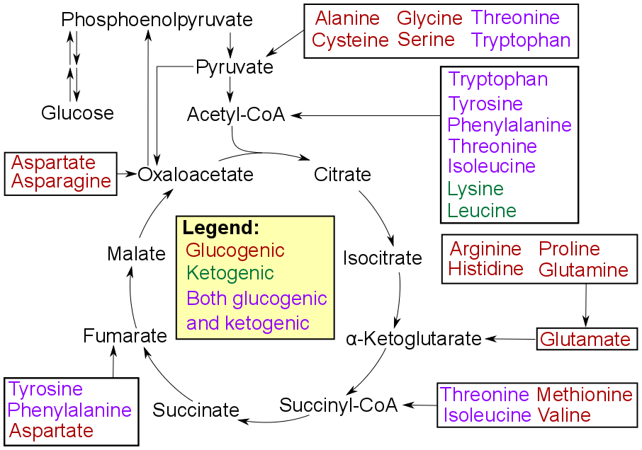
Gluconeogenesis (GNG) is a metabolic pathway that results in the biosynthesis of glucose from certain non-carbohydrate carbon substrates. It is a ubiquitous process, present in plants, animals, fungi, bacteria, and other microorganisms. In vertebrates, gluconeogenesis occurs mainly in the liver and, to a lesser extent, in the cortex of the kidneys. It is one of two primary mechanisms – the other being degradation of glycogen ( glycogenolysis) – used by humans and many other animals to maintain blood sugar levels, avoiding low levels (hypoglycemia). In ruminants, because dietary carbohydrates tend to be metabolized by rumen organisms, gluconeogenesis occurs regardless of fasting, low-carbohydrate diets, exercise, etc. In many other animals, the process occurs during periods of fasting, starvation, low-carbohydrate diets, or intense exercise. In humans, substrates for gluconeogenesis may come from any non-carbohydrate sources that can be converted to pyruvate or inter ... [...More Info...] [...Related Items...] OR: [Wikipedia] [Google] [Baidu] |
PPAR Gamma
Peroxisome proliferator-activated receptor gamma (PPAR-γ or PPARG), also known as the glitazone reverse insulin resistance receptor, or NR1C3 (nuclear receptor subfamily 1, group C, member 3) is a type II nuclear receptor functioning as a transcription factor that in humans is encoded by the ''PPARG'' gene. Tissue distribution PPARG is mainly present in adipose tissue, colon and macrophages. Two isoforms of PPARG are detected in the human and in the mouse: PPAR-γ1 (found in nearly all tissues except muscle) and PPAR-γ2 (mostly found in adipose tissue and the intestine). Gene expression This gene encodes a member of the peroxisome proliferator-activated receptor (PPAR) subfamily of nuclear receptors. PPARs form heterodimers with retinoid X receptors (RXRs) and these heterodimers regulate transcription of various genes. Three subtypes of PPARs are known: PPAR-alpha, PPAR-delta, and PPAR-gamma. The protein encoded by this gene is PPAR-gamma and is a regulator of adipocyte dif ... [...More Info...] [...Related Items...] OR: [Wikipedia] [Google] [Baidu] |
Preadipocyte
A lipoblast is a precursor cell for an adipocyte. Alternate terms include adipoblast and preadipocyte. Early stages are almost indistinguishable from fibroblasts. File:Lipoblasts and lipocytes.jpg, Lipoblasts (white arrow) and lipocytes (black arrow), in a case of lipoblastoma File:Dedifferentiated liposarcoma - cropped - very high mag.jpg, Micrograph showing a lipoblast (left-bottom of image) in a liposarcoma. H&E stain. File:Histopathology of liposarcoma, annotated.jpg, Histopathology of liposarcoma, H&E stain, with the main features: Topic Completed: 1 November 2017. Minor changes: 11 May 2021- Spindle cells with enlarged, hyperchromatic nuclei.- Apparently univacuolated adipocytes (may look normal).- Lipoblasts (multivacuolated), but neither necessary nor sufficient for diagnosis of liposarcoma. File:Histology of a lipoblast-like histiocyte in fat necrosis.jpg, Lipid-laden histiocytes may mimic lipoblasts, but have lightly eosinophilic cytoplasm and a small normochromatic ... [...More Info...] [...Related Items...] OR: [Wikipedia] [Google] [Baidu] |
Adipocyte
Adipocytes, also known as lipocytes and fat cells, are the cell (biology), cells that primarily compose adipose tissue, specialized in storing energy as fat. Adipocytes are derived from mesenchymal stem cells which give rise to adipocytes through adipogenesis. In cell culture, adipocyte progenitors can also form osteoblasts, myocytes and other cell types. There are two types of adipose tissue, white adipose tissue (WAT) and brown adipose tissue (BAT), which are also known as white and brown fat, respectively, and comprise two types of fat cells. Structure White fat cells White fat cells contain a single large lipid droplet surrounded by a layer of cytoplasm, and are known as unilocular. The Cell nucleus, nucleus is flattened and pushed to the periphery. A typical fat cell is 0.1 mm in diameter with some being twice that size, and others half that size. However, these numerical estimates of fat cell size depend largely on the measurement method and the location of the adi ... [...More Info...] [...Related Items...] OR: [Wikipedia] [Google] [Baidu] |
Visceral Adiposity
Abdominal obesity, also known as central obesity and truncal obesity, is the human condition of an excessive concentration of visceral fat around the stomach and abdomen to such an extent that it is likely to harm its bearer's health. Abdominal obesity has been strongly linked to cardiovascular disease, Alzheimer's disease, and other metabolic and vascular diseases. Visceral fat, central abdominal fat, and waist circumference show a strong association with type 2 diabetes. Visceral fat, also known as organ fat or ''intra-abdominal fat'', is located inside the peritoneal cavity, packed in between internal organs and torso, as opposed to subcutaneous fat, which is found underneath the skin, and intramuscular fat, which is found interspersed in skeletal muscle. Visceral fat is composed of several adipose depots including mesenteric, epididymal white adipose tissue (EWAT), and perirenal fat. An excess of adipose visceral fat is known as central obesity, the "pot belly" or "bee ... [...More Info...] [...Related Items...] OR: [Wikipedia] [Google] [Baidu] |
Abdominal Cavity
The abdominal cavity is a large body cavity in humans and many other animals that contain Organ (anatomy), organs. It is a part of the abdominopelvic cavity. It is located below the thoracic cavity, and above the pelvic cavity. Its dome-shaped roof is the thoracic diaphragm, a thin sheet of muscle under the lungs, and its floor is the pelvic inlet, opening into the pelvis. Structure Organs Organs of the abdominal cavity include the stomach, liver, gallbladder, spleen, pancreas, small intestine, kidneys, large intestine, and adrenal glands. Peritoneum The abdominal cavity is lined with a protective membrane termed the peritoneum. The inside wall is covered by the parietal peritoneum. The kidneys are located behind the peritoneum, in the retroperitoneum, outside the abdominal cavity. The viscera are also covered by visceral peritoneum. Between the visceral and parietal peritoneum is the peritoneal cavity, which is a potential space. It contains a serous fluid called peritoneal ... [...More Info...] [...Related Items...] OR: [Wikipedia] [Google] [Baidu] |
Thoracic Cavity
The thoracic cavity (or chest cavity) is the chamber of the body of vertebrates that is protected by the thoracic wall (rib cage and associated skin, muscle, and fascia). The central compartment of the thoracic cavity is the mediastinum. There are two openings of the thoracic cavity, a superior thoracic aperture known as the thoracic inlet and a lower inferior thoracic aperture known as the thoracic outlet. The thoracic cavity includes the tendons as well as the cardiovascular system which could be damaged from injury to the back, spine or the neck. Structure Structures within the thoracic cavity include: * structures of the cardiovascular system, including the heart and great vessels, which include the thoracic aorta, the pulmonary artery and all its branches, the superior and inferior vena cava, the pulmonary veins, and the azygos vein * structures of the respiratory system, including the diaphragm, trachea, bronchi and lungs * structures of the digestive system, incl ... [...More Info...] [...Related Items...] OR: [Wikipedia] [Google] [Baidu] |
Subcutaneous Adipose Tissue
Adipose tissue (also known as body fat or simply fat) is a loose connective tissue composed mostly of adipocytes. It also contains the stromal vascular fraction (SVF) of cells including preadipocytes, fibroblasts, vascular endothelial cells and a variety of immune cells such as adipose tissue macrophages. Its main role is to store energy in the form of lipids, although it also cushions and insulates the body. Previously treated as being hormonally inert, in recent years adipose tissue has been recognized as a major endocrine organ, as it produces hormones such as leptin, estrogen, resistin, and cytokines (especially TNFα). In obesity, adipose tissue is implicated in the chronic release of pro-inflammatory markers known as adipokines, which are responsible for the development of metabolic syndromea constellation of diseases including type 2 diabetes, cardiovascular disease and atherosclerosis. Adipose tissue is derived from preadipocytes and its formation appears to be cont ... [...More Info...] [...Related Items...] OR: [Wikipedia] [Google] [Baidu] |
White Adipose Distribution In The Body
White is the lightest color and is achromatic (having no chroma). It is the color of objects such as snow, chalk, and milk, and is the opposite of black. White objects fully (or almost fully) reflect and scatter all the visible wavelengths of light. White on television and computer screens is created by a mixture of red, blue, and green light. The color white can be given with white pigments, especially titanium dioxide. In ancient Egypt and ancient Rome, priestesses wore white as a symbol of purity, and Romans wore white togas as symbols of citizenship. In the Middle Ages and Renaissance a white unicorn symbolized chastity, and a white lamb sacrifice and purity. It was the royal color of the kings of France as well as the flag of monarchist France from 1815 to 1830, and of the monarchist movement that opposed the Bolsheviks during the Russian Civil War (1917–1922). Greek temples and Roman temples were faced with white marble, and beginning in the 18th century, wi ... [...More Info...] [...Related Items...] OR: [Wikipedia] [Google] [Baidu] |
Asprosin
Asprosin is a fasting-induced hormone encoded by the ''FBN1'' gene and derived from the cleavage of the fibrillin-1 protein, a structural component of the extracellular matrix. It is primarily produced and secreted by white adipose tissue. As a peripherally derived hormone, asprosin actively crosses the blood-brain barrier (BBB) to exert central effects on metabolic and behavioral regulation. It stimulates the liver to release glucose into the bloodstream during fasting, ensuring energy availability, and influences appetite and body weight regulation by acting on hypothalamic neurons. Dysregulation of asprosin levels has been implicated in metabolic disorders such as obesity and diabetes, making it a promising target for therapeutic interventions. Discovery Asprosin was first identified by Dr. Atul Chopra and colleagues at Baylor College of Medicine during their study of Marfanoid–progeroid–lipodystrophy syndrome (MPL), also known as neonatal progeroid syndrome (NPS), a rare g ... [...More Info...] [...Related Items...] OR: [Wikipedia] [Google] [Baidu] |
Leptin
Leptin (from Ancient Greek, Greek λεπτός ''leptos'', "thin" or "light" or "small"), also known as obese protein, is a protein hormone predominantly made by adipocytes (cells of adipose tissue). Its primary role is likely to regulate long-term Energy homeostasis, energy balance. As one of the major signals of energy status, leptin levels influence appetite, Hunger (physiology), satiety, and motivated behaviors oriented toward the maintenance of energy reserves (e.g., feeding, foraging behaviors). The amount of circulating leptin correlates with the amount of energy reserves, mainly Triglyceride, triglycerides stored in adipose tissue. High leptin levels are interpreted by the brain that energy reserves are high, whereas low leptin levels indicate that energy reserves are low, in the process adapting the organism to Starvation response, starvation through a variety of metabolic, endocrine, neurobiochemical, and behavioral changes. Leptin is coded for by the ''LEP'' gene ... [...More Info...] [...Related Items...] OR: [Wikipedia] [Google] [Baidu] |


