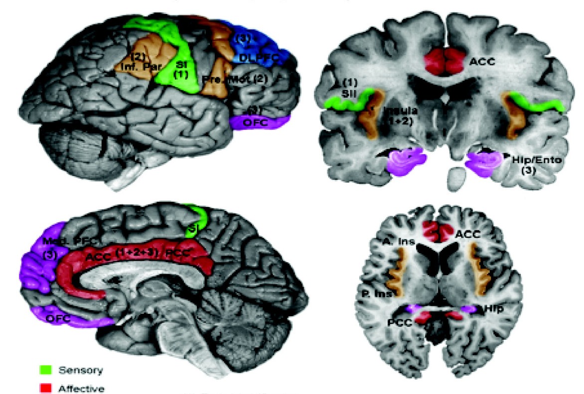
|
Ventral Posterolateral Nucleus
The ventral posterolateral nucleus (VPL) is one of the subdivisions of the ventral posterior nucleus in the ventral nuclear group of the thalamus. It relays sensory information from the second-order neurons of the neospinothalamic tract and medial lemniscus (of the dorsal column-medial lemniscus pathway) which synapse with the third-order neurons in the nucleus. These then project to the primary somatosensory cortex in the postcentral gyrus. There is uncertainty regarding the location of VMpo (posterior part of ventral medial nucleus), as determined by spinothalamic tract (STT) terminations and staining for calcium-binding proteins, and several authorities do not consider its existence as being proved. The term "ventral posterolateral nucleus" was introduced by Le Gros Clark in 1930. Anatomy Subdivisions The oral part of the ventral posterolateral nucleus (nucleus ventrointermedius) in the human, (VPLO) is a subdivision of the VPL with projections to the motor cortex ... [...More Info...] [...Related Items...] OR: [Wikipedia] [Google] [Baidu] |
Midline Nuclear Group
The midline nuclear group (or midline thalamic nuclei) is a region of the thalamus consisting of the following nuclei: * Paraventricular thalamus, paraventricular nucleus of thalamus (''nucleus paraventricularis thalami'') - not to be confused with paraventricular nucleus of hypothalamus * paratenial nucleus (''nucleus parataenialis'') * nucleus reuniens (also known as the medioventral nucleus) * rhomboidal nucleus (''nucleus commissuralis rhomboidalis'') * subfascicular nucleus (''nucleus subfascicularis'') The List of regions in the human brain#Thalamus, midline nuclei are often called "nonspecific" in that they project widely to the cortex and elsewhere. This has led to the assumption that they may be involved in general functions such as alerting. However, anatomical connections might suggest more specific functions, with the paraventricular and paratenial nuclei involved in viscero-limbic functions, and the reuniens and rhomboid nuclei involved in multimodal sensory processin ... [...More Info...] [...Related Items...] OR: [Wikipedia] [Google] [Baidu] |
Ventral Nuclear Group
The ventral nuclear group is a collection of nuclei on the ventral side of the thalamus The thalamus (: thalami; from Greek language, Greek Wikt:θάλαμος, θάλαμος, "chamber") is a large mass of gray matter on the lateral wall of the third ventricle forming the wikt:dorsal, dorsal part of the diencephalon (a division of ..., it consists of the following: * ventral anterior nucleus * ventral lateral nucleus * ventral posterior nucleus – this is made up of two nuclei: the ventral posterolateral nucleus and the ventral posteromedial nucleus See also * Anterolateral region of the motor thalamus References Thalamus {{Neuroanatomy-stub ... [...More Info...] [...Related Items...] OR: [Wikipedia] [Google] [Baidu] |
Motor Cortex
The motor cortex is the region of the cerebral cortex involved in the planning, motor control, control, and execution of voluntary movements. The motor cortex is an area of the frontal lobe located in the posterior precentral gyrus immediately anterior to the central sulcus. Components The motor cortex can be divided into three areas: 1. The primary motor cortex is the main contributor to generating neural impulses that pass down to the spinal cord and control the execution of movement. However, some of the other motor areas in the brain also play a role in this function. It is located on the anterior paracentral lobule on the medial surface. 2. The premotor cortex is responsible for some aspects of motor control, possibly including the preparation for movement, the sensory guidance of movement, the spatial guidance of reaching, or the direct control of some movements with an emphasis on control of proximal and trunk muscles of the body. Located anterior to the primary mo ... [...More Info...] [...Related Items...] OR: [Wikipedia] [Google] [Baidu] |
Postcentral Gyrus
In neuroanatomy, the postcentral gyrus is a prominent gyrus in the lateral parietal lobe of the human brain. It is the location of the primary somatosensory cortex, the main sensory receptive area for the sense of touch. Like other sensory areas, there is a map of sensory space in this location, called the '' sensory homunculus''. The primary somatosensory cortex was initially defined from surface stimulation studies of Wilder Penfield, and parallel surface potential studies of Bard, Woolsey, and Marshall. Although initially defined to be roughly the same as Brodmann areas 3, 1, and 2, more recent work by Kaas has suggested that for homogeny with other sensory fields only area 3 should be referred to as "primary somatosensory cortex", as it receives the bulk of the thalamocortical projections from the sensory input fields. Structure The lateral postcentral gyrus is bounded by: * medial longitudinal fissure medially (to the middle) * central sulcus rostrally (in front) ... [...More Info...] [...Related Items...] OR: [Wikipedia] [Google] [Baidu] |
Primary Somatosensory Cortex
In neuroanatomy, the primary somatosensory cortex is located in the postcentral gyrus of the brain's parietal lobe, and is part of the somatosensory system. It was initially defined from surface stimulation studies of Wilder Penfield, and parallel surface potential studies of Bard, Woolsey, and Marshall. Although initially defined to be roughly the same as Brodmann areas 3, 1 and 2, more recent work by Kaas has suggested that for homogeny with other sensory fields only area 3 should be referred to as "primary somatosensory cortex", as it receives the bulk of the thalamocortical projections from the sensory input fields. At the primary somatosensory cortex, tactile representation is orderly arranged (in an inverted fashion) from the toe (at the top of the cerebral hemisphere) to mouth (at the bottom). However, some body parts may be controlled by partially overlapping regions of cortex. Each cerebral hemisphere of the primary somatosensory cortex only contains a tactile representatio ... [...More Info...] [...Related Items...] OR: [Wikipedia] [Google] [Baidu] |
Nucleus (neuroanatomy)
In neuroanatomy, a nucleus (: nuclei) is a cluster of neurons in the central nervous system, located deep within the cerebral hemispheres and brainstem. The neurons in one nucleus usually have roughly similar connections and functions. Nuclei are connected to other nuclei by tracts, the bundles (fascicles) of axons (nerve fibers) extending from the cell bodies. A nucleus is one of the two most common forms of nerve cell organization, the other being layered structures such as the cerebral cortex or cerebellar cortex. In anatomical sections, a nucleus shows up as a region of gray matter, often bordered by white matter. The vertebrate brain contains hundreds of distinguishable nuclei, varying widely in shape and size. A nucleus may itself have a complex internal structure, with multiple types of neurons arranged in clumps (subnuclei) or layers. The term "nucleus" is in some cases used rather loosely, to mean simply an identifiably distinct group of neurons, even if they are sprea ... [...More Info...] [...Related Items...] OR: [Wikipedia] [Google] [Baidu] |
Dorsal Column-medial Lemniscus Pathway
Dorsal (from Latin ''dorsum'' ‘back’) may refer to: * Dorsal (anatomy), an anatomical term of location referring to the back or upper side of an organism or parts of an organism * Dorsal, positioned on top of an aircraft's fuselage The fuselage (; from the French language, French ''fuselé'' "spindle-shaped") is an aircraft's main body section. It holds Aircrew, crew, passengers, or cargo. In single-engine aircraft, it will usually contain an Aircraft engine, engine as wel ... * Dorsal consonant, a consonant articulated with the back of the tongue * Dorsal fin, the fin located on the back of a fish or aircraft * Dorsal transcription factor, a maternally synthesized transcription factor {{disambig ... [...More Info...] [...Related Items...] OR: [Wikipedia] [Google] [Baidu] |
Medial Lemniscus
The medial lemniscus, also known as Reil's band or Reil's ribbon (for German anatomist Johann Christian Reil), is a large ascending bundle of heavily myelinated axons that decussate in the brainstem The brainstem (or brain stem) is the posterior stalk-like part of the brain that connects the cerebrum with the spinal cord. In the human brain the brainstem is composed of the midbrain, the pons, and the medulla oblongata. The midbrain is conti ..., specifically in the medulla oblongata. The medial lemniscus is formed by the crossings of the internal arcuate fibers. The internal arcuate fibers are composed of axons of the gracile nucleus and the cuneate nucleus. The cell bodies of the nuclei lie contralaterally. The medial lemniscus is part of the somatosensory dorsal column–medial lemniscus pathway, which ascends in the spinal cord to the thalamus.Kamali A, Kramer LA, Butler IJ, Hasan KM. Diffusion tensor tractography of the somatosensory system in the human brains ... [...More Info...] [...Related Items...] OR: [Wikipedia] [Google] [Baidu] |
Neospinothalamic Tract
Pain is a distressing feeling often caused by intense or damaging stimuli. The International Association for the Study of Pain defines pain as "an unpleasant sensory and emotional experience associated with, or resembling that associated with, actual or potential tissue damage." Pain motivates organisms to withdraw from damaging situations, to protect a damaged body part while it heals, and to avoid similar experiences in the future. Congenital insensitivity to pain may result in reduced life expectancy. Most pain resolves once the noxious stimulus is removed and the body has healed, but it may persist despite removal of the stimulus and apparent healing of the body. Sometimes pain arises in the absence of any detectable stimulus, damage or disease. Pain is the most common reason for physician consultation in most developed countries. It is a major symptom in many medical conditions, and can interfere with a person's quality of life and general functioning. People in pain ... [...More Info...] [...Related Items...] OR: [Wikipedia] [Google] [Baidu] |
Second-order Neuron
Second-order may refer to: Mathematics * Second order approximation, an approximation that includes quadratic terms * Second-order arithmetic, an axiomatization allowing quantification of sets of numbers * Second-order differential equation, a differential equation in which the highest derivative is the second * Second-order logic, an extension of predicate logic * Second-order perturbation, in perturbation theory Science and technology * Second-order cybernetics, the recursive application of cybernetics to itself and the reflexive practice of cybernetics according to this critique. * Second-order fluid Second-order may refer to: Mathematics * Second order approximation, an approximation that includes quadratic terms * Second-order arithmetic, an axiomatization allowing quantification of sets of numbers * Second-order differential equation, a di ..., an extension of fluid dynamics * Second order Fresnel lens, a size of lighthouse lens * Second-order reaction, a reaction in w ... [...More Info...] [...Related Items...] OR: [Wikipedia] [Google] [Baidu] |
Thalamus
The thalamus (: thalami; from Greek language, Greek Wikt:θάλαμος, θάλαμος, "chamber") is a large mass of gray matter on the lateral wall of the third ventricle forming the wikt:dorsal, dorsal part of the diencephalon (a division of the forebrain). Nerve fibers project out of the thalamus to the cerebral cortex in all directions, known as the thalamocortical radiations, allowing hub (network science), hub-like exchanges of information. It has several functions, such as the relaying of sensory neuron, sensory and motor neuron, motor signals to the cerebral cortex and the regulation of consciousness, sleep, and alertness. Anatomically, the thalami are paramedian symmetrical structures (left and right), within the vertebrate brain, situated between the cerebral cortex and the midbrain. It forms during embryonic development as the main product of the diencephalon, as first recognized by the Swiss embryologist and anatomist Wilhelm His Sr. in 1893. Anatomy The thalami ar ... [...More Info...] [...Related Items...] OR: [Wikipedia] [Google] [Baidu] |


