|
Uterine Artery Embolization
Uterine artery embolization (UAE, uterine fibroid embolization, or UFE) is a procedure in which an interventional radiologist uses a catheter to deliver small particles that block the blood supply to the uterine body. The procedure is primarily done for the treatment of uterine fibroids and adenomyosis. Compared to surgical treatment for fibroids such as a hysterectomy, in which a woman's uterus is removed, uterine artery embolization may be beneficial in women who wish to retain their uterus. Other reasons for uterine artery embolization are postpartum hemorrhage and uterine arteriovenous malformations. Medical uses Uterine fibroids are the most common type of benign uterine tumor and are composed of smooth muscle. They often cause bulk-related symptoms, which can be characterized by back pain, heaviness in the pelvic area, abdominal bloating. Uterine artery embolization may be done to treat bothersome bulk-related symptoms as well as abnormal or heavy uterine bleeding due to ute ... [...More Info...] [...Related Items...] OR: [Wikipedia] [Google] [Baidu] |
Posterior (anatomy)
Standard anatomical terms of location are used to describe unambiguously the anatomy of humans and other animals. The terms, typically derived from Latin or Greek language, Greek roots, describe something in its standard anatomical position. This position provides a definition of what is at the front ("anterior"), behind ("posterior") and so on. As part of defining and describing terms, the body is described through the use of anatomical planes and anatomical axes, axes. The meaning of terms that are used can change depending on whether a vertebrate is a biped or a quadruped, due to the difference in the neuraxis, or if an invertebrate is a non-bilaterian. A non-bilaterian has no anterior or posterior surface for example but can still have a descriptor used such as proximal or distal in relation to a body part that is nearest to, or furthest from its middle. International organisations have determined vocabularies that are often used as standards for subdisciplines of anatomy. ... [...More Info...] [...Related Items...] OR: [Wikipedia] [Google] [Baidu] |
Uterine Balloon Tamponade
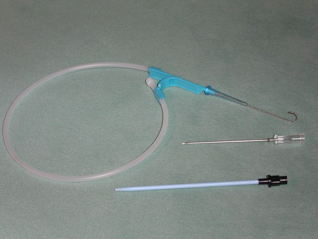
Uterine balloon tamponade (UBT) is a non-surgical method of treating refractory postpartum hemorrhage. Once postpartum hemorrhage has been identified and medical management given (including agents such as uterotonics and tranexamic acid), UBT may be employed to tamponade uterine bleeding without the need to pursue operative intervention. Numerous studies have supported the efficacy of UBT as a means of managing refractory postpartum hemorrhage. The International Federation of Gynecology and Obstetrics (FIGO) and the World Health Organization (WHO) recommend UBT as second-line treatment for severe postpartum hemorrhage. Method Regardless of which device is used, all share the same basic components and method of application. The UBT generally consists of a balloon, a catheter or some form of tubing to inflate the balloon, and a syringe to inflate the balloon. Balloons range from home-grown interventions such as a condom or glove, to custom made silicone balloons. After performing ut ... [...More Info...] [...Related Items...] OR: [Wikipedia] [Google] [Baidu] |
Quality Of Life (healthcare)
In healthcare, quality of life is an assessment of how the individual's well-being may be affected over time by a disease, disability or disorder. Measurement Early versions of healthcare-related quality of life measures referred to simple assessments of physical abilities by an external rater (for example, the patient is able to get up, eat and drink, and take care of personal hygiene without any help from others) or even to a single measurement (for example, the angle to which a limb could be flexed). The current concept of health-related quality of life acknowledges that subjects put their actual situation in relation to their personal expectation. The latter can vary over time, and react to external influences such as length and severity of illness, family support, etc. As with any situation involving multiple perspectives, patients' and physicians' rating of the same objective situation have been found to differ significantly. Consequently, health-related quality of life is ... [...More Info...] [...Related Items...] OR: [Wikipedia] [Google] [Baidu] |
Angiography
Angiography or arteriography is a medical imaging technique used to visualize the inside, or lumen, of blood vessels and organs of the body, with particular interest in the arteries, veins, and the heart chambers. Modern angiography is performed by injecting a radio-opaque contrast agent into the blood vessel and imaging using X-ray based techniques such as fluoroscopy. With time-of-flight (TOF) magnetic resonance it is no longer necessary to use a contrast. The word itself comes from the Greek words ἀνγεῖον ''angeion'' 'vessel' and γράφειν ''graphein'' 'to write, record'. The film or image of the blood vessels is called an ''angiograph'', or more commonly an ''angiogram''. Though the word can describe both an arteriogram and a venogram, in everyday usage the terms angiogram and arteriogram are often used synonymously, whereas the term venogram is used more precisely. The term angiography has been applied to radionuclide angiography and newer vascular ima ... [...More Info...] [...Related Items...] OR: [Wikipedia] [Google] [Baidu] |
Fluoroscopy
Fluoroscopy (), informally referred to as "fluoro", is an imaging technique that uses X-rays to obtain real-time moving images of the interior of an object. In its primary application of medical imaging, a fluoroscope () allows a surgeon to see the internal anatomy, structure and physiology, function of a patient, so that the pumping action of the heart or the motion of swallowing, for example, can be watched. This is useful for both medical diagnosis, diagnosis and therapy and occurs in general radiology, interventional radiology, and image-guided surgery. In its simplest form, a fluoroscope consists of an X-ray generator, X-ray source and a fluorescence, fluorescent screen, between which a patient is placed. However, since the 1950s most fluoroscopes have included X-ray image intensifiers and cameras as well, to improve the image's visibility and make it available on a remote display screen. For many decades, fluoroscopy tended to produce live pictures that were not recorded, bu ... [...More Info...] [...Related Items...] OR: [Wikipedia] [Google] [Baidu] |
Seldinger Technique
The Seldinger technique, also known as Seldinger wire technique, is a medical procedure to obtain safe access to blood vessels and other hollow organ (anatomy), organs. It is eponym, named after Sven Ivar Seldinger (1921–1998), a Sweden, Swedish radiology, radiologist who introduced the procedure in 1953. Uses The Seldinger technique is used for angiography, insertion of chest drains and central venous catheters, insertion of percutaneous endoscopic gastrostomy, PEG tubes using the push technique, insertion of the leads for an artificial pacemaker or implantable cardioverter-defibrillator, and numerous other interventional medical procedures. Complications The initial puncture is with a sharp instrument, and this may lead to hemorrhage or perforation of the organ in question. Infection is a possible complication, and hence asepsis is practiced during most Seldinger procedures. Loss of the guidewire into the cavity or blood vessel is a significant and generally preventable com ... [...More Info...] [...Related Items...] OR: [Wikipedia] [Google] [Baidu] |
Femoral Artery
The femoral artery is a large artery in the thigh and the main arterial supply to the thigh and leg. The femoral artery gives off the deep femoral artery and descends along the anteromedial part of the thigh in the femoral triangle. It enters and passes through the adductor canal, and becomes the popliteal artery as it passes through the adductor hiatus in the adductor magnus near the junction of the middle and distal thirds of the thigh. The femoral artery proximal to the origin of the deep femoral artery is referred to as the ''common femoral artery'', whereas the femoral artery distal to this origin is referred to as the ''superficial femoral artery''. Structure The femoral artery represents the continuation of the external iliac artery beyond the inguinal ligament underneath which the vessel passes to enter the thigh. The vessel passes under the inguinal ligament just medial of the midpoint of this ligament, midway between the anterior superior iliac spine and ... [...More Info...] [...Related Items...] OR: [Wikipedia] [Google] [Baidu] |
Radial Artery
In human anatomy, the radial artery is the main artery of the lateral aspect of the forearm. Structure The radial artery arises from the bifurcation of the brachial artery in the antecubital fossa. It runs distally on the anterior part of the forearm. There, it serves as a landmark for the division between the anterior compartment of the forearm, anterior and posterior compartment of the forearm, posterior compartments of the forearm, with the posterior compartment beginning just lateral to the artery. The artery winds laterally around the wrist, passing through the anatomical snuff box and between the heads of the first dorsal interossei of the hand, dorsal interosseous muscle. It passes anteriorly between the heads of the adductor pollicis, and becomes the deep palmar arch, which joins with the deep branch of the ulnar artery. Along its course, it is accompanied by a similarly named vein, the radial vein. Branches The named branches of the radial artery may be divided int ... [...More Info...] [...Related Items...] OR: [Wikipedia] [Google] [Baidu] |
Magnetic Resonance Imaging
Magnetic resonance imaging (MRI) is a medical imaging technique used in radiology to generate pictures of the anatomy and the physiological processes inside the body. MRI scanners use strong magnetic fields, magnetic field gradients, and radio waves to form images of the organs in the body. MRI does not involve X-rays or the use of ionizing radiation, which distinguishes it from computed tomography (CT) and positron emission tomography (PET) scans. MRI is a medical application of nuclear magnetic resonance (NMR) which can also be used for imaging in other NMR applications, such as NMR spectroscopy. MRI is widely used in hospitals and clinics for medical diagnosis, staging and follow-up of disease. Compared to CT, MRI provides better contrast in images of soft tissues, e.g. in the brain or abdomen. However, it may be perceived as less comfortable by patients, due to the usually longer and louder measurements with the subject in a long, confining tube, although ... [...More Info...] [...Related Items...] OR: [Wikipedia] [Google] [Baidu] |
Obstetrics And Gynaecology
Obstetrics and gynaecology (also spelled as obstetrics and gynecology; abbreviated as Obst and Gynae, O&G, OB-GYN and OB/GYN) is the medical specialty that encompasses the two subspecialties of obstetrics (covering pregnancy, childbirth, and the postpartum period) and gynaecology (covering the health of the female reproductive system – vagina, uterus, ovaries, and breasts). The specialization is an important part of care for women's health. Postgraduate training programs for both fields are usually combined, preparing the practising obstetrician-gynecologist to be adept both at the care of female reproductive organs' health and at the management of pregnancy, although many doctors go on to develop subspecialty interests in one field or the other. Scope United States According to the American Board of Obstetrics and Gynecology (ABOG), which is responsible for issuing OB-GYN certifications in the United States, the first step to OB-GYN certification is completing medica ... [...More Info...] [...Related Items...] OR: [Wikipedia] [Google] [Baidu] |
Sepsis
Sepsis is a potentially life-threatening condition that arises when the body's response to infection causes injury to its own tissues and organs. This initial stage of sepsis is followed by suppression of the immune system. Common signs and symptoms include fever, tachycardia, increased heart rate, hyperventilation, increased breathing rate, and mental confusion, confusion. There may also be symptoms related to a specific infection, such as a cough with pneumonia, or dysuria, painful urination with a pyelonephritis, kidney infection. The very young, old, and people with a immunodeficiency, weakened immune system may not have any symptoms specific to their infection, and their hypothermia, body temperature may be low or normal instead of constituting a fever. Severe sepsis may cause organ dysfunction and significantly reduced blood flow. The presence of Hypotension, low blood pressure, high blood Lactic acid, lactate, or Oliguria, low urine output may suggest poor blood flow. Se ... [...More Info...] [...Related Items...] OR: [Wikipedia] [Google] [Baidu] |


