|
Urea Transporter Family
A urea transporter is a membrane transport protein, transporting urea. Humans and other mammals have two types of urea transport proteins, UT-A and UT-B. The UT-A proteins are important for renal urea handling and are produced by alternative splicing of the SLC14A2 gene. Urea transport in the kidney is regulated by vasopressin. The structure of a urea transport family protein from '' Desulfovibrio vulgaris'' was determined by x-ray crystallography. The structure has a pathway through the membrane that is similar to that of ion channel proteins, accounting for the ability of urea transport proteins to move up to one million urea molecules per second across the membrane. Urea transporters can be inhibited by the action of urea analogues like thiourea and glycosides like phloretin. Their inhibition results in increased diuresis due to urea induced osmosis in the collecting ducts of the kidney. Types In mammals, there are two urea transporter genes: UT-A ('' SLC14A2'') and UT-B ... [...More Info...] [...Related Items...] OR: [Wikipedia] [Google] [Baidu] |
Membrane Transport Protein
A membrane transport protein is a membrane protein involved in the movement of ions, small molecules, and macromolecules, such as another protein, across a biological membrane. Transport proteins are integral membrane proteins, integral transmembrane proteins; that is they exist permanently within and span the membrane across which they transport substances. The proteins may assist in the movement of substances by facilitated diffusion, active transport, osmosis, or reverse diffusion. The two main types of proteins involved in such transport are broadly categorized as either ''channels'' or ''carriers'' (a.k.a. transporters, or permeases). Examples of channel/carrier proteins include the GLUT1, GLUT 1 uniporter, sodium channels, and potassium channels. The Solute carrier family, solute carriers and atypical SLCs are secondary active or facilitative transporters in humans. Collectively membrane transporters and channels are known as the transportome. Transportomes govern cellular i ... [...More Info...] [...Related Items...] OR: [Wikipedia] [Google] [Baidu] |
Promoter (biology)
In genetics, a promoter is a sequence of DNA to which proteins bind to initiate transcription (genetics), transcription of a single RNA transcript from the DNA downstream of the promoter. The RNA transcript may encode a protein (mRNA), or can have a function in and of itself, such as tRNA or rRNA. Promoters are located near the transcription start sites of genes, Upstream and downstream (DNA), upstream on the DNA (towards the Directionality (molecular biology)#5′-end, 5' region of the sense strand). Promoters can be about 100–1000 base pairs long, the sequence of which is highly dependent on the gene and product of transcription, type or class of RNA polymerase recruited to the site, and species of organism. Overview For transcription to take place, the enzyme that synthesizes RNA, known as RNA polymerase, must attach to the DNA near a gene. Promoters contain specific DNA sequences such as response elements that provide a secure initial binding site for RNA polymerase and for ... [...More Info...] [...Related Items...] OR: [Wikipedia] [Google] [Baidu] |
Transmembrane Transporters
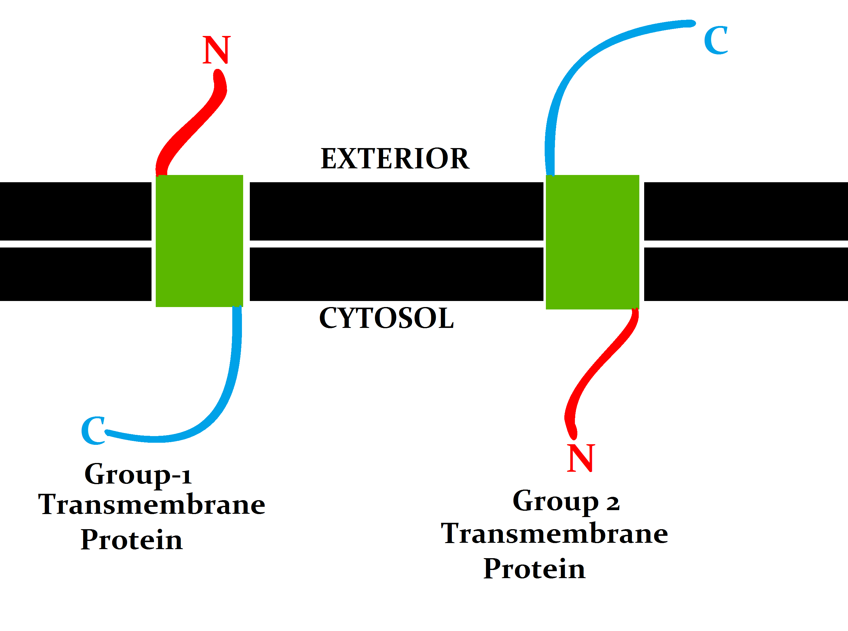
A transmembrane protein is a type of integral membrane protein that spans the entirety of the cell membrane. Many transmembrane proteins function as gateways to permit the transport of specific substances across the membrane. They frequently undergo significant conformational changes to move a substance through the membrane. They are usually highly hydrophobic and aggregate and precipitate in water. They require detergents or nonpolar solvents for extraction, although some of them (beta-barrels) can be also extracted using denaturing agents. The peptide sequence that spans the membrane, or the transmembrane segment, is largely hydrophobic and can be visualized using the hydropathy plot. Depending on the number of transmembrane segments, transmembrane proteins can be classified as single-pass membrane proteins, or as multipass membrane proteins. Some other integral membrane proteins are called monotopic, meaning that they are also permanently attached to the membrane, but do ... [...More Info...] [...Related Items...] OR: [Wikipedia] [Google] [Baidu] |
Straight Arterioles Of Kidney
The vasa recta of the kidney, (vasa recta renis) are the straight arterioles, and the straight venules of the kidney, – a series of blood vessels in the blood supply of the kidney that enter the medulla as the straight arterioles, and leave the medulla to ascend to the cortex as the straight venules. (Latin: ''vās'', "vessel"; ''rēctus'', "straight"). They lie parallel to the loop of Henle. These vessels branch off the efferent arterioles of juxtamedullary nephrons (those nephrons closest to the medulla). They enter the medulla, and surround the loop of Henle. Whereas the peritubular capillaries surround the cortical parts of the tubules, the vasa recta go into the medulla and are closer to the loop of Henle, and leave to ascend to the cortex. Terminations of the vasa recta form the straight venules, branches from the plexuses at the apices of the medullary pyramids. They run outward in a straight course between the tubes of the medullary substance and join the inter ... [...More Info...] [...Related Items...] OR: [Wikipedia] [Google] [Baidu] |
Blood–brain Barrier
The blood–brain barrier (BBB) is a highly selective semipermeable membrane, semipermeable border of endothelium, endothelial cells that regulates the transfer of solutes and chemicals between the circulatory system and the central nervous system, thus protecting the brain from harmful or unwanted substances in the blood. The blood–brain barrier is formed by endothelial cells of the Capillary, capillary wall, astrocyte end-feet ensheathing the capillary, and pericytes embedded in the capillary basement membrane. This system allows the passage of some small molecules by passive transport, passive diffusion, as well as the selective and active transport of various nutrients, ions, organic anions, and macromolecules such as glucose and amino acids that are crucial to neural function. The blood–brain barrier restricts the passage of pathogens, the diffusion of solutes in the blood, and Molecular mass, large or Hydrophile, hydrophilic molecules into the cerebrospinal fluid, while a ... [...More Info...] [...Related Items...] OR: [Wikipedia] [Google] [Baidu] |
Intestine
The gastrointestinal tract (GI tract, digestive tract, alimentary canal) is the tract or passageway of the digestive system that leads from the mouth to the anus. The tract is the largest of the body's systems, after the cardiovascular system. The GI tract contains all the major organs of the digestive system, in humans and other animals, including the esophagus, stomach, and intestines. Food taken in through the mouth is digested to extract nutrients and absorb energy, and the waste expelled at the anus as feces. ''Gastrointestinal'' is an adjective meaning of or pertaining to the stomach and intestines. Most animals have a "through-gut" or complete digestive tract. Exceptions are more primitive ones: sponges have small pores ( ostia) throughout their body for digestion and a larger dorsal pore ( osculum) for excretion, comb jellies have both a ventral mouth and dorsal anal pores, while cnidarians and acoels have a single pore for both digestion and excretion. The human gas ... [...More Info...] [...Related Items...] OR: [Wikipedia] [Google] [Baidu] |
Erythrocyte
Red blood cells (RBCs), referred to as erythrocytes (, with -''cyte'' translated as 'cell' in modern usage) in academia and medical publishing, also known as red cells, erythroid cells, and rarely haematids, are the most common type of blood cell and the vertebrate's principal means of delivering oxygen () to the body tissues—via blood flow through the circulatory system. Erythrocytes take up oxygen in the lungs, or in fish the gills, and release it into tissues while squeezing through the body's capillaries. The cytoplasm of a red blood cell is rich in hemoglobin (Hb), an iron-containing biomolecule that can bind oxygen and is responsible for the red color of the cells and the blood. Each human red blood cell contains approximately 270 million hemoglobin molecules. The cell membrane is composed of proteins and lipids, and this structure provides properties essential for physiological cell function such as deformability and stability of the blood cell while traversing ... [...More Info...] [...Related Items...] OR: [Wikipedia] [Google] [Baidu] |
Thin Descending Loop Of Henle
Thin may refer to: * ''Thin'' (film), a 2006 documentary about eating disorders * Thin, a web server based on Mongrel * Thin (name), including a list of people with the name * Mal language, also known as Thin See also * * * Body shape * Emaciation * Underweight * Paper Thin (other) * Thin capitalisation * Thin client, a computer in a client-server architecture network. * Thin film, a material layer of about 1 μm thickness. * Thin-layer chromatography (TLC), a chromatography technique used in chemistry to separate chemical compounds * Thin layers (oceanography), congregations of phytoplankton and zooplankton in the water column * Thin lens, lens with a thickness that is negligible compared to the focal length of the lens in optics * Thin Lizzy, Irish rock band formed in Dublin in 1969 * Thin Man (other) * The Thin Blue Line (other) The thin blue line is a colloquial term for police forces. __NOTOC__ The Thin Blue Line or Thin Blue Line may als ... [...More Info...] [...Related Items...] OR: [Wikipedia] [Google] [Baidu] |
Vasopressin
Mammalian vasopressin, also called antidiuretic hormone (ADH), arginine vasopressin (AVP) or argipressin, is a hormone synthesized from the ''AVP'' gene as a peptide prohormone in neurons in the hypothalamus, and is converted to AVP. It then travels down the axon terminating in the posterior pituitary, and is released from vesicles into the circulation in response to extracellular fluid hypertonicity ( hyperosmolality). AVP has two primary functions. First, it increases the amount of solute-free water reabsorbed back into the circulation from the filtrate in the kidney tubules of the nephrons. Second, AVP constricts arterioles, which increases peripheral vascular resistance and raises arterial blood pressure. A third function is possible. Some AVP may be released directly into the brain from the hypothalamus, and may play an important role in social behavior, sexual motivation and pair bonding, and maternal responses to stress. Vasopressin induces differentiation o ... [...More Info...] [...Related Items...] OR: [Wikipedia] [Google] [Baidu] |
Kidneys
In humans, the kidneys are two reddish-brown bean-shaped blood-filtering organs that are a multilobar, multipapillary form of mammalian kidneys, usually without signs of external lobulation. They are located on the left and right in the retroperitoneal space, and in adult humans are about in length. They receive blood from the paired renal arteries; blood exits into the paired renal veins. Each kidney is attached to a ureter, a tube that carries excreted urine to the bladder. The kidney participates in the control of the volume of various body fluids, fluid osmolality, acid-base balance, various electrolyte concentrations, and removal of toxins. Filtration occurs in the glomerulus: one-fifth of the blood volume that enters the kidneys is filtered. Examples of substances reabsorbed are solute-free water, sodium, bicarbonate, glucose, and amino acids. Examples of substances secreted are hydrogen, ammonium, potassium and uric acid. The nephron is the structural and functi ... [...More Info...] [...Related Items...] OR: [Wikipedia] [Google] [Baidu] |
Inner Medullary Collecting Duct
The collecting duct system of the kidney consists of a series of tubules and ducts that physically connect nephrons to a minor calyx or directly to the renal pelvis. The collecting duct participates in electrolyte and fluid balance through reabsorption and excretion, processes regulated by the hormones aldosterone and vasopressin (antidiuretic hormone). There are several components of the collecting duct system, including the connecting tubules, cortical collecting ducts, and medullary collecting ducts. Structure Segments The segments of the system are as follows: Connecting tubule With respect to the renal corpuscle, the connecting tubule (CNT, or junctional tubule, or arcuate renal tubule) is the most proximal part of the collecting duct system. It is adjacent to the distal convoluted tubule, the most distal segment of the renal tubule. Connecting tubules from several adjacent nephrons merge to form cortical collecting tubules, and these may join to form cortical col ... [...More Info...] [...Related Items...] OR: [Wikipedia] [Google] [Baidu] |
Intracellular Space
Intracellular space is the interior space of the plasma membrane The cell membrane (also known as the plasma membrane or cytoplasmic membrane, and historically referred to as the plasmalemma) is a biological membrane that separates and protects the interior of a cell from the outside environment (the extr .... It contains about two-thirds of TBW. Cellular rupture may occur if the intracellular space becomes dehydrated, or if the opposite happens, where it becomes too bloated. Thus it is important for the liquid to stay in optimal quantity. See also * Extracellular space References {{Cellular structures Cell anatomy Cell biology ... [...More Info...] [...Related Items...] OR: [Wikipedia] [Google] [Baidu] |


