|
Tongue
The tongue is a Muscle, muscular organ (anatomy), organ in the mouth of a typical tetrapod. It manipulates food for chewing and swallowing as part of the digestive system, digestive process, and is the primary organ of taste. The tongue's upper surface (dorsum) is covered by taste buds housed in numerous lingual papillae. It is sensitive and kept moist by saliva and is richly supplied with nerves and blood vessels. The tongue also serves as a natural means of cleaning the teeth. A major function of the tongue is to enable speech in humans and animal communication, vocalization in other animals. The human tongue is divided into two parts, an oral cavity, oral part at the front and a pharynx, pharyngeal part at the back. The left and right sides are also separated along most of its length by a vertical section of connective tissue, fibrous tissue (the lingual septum) that results in a groove, the median sulcus, on the tongue's surface. There are two groups of glossal muscles. The f ... [...More Info...] [...Related Items...] OR: [Wikipedia] [Google] [Baidu] |
Taste Buds
Taste buds are clusters of taste receptor cells, which are also known as gustatory cells. The taste receptors are located around the small structures known as papillae found on the upper surface of the tongue, soft palate, upper esophagus, the cheek, and epiglottis. These structures are involved in detecting the five elements of taste perception: saltiness, sourness, bitterness, sweetness and savoriness (umami). A popular assumption assigns these different tastes to different regions of the tongue; in actuality, these tastes can be detected by any area of the tongue. Via small openings in the tongue epithelium, called taste pores, parts of the food dissolved in saliva come into contact with the taste receptors. These are located on top of the taste receptor cells that constitute the taste buds. The taste receptor cells send information detected by clusters of various receptors and ion channels to the gustatory areas of the brain via the seventh, ninth and tenth cranial ... [...More Info...] [...Related Items...] OR: [Wikipedia] [Google] [Baidu] |
Gustatory System
The gustatory system or sense of taste is the sensory system that is partially responsible for the perception of taste. Taste is the perception stimulated when a substance in the mouth reacts chemically with taste receptor cells located on taste buds in the oral cavity, mostly on the tongue. Taste, along with the sense of smell and trigeminal nerve stimulation (registering texture, pain, and temperature), determines flavors of food and other substances. Humans have taste receptors on taste buds and other areas, including the upper surface of the tongue and the epiglottis. The gustatory cortex is responsible for the perception of taste. The tongue is covered with thousands of small bumps called papillae, which are visible to the naked eye. Within each papilla are hundreds of taste buds. The exceptions to this is the filiform papillae that do not contain taste buds. There are between 2000 and 5000Boron, W.F., E.L. Boulpaep. 2003. Medical Physiology. 1st ed. Elsevier Scie ... [...More Info...] [...Related Items...] OR: [Wikipedia] [Google] [Baidu] |
Tuberculum Impar
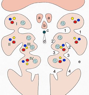
The median tongue bud (also tuberculum impar) marks the beginning of the development of the tongue. It appears as a midline swelling from the first pharyngeal arch late in the fourth week of embryogenesis. In the fifth week, a pair of lateral lingual swelling The tongue is a Muscle, muscular organ (anatomy), organ in the mouth of a typical tetrapod. It manipulates food for chewing and swallowing as part of the digestive system, digestive process, and is the primary organ of taste. The tongue's upper s ...s (or ''distal tongue buds'') develop above and in line with the median tongue bud. These swellings grow downwards towards each other, quickly overgrowing the median tongue bud. The line of the fusion of the distal tongue buds is marked by the median sulcus. References External links * Embryology {{Portal bar, Anatomy ... [...More Info...] [...Related Items...] OR: [Wikipedia] [Google] [Baidu] |
Digestive System
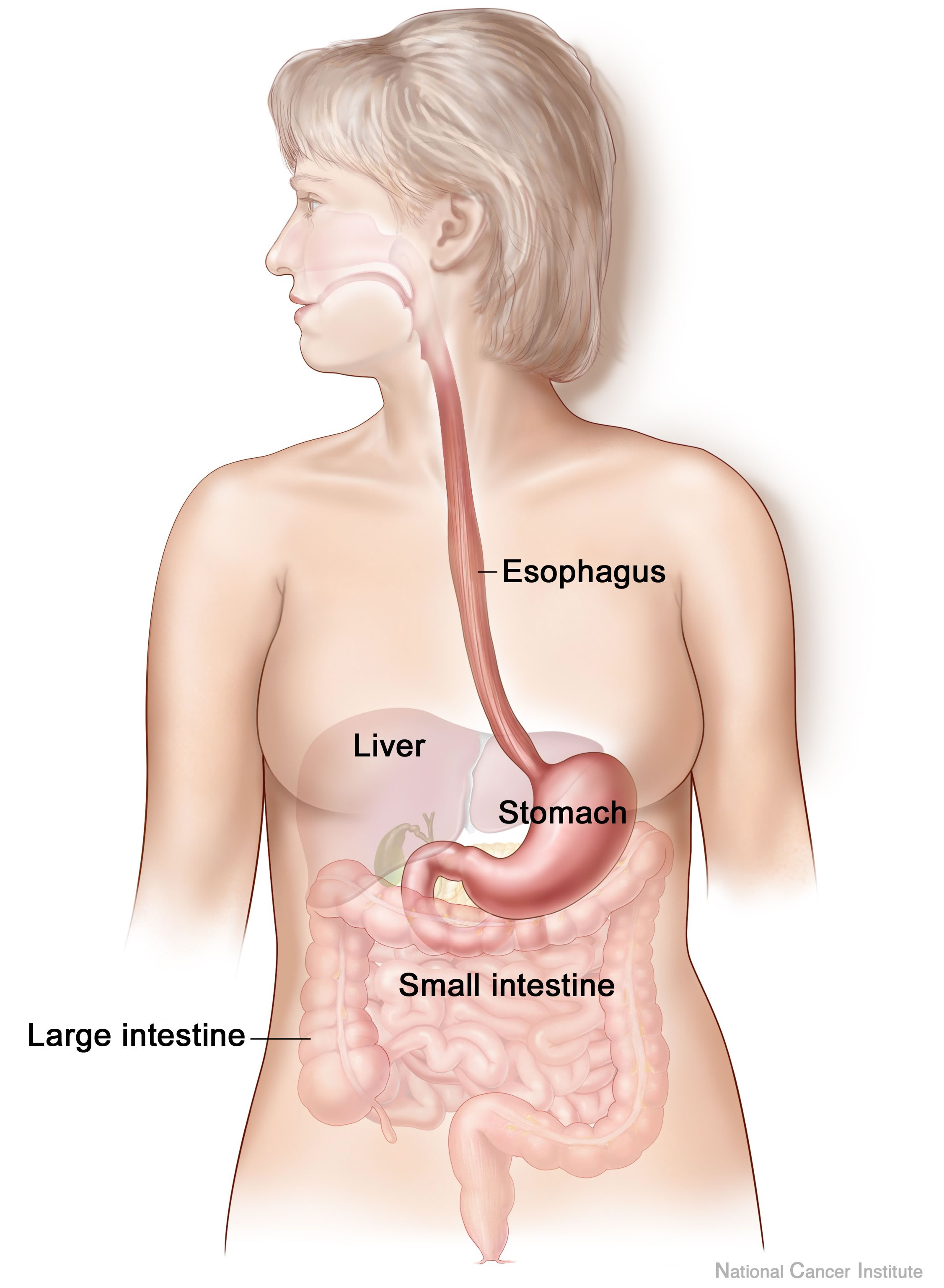
The human digestive system consists of the gastrointestinal tract plus the accessory organs of digestion (the tongue, salivary glands, pancreas, liver, and gallbladder). Digestion involves the breakdown of food into smaller and smaller components, until they can be absorbed and assimilated into the body. The process of digestion has three stages: the cephalic phase, the gastric phase, and the intestinal phase. The first stage, the cephalic phase of digestion, begins with secretions from gastric glands in response to the sight and smell of food, and continues in the human mouth, mouth with the mechanical breakdown of food by chewing, and the chemical breakdown by digestive enzymes in the saliva. Saliva contains amylase, and lingual lipase, secreted by the salivary glands, and serous glands on the tongue. Chewing mixes the food with saliva to produce a Bolus (digestion), bolus to be Swallowing, swallowed down the esophagus to enter the stomach. The second stage, the gastric phase ... [...More Info...] [...Related Items...] OR: [Wikipedia] [Google] [Baidu] |
Lingual Papilla
Lingual papillae (: papilla, ) are small structures on the upper surface of the tongue that give it its characteristic rough texture. The four types of papillae on the human tongue have different structures and are accordingly classified as circumvallate (or vallate), fungiform, filiform, and foliate. All except the filiform papillae are associated with taste buds. Structure In living subjects, lingual papillae are more readily seen when the tongue is dry. There are four types of papillae present on the tongue in humans: Filiform papillae Filiform papillae () are the most numerous of the lingual papillae. They are fine, small, cone-shaped papillae found on the anterior surface of the tongue. They are responsible for giving the tongue its texture and are responsible for the sensation of touch. Unlike the other kinds of papillae, filiform papillae do not contain taste buds. They cover most of the front two-thirds of the tongue's surface. They appear as very small, conical or cyl ... [...More Info...] [...Related Items...] OR: [Wikipedia] [Google] [Baidu] |
Palatoglossus Muscle
The palatoglossal muscle is a muscle of the soft palate and an extrinsic muscle of the tongue. Its surface is covered by oral mucosa and forms the visible palatoglossal arch. Structure From its origin, it passes anteroinferiorly and laterally. It passes anterior to the palatine tonsil. Origin The palatoglossus arises (the oral aspect of) the palatine aponeurosis of the soft palate, where it is continuous with its contralateral partner (i.e. the same muscle of the opposite side). Insertion It inserts onto the side of the tongue; some of its fibers extend over the dorsum of the tongue, and some pass into the substance of the tongue to intermingle with the transverse muscle of tongue. Innervation The palatoglossus muscle receives motor innervation from the pharyngeal plexus of vagus nerve. It is the only muscle of the tongue ''not'' innervated by the hypoglossal nerve (CN XII). Controversy Some sources state that the palatoglossus is innervated by fibers from the crani ... [...More Info...] [...Related Items...] OR: [Wikipedia] [Google] [Baidu] |
Hypoglossal Nerve
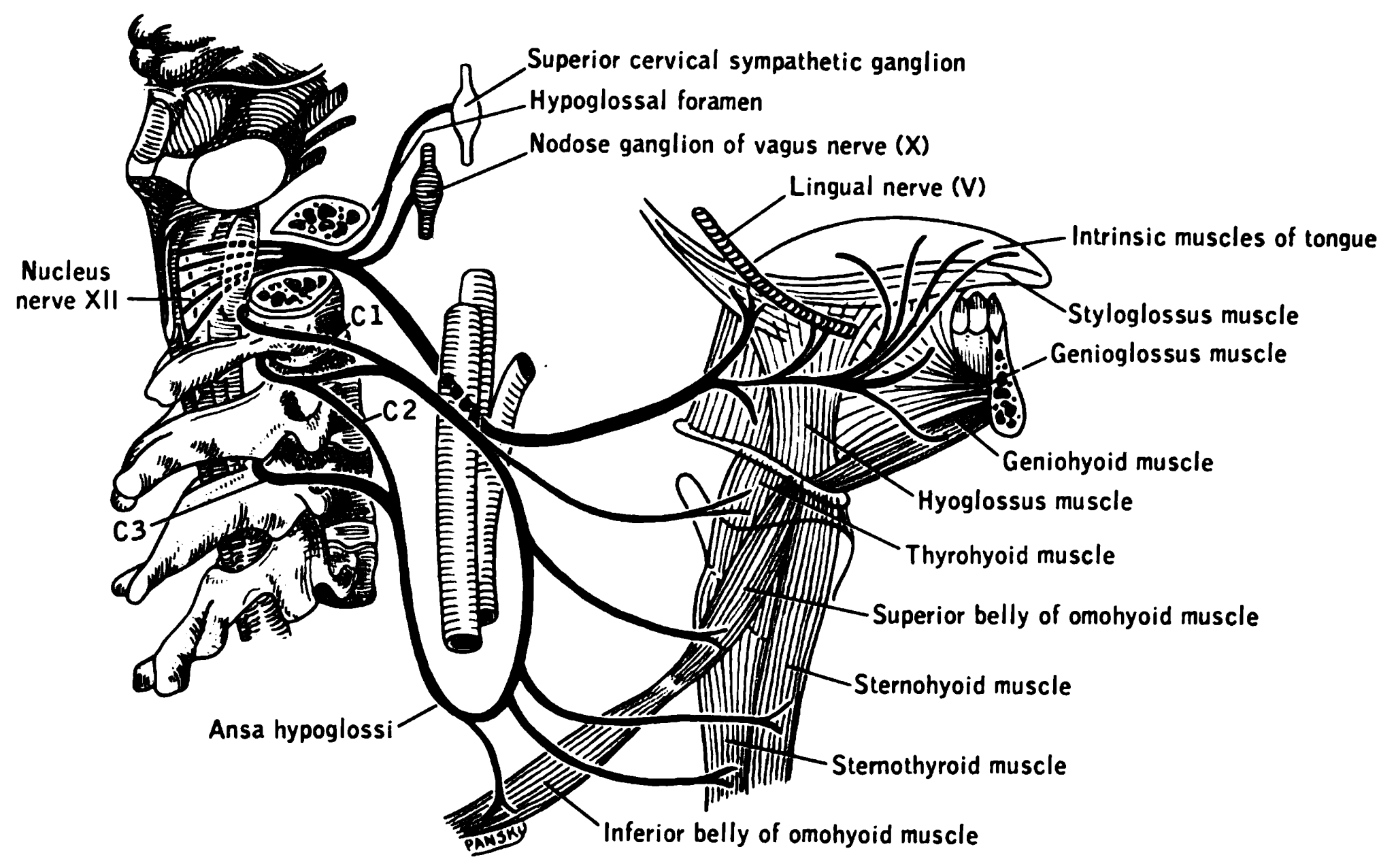
The hypoglossal nerve, also known as the twelfth cranial nerve, cranial nerve XII, or simply CN XII, is a cranial nerve that innervates all the extrinsic and intrinsic muscles of the tongue except for the palatoglossus, which is innervated by the vagus nerve. CN XII is a nerve with a sole motor function. The nerve arises from the hypoglossal nucleus in the medulla as a number of small rootlets, pass through the hypoglossal canal and down through the neck, and eventually passes up again over the tongue muscles it supplies into the tongue. The nerve is involved in controlling tongue movements required for speech and swallowing, including sticking out the tongue and moving it from side to side. Damage to the nerve or the neural pathways which control it can affect the ability of the tongue to move and its appearance, with the most common sources of damage being injury from trauma or surgery, and motor neuron disease. The first recorded description of the nerve was by Her ... [...More Info...] [...Related Items...] OR: [Wikipedia] [Google] [Baidu] |
Pharyngeal Arches
The pharyngeal arches, also known as visceral arches'','' are transient structures seen in the Animal embryonic development, embryonic development of humans and other vertebrates, that are recognisable precursors for many structures. In fish, the arches support the Fish gill, gills and are known as the branchial arches, or gill arches. In the human embryo, the arches are first seen during the fourth week of human embryonic development, development. They appear as a series of outpouchings of mesoderm on both sides of the developing pharynx. The vasculature of the pharyngeal arches are the aortic arches that arise from the aortic sac. Structure In humans and other vertebrates, the pharyngeal arches are derived from all three germ layers (the primary layers of cells that form during embryonic development). Neural crest cells enter these arches where they contribute to features of the skull and facial skeleton such as bone and cartilage. However, the existence of pharyngeal structu ... [...More Info...] [...Related Items...] OR: [Wikipedia] [Google] [Baidu] |
Chorda Tympani
Chorda tympani is a branch of the facial nerve that carries gustatory (taste) sensory innervation from the front of the tongue and parasympathetic ( secretomotor) innervation to the submandibular and sublingual salivary glands. Chorda tympani has a complex course from the brainstem, through the temporal bone and middle ear, into the infratemporal fossa, and ending in the oral cavity. Structure Chorda tympani fibers emerge from the pons of the brainstem as part of the intermediate nerve of the facial nerve. The facial nerve exits the cranial cavity through the internal acoustic meatus and enters the facial canal. In the facial canal, the chorda tympani branches off the facial nerve and enters the lateral wall of the tympanic cavity inside the middle ear where it runs across the tympanic membrane (from posterior to anterior) and medial to the neck of the malleus. The chorda then exits the skull by descending through the petrotympanic fissure into the infrate ... [...More Info...] [...Related Items...] OR: [Wikipedia] [Google] [Baidu] |
Lingual Nerve
The lingual nerve carries sensory innervation from the anterior two-thirds of the tongue. It contains fibres from both the mandibular division of the trigeminal nerve (CN V) and from the facial nerve (CN VII). The fibres from the trigeminal nerve are for touch, pain and temperature (general sensation), and the ones from the facial nerve are for taste (special sensation). Structure Origin The lingual nerve arises from the posterior trunk of mandibular nerve (CN V) within the infratemporal fossa. Course The lingual nerve first courses deep to the lateral pterygoid muscle and superior to the tensor veli palatini muscle; while passing between these two muscle, it is joined by the chorda tympani, and often by a communicating branch from the inferior alveolar nerve. The nerve then comes to pass inferoanteriorly upon the medial pterygoid muscle towards the medial aspect of the ramus of mandible, eventually meeting the mandible at the junction of the ramus and body of mandibl ... [...More Info...] [...Related Items...] OR: [Wikipedia] [Google] [Baidu] |
Lingual Veins
The lingual veins are veins of the tongue with two distinct courses: one group drains into the lingual vein, while another group drains either into the lingual artery, (common) facial vein, or internal jugular vein. Clinical significance The lingual veins are clinically significant due to their ability to rapidly absorb drugs. For this reason, nitroglycerin is administered sublingually to patients experiencing angina pectoris Angina, also known as angina pectoris, is chest pain or pressure, usually caused by insufficient blood flow to the heart muscle (myocardium). It is most commonly a symptom of coronary artery disease. Angina is typically the result of part .... See also * Deep lingual vein * Dorsal lingual veins External links Photo of model (frog) References * Moore NA and Roy W. Rapid Review: Gross Anatomy. Elsevier, 2010. Veins of the head and neck {{circulatory-stub ... [...More Info...] [...Related Items...] OR: [Wikipedia] [Google] [Baidu] |


