|
Third Ventricle
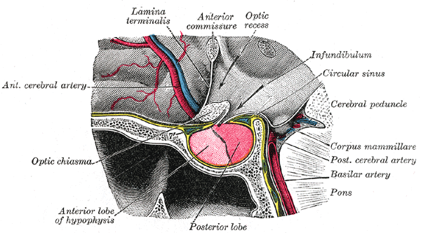
The third ventricle is one of the four connected cerebral ventricles of the ventricular system within the mammalian brain. It is a slit-like cavity formed in the diencephalon between the two thalami, in the midline between the right and left lateral ventricles, and is filled with cerebrospinal fluid (CSF). Running through the third ventricle is the interthalamic adhesion, which contains thalamic neurons and fibers that may connect the two thalami. Structure The third ventricle is a narrow, laterally flattened, vaguely rectangular region, filled with cerebrospinal fluid, and lined by ependyma. It is connected at the superior anterior corner to the lateral ventricles, by the interventricular foramina, and becomes the cerebral aqueduct (''aqueduct of Sylvius'') at the posterior caudal corner. Since the interventricular foramina are on the lateral edge, the corner of the third ventricle itself forms a bulb, known as the ''anterior recess'' (it is also known as the ''bulb ... [...More Info...] [...Related Items...] OR: [Wikipedia] [Google] [Baidu] |
Lateral Ventricle
The lateral ventricles are the two largest ventricular system, ventricles of the brain and contain cerebrospinal fluid. Each cerebral hemisphere contains a lateral ventricle, known as the left or right lateral ventricle, respectively. Each lateral ventricle resembles a C-shaped cavity that begins at an inferior horn in the temporal lobe, travels through a body in the parietal lobe and frontal lobe, and ultimately terminates at the Interventricular foramina (neural anatomy), interventricular foramina where each lateral ventricle connects to the single, central third ventricle. Along the path, a posterior horn extends backward into the occipital lobe, and an anterior horn of lateral ventricle, anterior horn extends farther into the frontal lobe. Structure Each lateral ventricle takes the form of an elongated curve, with an additional anterior-facing continuation emerging inferiorly from a point near the posterior end of the curve; the junction is known as the ''trigone of the lat ... [...More Info...] [...Related Items...] OR: [Wikipedia] [Google] [Baidu] |
Interventricular Foramina (neuroanatomy)
In the brain, the interventricular foramina (foramina of Monro) are channels that connect the paired lateral ventricles with the third ventricle at the midline of the brain. As channels, they allow cerebrospinal fluid (CSF) produced in the lateral ventricles to reach the third ventricle and then the rest of the brain's ventricular system. The walls of the interventricular foramina also contain choroid plexus, a specialized CSF-producing structure, that is continuous with that of the lateral and third ventricles above and below it. Structure The interventricular foramina are two holes (, pl. ''foramina'') that connect the left and the right lateral ventricles to the third ventricle. They are located on the underside near the midline of the lateral ventricles, and join the third ventricle where its roof meets its anterior surface. In front of the foramen is the fornix and behind is the thalamus. The foramen is normally crescent-shaped, but rounds and increases in size depending o ... [...More Info...] [...Related Items...] OR: [Wikipedia] [Google] [Baidu] |
Posterior Commissure
The posterior commissure (also known as the epithalamic commissure) is a rounded band of white fibers crossing the middle line on the dorsal aspect of the rostral end of the cerebral aqueduct. It is important in the bilateral pupillary light reflex. It constitutes part of the epithalamus. Its fibers acquire their medullary sheaths early, but their connections have not been definitively determined. Most of them have their origin in a nucleus, the ''nucleus of the posterior commissure'' (nucleus of Darkschewitsch), which lies in the periaqueductal grey at rostral end of the cerebral aqueduct, in front of the oculomotor nucleus. Some are thought to be derived from the posterior part of the thalamus and from the superior colliculus, whereas others are believed to be continued downward into the medial longitudinal fasciculus The medial longitudinal fasciculus (MLF) is a prominent bundle of nerve fibres which pass within the ventral/anterior portion of periaqueductal gray of ... [...More Info...] [...Related Items...] OR: [Wikipedia] [Google] [Baidu] |
Pineal Gland
The pineal gland (also known as the pineal body or epiphysis cerebri) is a small endocrine gland in the brain of most vertebrates. It produces melatonin, a serotonin-derived hormone, which modulates sleep, sleep patterns following the diurnal cycles. The shape of the gland resembles a pine cone, which gives it its name. The pineal gland is located in the epithalamus, near the center of the brain, between the two cerebral hemisphere, hemispheres, tucked in a groove where the two halves of the thalamus join. It is one of the neuroendocrinology, neuroendocrine Circumventricular organs, secretory circumventricular organs in which capillaries are mostly Vascular permeability, permeable to solutes in the blood. The pineal gland is present in almost all vertebrates, but is absent in Protochordata, protochordates in which there is a simple pineal homologue. The hagfish, archaic vertebrates, lack a pineal gland. In some species of amphibians and reptiles, the gland is linked to a light-s ... [...More Info...] [...Related Items...] OR: [Wikipedia] [Google] [Baidu] |
Habenular Commissure
The habenular commissure is a nerve tract of commissural fibers that connects the habenular nuclei on both sides of the habenular trigone in the epithalamus. The habenular commissure is part of the habenular trigone (a small depressed triangular area situated in front of the superior colliculus and on the lateral aspect of the posterior part of the taenia thalami). The habenulum trigone also contains the habenular nuclei. Fibers enter the habenular trigone from the stalk of the pineal gland, and the habenular commissure. Most of the habenular trigone's fibers are, however, directed downward and form a bundle, the fasciculus retroflexus, which passes medial to the red nucleus, and, after decussating with the corresponding fasciculus of the opposite side, ends in the interpeduncular nucleus. External links NIF Search - Habenular commissurevia the Neuroscience Information Framework The Neuroscience Information Framework is a repository of global neuroscience web resources, i ... [...More Info...] [...Related Items...] OR: [Wikipedia] [Google] [Baidu] |
Epithalamus
The epithalamus (: epithalami) is a posterior (dorsal) segment of the diencephalon. The epithalamus includes the habenular nuclei, the stria medullaris, the anterior and posterior paraventricular nuclei, the posterior commissure, and the pineal gland. Functions The function of the epithalamus is to connect the limbic system to other parts of the brain. The epithalamus also serves as a connecting point for the dorsal diencephalic conduction system, which is responsible for carrying information from the limbic forebrain to limbic midbrain structures. Some functions of its components include the secretion of melatonin from the pineal gland (circadian rhythms), regulation of motor pathways and emotions, and how energy is conserved in the body. A study has shown that the lateral habenula, in the epithalamus, produces spontaneous theta oscillatory activity that was correlated with theta oscillation in the hippocampus. The same study also found that the increase in theta waves in ... [...More Info...] [...Related Items...] OR: [Wikipedia] [Google] [Baidu] |
Commissure
A commissure () is the location at which two objects wikt:abut#Verb, abut or are joined. The term is used especially in the fields of anatomy and biology. * The most common usage of the term refers to the brain's commissures, of which there are at least nine. Such a commissure is a bundle of commissural fibers as a nerve tract, tract that crosses the midline at its level of origin or entry (as opposed to a decussation of fibers that cross obliquely). The nine are the anterior commissure, posterior commissure, corpus callosum, Fornix (neuroanatomy)#Commissure, commissure of fornix (hippocampal commissure), habenular commissure, ventral supraoptic decussation, Meynert's commissure, anterior hypothalamic commissure of Gasner, and the interthalamic adhesion. They consist of fibre Neural pathway, tracts that connect the two cerebral hemispheres and span the longitudinal fissure. In the spinal cord, there are the anterior white commissure, and the gray commissure. ''Commissural neurons'' ... [...More Info...] [...Related Items...] OR: [Wikipedia] [Google] [Baidu] |
Hernia
A hernia (: hernias or herniae, from Latin, meaning 'rupture') is the abnormal exit of tissue or an organ (anatomy), organ, such as the bowel, through the wall of the cavity in which it normally resides. The term is also used for the normal Development of the digestive system, development of the intestinal tract, referring to the retraction of the intestine from the extra-embryonal navel coelom into the abdomen in the healthy embryo at about 7 weeks. Various types of hernias can occur, most commonly involving the abdomen, and specifically the groin. Groin hernias are most commonly inguinal hernia, inguinal hernias but may also be femoral hernias. Other types of hernias include Hiatal hernia, hiatus, incisional hernia, incisional, and umbilical hernias. Symptoms are present in about 66% of people with groin hernias. This may include pain or discomfort in the lower abdomen, especially with coughing, exercise, or Urination, urinating or Defecation, defecating. Often, it gets worse th ... [...More Info...] [...Related Items...] OR: [Wikipedia] [Google] [Baidu] |
Hypothalamus
The hypothalamus (: hypothalami; ) is a small part of the vertebrate brain that contains a number of nucleus (neuroanatomy), nuclei with a variety of functions. One of the most important functions is to link the nervous system to the endocrine system via the pituitary gland. The hypothalamus is located below the thalamus and is part of the limbic system. It forms the Basal (anatomy), basal part of the diencephalon. All vertebrate brains contain a hypothalamus. In humans, it is about the size of an Almond#Nut, almond. The hypothalamus has the function of regulating certain metabolic biological process, processes and other activities of the autonomic nervous system. It biosynthesis, synthesizes and secretes certain neurohormones, called releasing hormones or hypothalamic hormones, and these in turn stimulate or inhibit the secretion of hormones from the pituitary gland. The hypothalamus controls thermoregulation, body temperature, hunger (physiology), hunger, important aspects o ... [...More Info...] [...Related Items...] OR: [Wikipedia] [Google] [Baidu] |
Thalamus
The thalamus (: thalami; from Greek language, Greek Wikt:θάλαμος, θάλαμος, "chamber") is a large mass of gray matter on the lateral wall of the third ventricle forming the wikt:dorsal, dorsal part of the diencephalon (a division of the forebrain). Nerve fibers project out of the thalamus to the cerebral cortex in all directions, known as the thalamocortical radiations, allowing hub (network science), hub-like exchanges of information. It has several functions, such as the relaying of sensory neuron, sensory and motor neuron, motor signals to the cerebral cortex and the regulation of consciousness, sleep, and alertness. Anatomically, the thalami are paramedian symmetrical structures (left and right), within the vertebrate brain, situated between the cerebral cortex and the midbrain. It forms during embryonic development as the main product of the diencephalon, as first recognized by the Swiss embryologist and anatomist Wilhelm His Sr. in 1893. Anatomy The thalami ar ... [...More Info...] [...Related Items...] OR: [Wikipedia] [Google] [Baidu] |
Hypothalamic Sulcus
The hypothalamic sulcus (sulcus of Monro) is a groove in the lateral wall of the third ventricle, marking the boundary between the thalamus and hypothalamus. The upper and lower portions of the lateral wall of the third ventricle correspond to the alar lamina and basal lamina, respectively, of the lateral wall of the fore-brain vesicle and are separated from each other by a furrow, the hypothalamic sulcus, which extends from the interventricular foramen to the cerebral aqueduct The cerebral aqueduct (aqueduct of the midbrain, aqueduct of Sylvius, Sylvian aqueduct, mesencephalic duct) is a small, narrow tube connecting the third and fourth ventricles of the brain. The cerebral aqueduct is a midline structure that passe .... References External links * Diencephalon Ventricular system {{Neuroanatomy-stub ... [...More Info...] [...Related Items...] OR: [Wikipedia] [Google] [Baidu] |
Sulcus (neuroanatomy)
In neuroanatomy, a sulcus (Latin: "furrow"; : sulci) is a shallow Sulcus (morphology), depression or groove in the cerebral cortex. One or more sulci surround a gyrus (pl. gyri), a ridge on the surface of the cortex, creating the characteristic folded appearance of the brain in humans and most other mammals. The larger sulci are also called Sulcus (morphology)#Brain, fissures. The cortex develops in the fetal stage of corticogenesis, preceding the cortical folding stage known as gyrification. The large fissures and main sulci are the first to develop. Mammals that have a folded cortex are known as ''gyrencephalic'', and the small-brained mammals that have a smooth cortex, such as rats and mice are termed lissencephaly, lissencephalic. Structure Sulci, the grooves, and gyri, the folds or ridges, make up the gyrification, folded surface of the cerebral cortex. Larger or deeper sulci are also often termed fissures. The folded cortex creates a larger surface area for the brain in h ... [...More Info...] [...Related Items...] OR: [Wikipedia] [Google] [Baidu] |


