|
Third Occipital Nerve
While under the Trapezius, the medial branch of the posterior division of the third cervical nerve gives off a branch called the third occipital nerve (also known as the least occipital nerve), which pierces the Trapezius and ends in the skin of the lower part of the back of the head. It lies medial to the greater occipital The greater occipital nerve is a nerve of the head. It is a spinal nerve, specifically the medial branch of the dorsal primary ramus of cervical spinal nerve 2. It arises from between the first and second cervical vertebrae, ascends, and then p ... nerve and communicates with it. Additional images File:Gray784.png, Dermatome distribution of the trigeminal nerve External links * http://www.dartmouth.edu/~humananatomy/figures/chapter_47/47-2.HTM * http://www.dartmouth.edu/~humananatomy/figures/chapter_47/47-6.HTM Spinal nerves {{Neuroanatomy-stub ... [...More Info...] [...Related Items...] OR: [Wikipedia] [Google] [Baidu] |
Cervical Nerves
A spinal nerve is a mixed nerve, which carries motor, sensory, and autonomic signals between the spinal cord and the body. In the human body there are 31 pairs of spinal nerves, one on each side of the vertebral column. These are grouped into the corresponding cervical, thoracic, lumbar, sacral and coccygeal regions of the spine. There are eight pairs of cervical nerves, twelve pairs of thoracic nerves, five pairs of lumbar nerves, five pairs of sacral nerves, and one pair of coccygeal nerves. The spinal nerves are part of the peripheral nervous system. Structure Each spinal nerve is a mixed nerve, formed from the combination of nerve fibers from its dorsal and ventral roots. The dorsal root is the afferent sensory root and carries sensory information to the brain. The ventral root is the efferent motor root and carries motor information from the brain. The spinal nerve emerges from the spinal column through an opening (intervertebral foramen) between adjacent verteb ... [...More Info...] [...Related Items...] OR: [Wikipedia] [Google] [Baidu] |
Trapezius
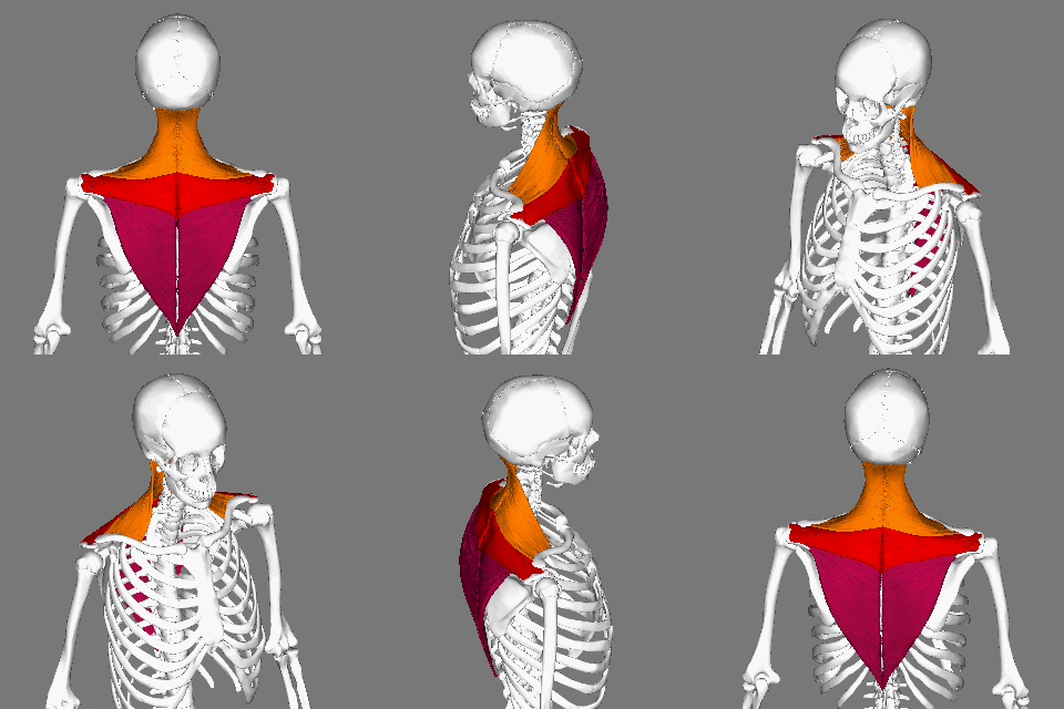
The trapezius is a large paired trapezoid-shaped surface muscle that extends longitudinally from the occipital bone to the lower thoracic vertebrae of the spine and laterally to the spine of the scapula. It moves the scapula and supports the arm. The trapezius has three functional parts: an upper (descending) part which supports the weight of the arm; a middle region (transverse), which retracts the scapula; and a lower (ascending) part which medially rotates and depresses the scapula. Name and history The trapezius muscle resembles a trapezium, also known as a trapezoid, or diamond-shaped quadrilateral. The word "spinotrapezius" refers to the human trapezius, although it is not commonly used in modern texts. In other mammals, it refers to a portion of the analogous muscle. Similarly, the term "tri-axle back plate" was historically used to describe the trapezius muscle. Structure The ''superior'' or ''upper'' (or descending) fibers of the trapezius originate from t ... [...More Info...] [...Related Items...] OR: [Wikipedia] [Google] [Baidu] |
Posterior Division
The dorsal ramus of spinal nerve (or posterior ramus of spinal nerve, or posterior primary division) is the posterior division of a spinal nerve. The dorsal ramus (Latin for branch, plural ''rami'' ) is the dorsal branch of a spinal nerve that forms from the dorsal root of the nerve after it emerges from the spinal cord. The spinal nerve is formed from the dorsal and ventral rami. The dorsal ramus carries information that supplies muscles and skin sensation to the human back. Structure Ventral root axons join with dorsal root ganglia to form mixed spinal nerves (below). These then merge to form peripheral nerves. Shortly after this spinal nerve forms, it then branches into the dorsal ramus and ventral ramus. Spinal nerves are mixed nerves that carry both sensory and motor information. It also branches to form the grey and the white rami communicantes which make connections with the sympathetic ganglia. After it is formed, the dorsal ramus of each spinal nerve travels backward, ... [...More Info...] [...Related Items...] OR: [Wikipedia] [Google] [Baidu] |
Cervical Nerve
A spinal nerve is a mixed nerve, which carries motor, sensory, and autonomic signals between the spinal cord and the body. In the human body there are 31 pairs of spinal nerves, one on each side of the vertebral column. These are grouped into the corresponding cervical, thoracic, lumbar, sacral and coccygeal regions of the spine. There are eight pairs of cervical nerves, twelve pairs of thoracic nerves, five pairs of lumbar nerves, five pairs of sacral nerves, and one pair of coccygeal nerves. The spinal nerves are part of the peripheral nervous system. Structure Each spinal nerve is a mixed nerve, formed from the combination of nerve fibers from its dorsal and ventral roots. The dorsal root is the afferent sensory root and carries sensory information to the brain. The ventral root is the efferent motor root and carries motor information from the brain. The spinal nerve emerges from the spinal column through an opening (intervertebral foramen) between adjacent vertebra ... [...More Info...] [...Related Items...] OR: [Wikipedia] [Google] [Baidu] |
Greater Occipital
The greater occipital nerve is a nerve of the head. It is a spinal nerve, specifically the medial branch of the dorsal primary ramus of cervical spinal nerve 2. It arises from between the first and second cervical vertebrae, ascends, and then passes through the semispinalis muscle. It ascends further to supply the skin along the posterior part of the scalp to the vertex. It supplies sensation the scalp at the top of the head, over the ear and over the parotid glands. Structure The greater occipital nerve is the medial branch of the dorsal primary ramus of cervical spinal nerve 2. It may also involve fibres from cervical spinal nerve 3. It arises from between the first and second cervical vertebrae, along with the lesser occipital nerve. It ascends after emerging from below the suboccipital triangle beneath the obliquus capitis inferior muscle. Just below the superior nuchal ridge, it pierces the fascia. It ascends further to supply the skin along the posterior pa ... [...More Info...] [...Related Items...] OR: [Wikipedia] [Google] [Baidu] |


