|
Syndesmotic Ankle Sprain
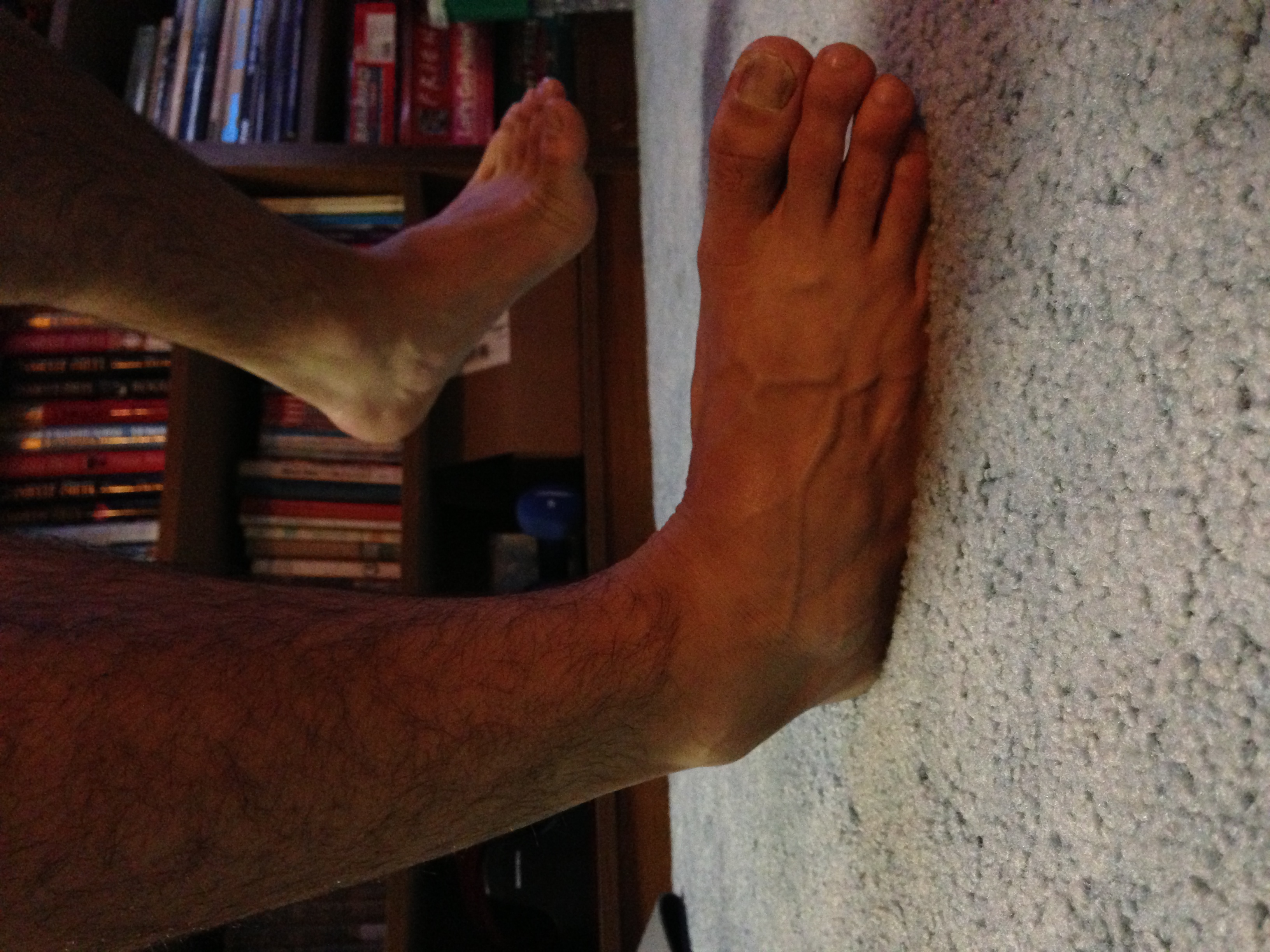
A high ankle sprain, also known as a syndesmotic ankle sprain (SAS), is a sprain of the Inferior tibiofibular articulation, syndesmotic ligaments that connect the tibia and fibula in the lower leg, thereby creating a mortise and tenon joint for the ankle. High ankle sprains are described as high because they are located above the ankle. They comprise approximately 15% of all ankle sprains. Unlike the common lateral ankle sprains, when ligaments around the ankle are injured through an inward twisting, high ankle sprains are caused when the lower leg and foot externally rotates (twists out). Mechanism The ankle joint consists of the talus bone, talus resting within the mortise created by the tibia and fibula as previously described. Since the talus is wider anteriorly (in the front) than posteriorly (at the back), as the front of the foot is raised Anatomical terms of motion, (dorsiflexed) reducing the angle between the foot and lower leg to less than 90°, then the mortise is confron ... [...More Info...] [...Related Items...] OR: [Wikipedia] [Google] [Baidu] |
Orthopedics
Orthopedic surgery or orthopedics (American and British English spelling differences, alternative spelling orthopaedics) is the branch of surgery concerned with conditions involving the musculoskeletal system. Orthopedic surgeons use both surgical and nonsurgical means to treat musculoskeletal Physical trauma, trauma, Spinal disease, spine diseases, Sports injury, sports injuries, degenerative diseases, infections, tumors and congenital disorders. Etymology Nicholas Andry coined the word in French as ', derived from the Ancient Greek words ("correct", "straight") and ("child"), and published ''Orthopedie'' (translated as ''Orthopædia: Or the Art of Correcting and Preventing Deformities in Children'') in 1741. The word was Assimilation (linguistics), assimilated into English as ''orthopædics''; the Typographic ligature, ligature ''æ'' was common in that era for ''ae'' in Greek- and Latin-based words. As the name implies, the discipline was initially developed with atte ... [...More Info...] [...Related Items...] OR: [Wikipedia] [Google] [Baidu] |
Anterior Tibiofibular Ligament
The anterior ligament of the lateral malleolus (anterior tibiofibular ligament or anterior inferior ligament) is a flat, trapezoidal band of fibers, broader below than above, which extends obliquely downward and lateralward between the adjacent margins of the tibia and fibula, on the front aspect of the syndesmosis. It is in relation, in front, with the fibularis tertius, the aponeurosis of the leg, and the integument; behind, with the interosseous ligament; and lies in contact with the cartilage covering the talus. References External links * - "Anterior inferior tibiofibular ligament" Additional images File:Slide2tat.JPG, Ankle joint. Deep dissection. Anterior view. File:Slide2coco.JPG, Dorsum of Foot. Ankle joint. Deep dissection File:Slide4CEC3.JPG, Ankle joint. Deep dissection. File:Slide8CEC7.JPG, Ankle joint. Deep dissection. Ligaments of the lower limb {{ligament-stub ... [...More Info...] [...Related Items...] OR: [Wikipedia] [Google] [Baidu] |
X-ray
An X-ray (also known in many languages as Röntgen radiation) is a form of high-energy electromagnetic radiation with a wavelength shorter than those of ultraviolet rays and longer than those of gamma rays. Roughly, X-rays have a wavelength ranging from 10 Nanometre, nanometers to 10 Picometre, picometers, corresponding to frequency, frequencies in the range of 30 Hertz, petahertz to 30 Hertz, exahertz ( to ) and photon energies in the range of 100 electronvolt, eV to 100 keV, respectively. X-rays were discovered in 1895 in science, 1895 by the German scientist Wilhelm Röntgen, Wilhelm Conrad Röntgen, who named it ''X-radiation'' to signify an unknown type of radiation.Novelline, Robert (1997). ''Squire's Fundamentals of Radiology''. Harvard University Press. 5th edition. . X-rays can penetrate many solid substances such as construction materials and living tissue, so X-ray radiography is widely used in medical diagnostics (e.g., checking for Bo ... [...More Info...] [...Related Items...] OR: [Wikipedia] [Google] [Baidu] |
Magnetic Resonance Imaging
Magnetic resonance imaging (MRI) is a medical imaging technique used in radiology to generate pictures of the anatomy and the physiological processes inside the body. MRI scanners use strong magnetic fields, magnetic field gradients, and radio waves to form images of the organs in the body. MRI does not involve X-rays or the use of ionizing radiation, which distinguishes it from computed tomography (CT) and positron emission tomography (PET) scans. MRI is a medical application of nuclear magnetic resonance (NMR) which can also be used for imaging in other NMR applications, such as NMR spectroscopy. MRI is widely used in hospitals and clinics for medical diagnosis, staging and follow-up of disease. Compared to CT, MRI provides better contrast in images of soft tissues, e.g. in the brain or abdomen. However, it may be perceived as less comfortable by patients, due to the usually longer and louder measurements with the subject in a long, confining tube, although ... [...More Info...] [...Related Items...] OR: [Wikipedia] [Google] [Baidu] |
Medical Ultrasonography
Medical ultrasound includes Medical diagnosis, diagnostic techniques (mainly medical imaging, imaging) using ultrasound, as well as therapeutic ultrasound, therapeutic applications of ultrasound. In diagnosis, it is used to create an image of internal body structures such as tendons, muscles, joints, blood vessels, and internal organs, to measure some characteristics (e.g., distances and velocities) or to generate an informative audible sound. The usage of ultrasound to produce visual images for medicine is called medical ultrasonography or simply sonography, or echography. The practice of examining pregnant women using ultrasound is called obstetric ultrasonography, and was an early development of clinical ultrasonography. The machine used is called an ultrasound machine, a sonograph or an echograph. The visual image formed using this technique is called an ultrasonogram, a sonogram or an echogram. Ultrasound is composed of sound waves with frequency, frequencies greater than ... [...More Info...] [...Related Items...] OR: [Wikipedia] [Google] [Baidu] |
Radiographs
Radiography is an imaging technique using X-rays, gamma rays, or similar ionizing radiation and non-ionizing radiation to view the internal form of an object. Applications of radiography include medical ("diagnostic" radiography and "therapeutic radiography") and industrial radiography. Similar techniques are used in airport security, (where "body scanners" generally use backscatter X-ray). To create an image in conventional radiography, a beam of X-rays is produced by an X-ray generator and it is projected towards the object. A certain amount of the X-rays or other radiation are absorbed by the object, dependent on the object's density and structural composition. The X-rays that pass through the object are captured behind the object by a detector (either photographic film or a digital detector). The generation of flat two-dimensional images by this technique is called projectional radiography. In computed tomography (CT scanning), an X-ray source and its associated de ... [...More Info...] [...Related Items...] OR: [Wikipedia] [Google] [Baidu] |
Maisonneuve Fracture
The Maisonneuve fracture is a spiral fracture of the proximal third of the fibula associated with a tear of the distal Inferior tibiofibular joint, tibiofibular syndesmosis and the interosseous membrane of the leg, interosseous membrane. There is an associated fracture of the medial malleolus or rupture of the deep medial ligament of talocrural joint, deltoid ligament of the ankle. This type of injury can be difficult to detect. The Maisonneuve fracture is typically a result of excessive, External rotation, external rotative force being applied to the deltoid and syndesmotic ligaments. Due to this, the Maisonneuve fracture is described as a pronation-external rotation injury according to the Lauge-Hansen classification, Lauge-Hansen classification system.Lauge-Hansen, N. (1950). Fractures of the ankle. II. Combined experimental-surgical and experimental-roentgenologic investigations. Arch Surg. 60(5): 957- 985.' It is also classified as a Type C ankle fracture according to the Dani ... [...More Info...] [...Related Items...] OR: [Wikipedia] [Google] [Baidu] |
Medial Malleolus
A malleolus is the bony prominence on each side of the human ankle. Each leg is supported by two bones, the tibia on the inner side (medial) of the leg and the fibula on the outer side (lateral) of the leg. The medial malleolus is the prominence on the inner side of the ankle, formed by the lower end of the tibia. The lateral malleolus is the prominence on the outer side of the ankle, formed by the lower end of the fibula. The word ''malleolus'' (), plural ''malleoli'' (), comes from Latin and means "small hammer". (It is cognate with '' mallet''.) Medial malleolus The medial malleolus is found at the foot end of the tibia. The medial surface of the lower extremity of tibia is prolonged downward to form a strong pyramidal process, flattened from without inward - the medial malleolus. * The ''medial surface'' of this process is convex and subcutaneous. * The ''lateral'' or ''articular surface'' is smooth and slightly concave, and articulates with the talus. * The ''anterio ... [...More Info...] [...Related Items...] OR: [Wikipedia] [Google] [Baidu] |
Anatomical Terms Of Motion
Motion, the process of movement, is described using specific anatomical terms. Motion includes movement of organs, joints, limbs, and specific sections of the body. The terminology used describes this motion according to its direction relative to the anatomical position of the body parts involved. Anatomists and others use a unified set of terms to describe most of the movements, although other, more specialized terms are necessary for describing unique movements such as those of the hands, feet, and eyes. In general, motion is classified according to the anatomical plane it occurs in. ''Flexion'' and ''extension'' are examples of ''angular'' motions, in which two axes of a joint are brought closer together or moved further apart. ''Rotational'' motion may occur at other joints, for example the shoulder, and are described as ''internal'' or ''external''. Other terms, such as ''elevation'' and ''depression'', describe movement above or below the horizontal plane. Many anatom ... [...More Info...] [...Related Items...] OR: [Wikipedia] [Google] [Baidu] |
Sprain
A sprain is a soft tissue injury of the ligaments within a joint, often caused by a sudden movement abruptly forcing the joint to exceed its functional range of motion. Ligaments are tough, inelastic fibers made of collagen that connect two or more bones to form a joint and are important for joint stability and proprioception, which is the body's sense of limb position and movement. Sprains may be mild (first degree), moderate (second degree), or severe (third degree), with the latter two classes involving some degree of tearing of the ligament. Sprains can occur at any joint but most commonly occur in the ankle, knee, or wrist. An equivalent injury to a muscle or tendon is known as a strain. The majority of sprains are mild, causing minor swelling and bruising that can be resolved with conservative treatment, typically summarized as RICE: rest, ice, compression, elevation. However, severe sprains involve complete tears, ruptures, or avulsion fractures, often leading to j ... [...More Info...] [...Related Items...] OR: [Wikipedia] [Google] [Baidu] |
Talus Bone
The talus (; Latin for ankle or ankle bone; : tali), talus bone, astragalus (), or ankle bone is one of the group of Foot#Structure, foot bones known as the tarsus (skeleton), tarsus. The tarsus forms the lower part of the ankle joint. It transmits the entire weight of the body from the lower legs to the foot.Platzer (2004), p 216 The talus has joints with the two bones of the lower leg, the tibia and thinner fibula. These leg bones have two prominences (the Lateral malleolus, lateral and Medial malleolus, medial malleoli) that articulation (anatomy), articulate with the talus. At the foot end, within the tarsus, the talus articulates with the calcaneus (heel bone) below, and with the curved navicular bone in front; together, these foot articulations form the Ball-and-socket joint, ball-and-socket-shaped talocalcaneonavicular joint. The talus is the second largest of the Tarsus (skeleton), tarsal bones; it is also one of the bones in the human body with the highest percentage of i ... [...More Info...] [...Related Items...] OR: [Wikipedia] [Google] [Baidu] |
Ankle Sprain
A sprained ankle (twisted ankle, rolled ankle, turned ankle, etc.) is an injury where sprain occurs on one or more ligaments of the ankle. It is the most commonly occurring injury in sports, mainly in ball sports (basketball, volleyball, and football) as well as racquet sports (tennis, badminton and pickleball). Signs and symptoms Knowing the symptoms that can be experienced with a sprain is important in determining that the injury is not really a break in the bone. When a sprain occurs, hematoma occurs within the tissue that surrounds the joint, causing a bruise. White blood cells responsible for inflammation migrate to the area, and blood flow increases as well. Along with this inflammation, swelling and pain is experienced. The nerves in the area become more sensitive when the injury is suffered, so pain is felt as throbbing and will worsen if there is pressure placed on the area. Warmth and redness are also seen as blood flow is increased. There is also decreased ability to ... [...More Info...] [...Related Items...] OR: [Wikipedia] [Google] [Baidu] |


