|
Suprachoroidal Hemorrhage
Intraocular hemorrhage (sometimes called hemophthalmos or hemophthalmia) is bleeding inside the eye (''oculus'' in Latin). Bleeding can occur from any structure of the eye where there is vasculature or blood flow, including the anterior chamber, vitreous cavity, retina, choroid, suprachoroidal space, or optic disc. Intraocular hemorrhage may be caused by physical trauma (direct injury to the eye); ocular surgery (such as to repair cataracts); or other diseases, injuries, or disorders (such as diabetes, hypertension, or shaken baby syndrome). Severe bleeding may cause high pressure inside the eye, leading to blindness. Types Intraocular hemorrhage is classified based on the location of the bleeding: * Hyphema (in the anterior chamber) * Suprachoroidal hemorrhage (SCH) is a rare complication of intraocular surgery in which blood from the ciliary arteries enters the space between the choroid and the sclera. It is potentially vision-threatening. *In the posterior segment of the ... [...More Info...] [...Related Items...] OR: [Wikipedia] [Google] [Baidu] |
Bleeding
Bleeding, hemorrhage, haemorrhage or blood loss, is blood escaping from the circulatory system from damaged blood vessels. Bleeding can occur internally, or externally either through a natural opening such as the mouth, nose, ear, urethra, vagina, or anus, or through a puncture in the skin. Hypovolemia is a massive decrease in blood volume, and death by excessive loss of blood is referred to as exsanguination. Typically, a healthy person can endure a loss of 10–15% of the total blood volume without serious medical difficulties (by comparison, blood donation typically takes 8–10% of the donor's blood volume). The stopping or controlling of bleeding is called hemostasis and is an important part of both first aid and surgery. Types * Upper head ** Intracranial hemorrhage — bleeding in the skull. ** Cerebral hemorrhage — a type of intracranial hemorrhage, bleeding within the brain tissue itself. ** Intracerebral hemorrhage — bleeding in the brain caused by ... [...More Info...] [...Related Items...] OR: [Wikipedia] [Google] [Baidu] |
Ciliary Arteries
The ciliary arteries are divisible into three groups, the long posterior, short posterior, and the anterior. * The short posterior ciliary arteries from six to twelve in number, arise from the ophthalmic artery as it crosses the optic nerve. * The long posterior ciliary arteries, two for each eye, pierce the posterior part of the sclera at some little distance from the optic nerve. * The anterior ciliary arteries The anterior ciliary arteries are seven arteries in each eye-socket that arise from muscular branches of the ophthalmic artery and supply the conjunctiva, sclera, rectus muscles, and the ciliary body. The arteries end by anastomosing with branc ... are derived from the muscular branches of the ophthalmic artery. Additional images File:Gray514.png, The ophthalmic artery and its branches File:Gray878.png, Iris, front view. File:Gray880.png, The terminal portion of the optic nerve and its entrance into the eyeball, in horizontal section. References Arteries ... [...More Info...] [...Related Items...] OR: [Wikipedia] [Google] [Baidu] |
Blunt Trauma
A blunt trauma, also known as a blunt force trauma or non-penetrating trauma, is a physical trauma due to a forceful impact without penetration of the body's surface. Blunt trauma stands in contrast with penetrating trauma, which occurs when an object pierces the skin, enters body tissue, and creates an open wound. Blunt trauma occurs due to direct physical trauma or impactful force to a body part. Such incidents often occur with road traffic collisions, assaults, and sports-related injuries, and are notably common among the elderly who experience falls. Blunt trauma can lead to a wide range of injuries including contusions, concussions, abrasions, lacerations, internal or external hemorrhages, and bone fractures. The severity of these injuries depends on factors such as the force of the impact, the area of the body affected, and the underlying comorbidities of the affected individual. In some cases, blunt force trauma can be life-threatening and may require immedia ... [...More Info...] [...Related Items...] OR: [Wikipedia] [Google] [Baidu] |
Blood Vessel
Blood vessels are the tubular structures of a circulatory system that transport blood throughout many Animal, animals’ bodies. Blood vessels transport blood cells, nutrients, and oxygen to most of the Tissue (biology), tissues of a Body (biology), body. They also take waste and carbon dioxide away from the tissues. Some tissues such as cartilage, epithelium, and the lens (anatomy), lens and cornea of the eye are not supplied with blood vessels and are termed ''avascular''. There are five types of blood vessels: the arteries, which carry the blood away from the heart; the arterioles; the capillaries, where the exchange of water and chemicals between the blood and the tissues occurs; the venules; and the veins, which carry blood from the capillaries back towards the heart. The word ''vascular'', is derived from the Latin ''vas'', meaning ''vessel'', and is mostly used in relation to blood vessels. Etymology * artery – late Middle English; from Latin ''arteria'', from Gree ... [...More Info...] [...Related Items...] OR: [Wikipedia] [Google] [Baidu] |
Cough
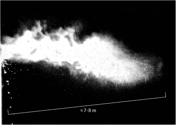
A cough is a sudden expulsion of air through the large breathing passages which can help clear them of fluids, irritants, foreign particles and Microorganism, microbes. As a protective reflex, coughing can be repetitive with the cough reflex following three phases: an inhalation, a forced exhalation against a closed glottis, and a violent release of air from the lungs following opening of the glottis, usually accompanied by a distinctive sound. Coughing into one's elbow or toward the ground—rather than forward at breathing height—can reduce the spread of infectious droplets in the air. Frequent coughing usually indicates the presence of a disease. Many viruses and bacteria benefit, from an evolutionary perspective, by causing the Host (biology), host to cough, which helps to spread the disease to new hosts. Irregular coughing is usually caused by a respiratory tract infection but can also be triggered by choking, smoking, air pollution, asthma, gastroesophageal reflux disease ... [...More Info...] [...Related Items...] OR: [Wikipedia] [Google] [Baidu] |
Sneeze
A sneeze (also known as sternutation) is a semi-autonomous, convulsive expulsion of air from the lungs through the nose and mouth, usually caused by foreign particles irritating the nasal mucosa. A sneeze expels air forcibly from the mouth and nose in an explosive, spasmodic involuntary action. This action allows for mucus to escape through the nasal cavity and saliva to escape from the oral cavity. Sneezing is possibly linked to sudden exposure to bright light (known as photic sneeze reflex), sudden change (drop) in temperature, breeze of cold air, a particularly full stomach, exposure to allergens, or viral infection. Because sneezes can spread disease through infectious aerosol droplets, it is recommended to cover one's mouth and nose with the forearm, the inside of the elbow, a tissue or a handkerchief while sneezing. In addition to covering the mouth, looking down is also recommended to change the direction of the droplets spread and avoid high concentration in the ... [...More Info...] [...Related Items...] OR: [Wikipedia] [Google] [Baidu] |
Conjunctiva
In the anatomy of the eye, the conjunctiva (: conjunctivae) is a thin mucous membrane that lines the inside of the eyelids and covers the sclera (the white of the eye). It is composed of non-keratinized, stratified squamous epithelium with goblet cells, stratified columnar epithelium and stratified cuboidal epithelium (depending on the zone). The conjunctiva is highly Angiogenesis, vascularised, with many microvessels easily accessible for imaging studies. Structure The conjunctiva is typically divided into three parts: Blood supply Blood to the bulbar conjunctiva is primarily derived from the ophthalmic artery. The blood supply to the palpebral conjunctiva (the eyelid) is derived from the external carotid artery. However, the circulations of the bulbar conjunctiva and palpebral conjunctiva are linked, so both bulbar conjunctival and palpebral conjunctival vessels are supplied by both the ophthalmic artery and the external carotid artery, to varying extents. Nerve supply Se ... [...More Info...] [...Related Items...] OR: [Wikipedia] [Google] [Baidu] |
Subconjunctival Hemorrhage
Subconjunctival bleeding, also known as subconjunctival hemorrhage or subconjunctival haemorrhage, is bleeding from a small blood vessel over the whites of the eye. It results in a red spot in the white of the eye. There is generally little to no pain and vision is not affected. Generally only one eye is affected. Causes can include coughing, vomiting, heavy lifting, straining during acute constipation or the act of "bearing down" during childbirth, as these activities can increase the blood pressure in the vascular systems supplying the conjunctiva. Other causes include blunt or penetrating trauma to the eye. Risk factors include hypertension, diabetes, old age, and blood thinners. Subconjunctival bleeding occurs in about 2% of newborns following a vaginal delivery. The blood accumulates between the conjunctiva and the episclera. Diagnosis is generally based on the appearance of the conjunctiva. The condition is relatively common, and both sexes are affected equally. Sponta ... [...More Info...] [...Related Items...] OR: [Wikipedia] [Google] [Baidu] |
Macula
The macula (/ˈmakjʊlə/) or macula lutea is an oval-shaped pigmented area in the center of the retina of the human eye and in other animals. The macula in humans has a diameter of around and is subdivided into the umbo, foveola, foveal avascular zone, fovea, parafovea, and perifovea areas. The anatomical macula at a size of is much larger than the clinical macula which, at a size of , corresponds to the anatomical fovea. The macula is responsible for the central, high-resolution, color vision that is possible in good light. This kind of vision is impaired if the macula is damaged, as in macular degeneration. The clinical macula is seen when viewed from the pupil, as in ophthalmoscopy or retinal photography. The term macula lutea comes from Latin ''macula'', "spot", and ''lutea'', "yellow". Structure The macula is an oval-shaped pigmented area in the center of the retina of the human eye and other animal eyes. Its center is shifted slightly away from the optical axi ... [...More Info...] [...Related Items...] OR: [Wikipedia] [Google] [Baidu] |
Vitreous Humour
The vitreous body (''vitreous'' meaning "glass-like"; , ) is the clear gel that fills the space between the lens and the retina of the eyeball (the vitreous chamber) in humans and other vertebrates. It is often referred to as the vitreous humor (also spelled humour), from Latin meaning liquid, or simply "the vitreous". Vitreous fluid or "liquid vitreous" is the liquid component of the vitreous gel, found after a vitreous detachment. It is not to be confused with the aqueous humor, the other fluid in the eye that is found between the cornea and lens. Structure The vitreous humor is a transparent, colorless, gelatinous mass that fills the space in the eye between the lens and the retina. It is surrounded by a layer of collagen called the vitreous membrane (or hyaloid membrane or vitreous cortex) separating it from the rest of the eye. It makes up four-fifths of the volume of the eyeball. The vitreous humour is fluid-like near the centre, and gel-like near the edges. The vit ... [...More Info...] [...Related Items...] OR: [Wikipedia] [Google] [Baidu] |


