|
Superior Ramus Of The Ischium
The ischium (; : ischia) is a paired bone forming the lower and back part of the . Situated below the ilium and behind the pubis, it is one of three regions whose fusion creates the . The superior portion of this region forms approximately one-third of the |
Superior Gemellus Muscle
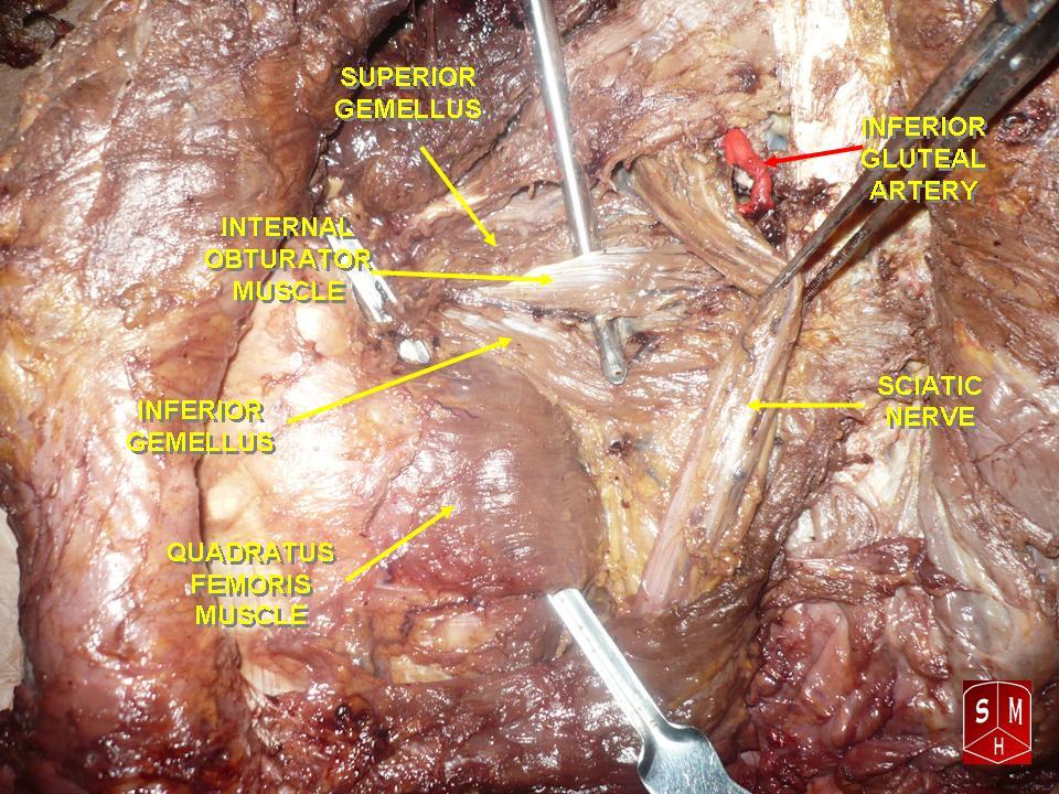
The gemelli muscles are the inferior gemellus muscle and the superior gemellus muscle, two small accessory fasciculi to the tendon of the internal obturator muscle. The gemelli muscles belong to the lateral rotator group of six muscles of the hip that rotate the femur in the hip joint. Superior gemellus muscle The gemelli muscles are two small muscular fasciculi, accessories to the tendon of the internal obturator muscle which is received into a groove between them. The superior gemellus muscle is the higher placed gemellus muscle that arises from the outer (gluteal) surface of the ischial spine, and blends with the upper part of the tendon of the internal obturator. It is smaller than the inferior gemellus. In some people, the fibres of the gemellus superior extend further than average, and are prolonged onto the medial surface of the greater trochanter of the femur. The superior and inferior gemelli are supplied by the inferior gluteal artery. Nerve supply to the superio ... [...More Info...] [...Related Items...] OR: [Wikipedia] [Google] [Baidu] |
Acetabular Fossa
The acetabular fossa is the non-articular depressed region at the centre of the floor of the acetabulum. It is surrounded by the articular lunate surface. The floor of the fossa is formed mostly by the ischium; it is rough and thin (often to the point of transparency). The space of the fossa is continuous inferiorly with the acetabular notch. The fossa does not contain any cartilage. It is occupied by the ligament of head of femur, and by fibroelastic adipose tissue (within which the acetabular branch of the obturator artery ramifies) that is mostly lined with synovial membrane Synovial () may refer to: * Synovial fluid * Synovial joint A synovial joint, also known as diarthrosis, joins bones or cartilage with a fibrous joint capsule that is continuous with the periosteum of the joined bones, constitutes the outer bou .... The acetabular "fat pad" is thought to contain abundant proprioceptive nerve endings that sense compression of the fat pad or its displacement through the ... [...More Info...] [...Related Items...] OR: [Wikipedia] [Google] [Baidu] |
Tuberosity Of The Ischium
The ischial tuberosity (or tuberosity of the ischium, tuber ischiadicum), also known colloquially as the sit bones or sitz bones, or as a pair the sitting bones, is a large posterior (anatomy), posterior bone, bony protuberance on the superior ramus of the ischium, superior ramus of the ischium. It marks the lateral boundary of the pelvic outlet. When sitting, the weight is frequently placed upon the ischial tuberosity. The Gluteus maximus muscle, gluteus maximus provides cover in the upright posture, but leaves it free in the seated position.Platzer (2004), p 236 The distance between a cyclist's ischial tuberosities is one of the factors in the choice of a bicycle saddle. Divisions The tuberosity is divided into two portions: a lower, rough, somewhat triangular part, and an upper, smooth, quadrilateral portion. * The ''lower portion'' is subdivided by a prominent longitudinal ridge, passing from base to apex, into two parts: ** The outer gives attachment to the adductor magnus ... [...More Info...] [...Related Items...] OR: [Wikipedia] [Google] [Baidu] |
Ischiocavernosus
The ischiocavernosus muscle (erectores penis ''or'' erector clitoridis in older texts) is a muscle just below the surface of the perineum, present in both men and women. Structure It arises by tendinous and fleshy fibers from the inner surface of the tuberosity of the ischium, behind the crus penis; and from the Inferior pubic ramus, inferior pubic rami and ischium on either side of the crus. From these points fleshy fibers succeed, and end in an aponeurosis which is inserted into the sides and under surface of the crus penis. Function In females, the ischiocavernosus muscle assists with clitoral erection. In males, it helps to stabilize the erect penis by compressing the crus penis and retarding the return of blood through the veins. Additional images File:Gray236.png, Right hip bone. Internal surface. File:Gray407.png, Coronal section of anterior part of pelvis, through the pubic arch. Seen from in front. File:Gray542.png, The superficial branches of the internal pudendal ... [...More Info...] [...Related Items...] OR: [Wikipedia] [Google] [Baidu] |
Superficial Transverse Perineal Muscle
The transverse perineal muscles (transversus perinei) are the superficial and the deep transverse perineal muscles. Superficial transverse perineal The superficial transverse perineal muscle (transversus superficialis perinei or Lloyd-Beanie muscle) is a narrow muscular slip, which passes more or less transversely across the perineal space in front of the anus. It arises by tendinous fibers from the inner and forepart of the ischial tuberosity and, running medially, is inserted into the central tendinous point of the perineum (perineal body), joining in this situation with the muscle of the opposite side, with the external anal sphincter muscle behind, and with the bulbospongiosus muscle in front. In some cases, the fibers of the deeper layer of the external anal sphincter cross over in front of the anus and are continued into thi ... [...More Info...] [...Related Items...] OR: [Wikipedia] [Google] [Baidu] |
Sacrotuberous Ligament
The sacrotuberous ligament (great or posterior sacrosciatic ligament) is situated at the lower and back part of the pelvis. It is flat, and triangular in form; narrower in the middle than at the ends. Structure It runs from the sacrum (the lower transverse sacral tubercles, the inferior margins sacrum and the upper coccyx) to the tuberosity of the ischium. It is a remnant of part of biceps femoris muscle. The sacrotuberous ligament is attached by its broad base to the posterior superior iliac spine, the posterior sacroiliac ligaments (with which it is partly blended), to the lower transverse sacral tubercles and the lateral margins of the lower sacrum and upper coccyx. Its oblique fibres descend laterally, converging to form a thick, narrow band that widens again below and is attached to the medial margin of the ischial tuberosity. It then spreads along the ischial ramus as the falciform process, whose concave edge blends with the fascial sheath of the internal pudendal vessels ... [...More Info...] [...Related Items...] OR: [Wikipedia] [Google] [Baidu] |
Falciform
{{Short pages monitor ... [...More Info...] [...Related Items...] OR: [Wikipedia] [Google] [Baidu] |
Pelvis
The pelvis (: pelves or pelvises) is the lower part of an Anatomy, anatomical Trunk (anatomy), trunk, between the human abdomen, abdomen and the thighs (sometimes also called pelvic region), together with its embedded skeleton (sometimes also called bony pelvis or pelvic skeleton). The pelvic region of the trunk includes the bony pelvis, the pelvic cavity (the space enclosed by the bony pelvis), the pelvic floor, below the pelvic cavity, and the perineum, below the pelvic floor. The pelvic skeleton is formed in the area of the back, by the sacrum and the coccyx and anteriorly and to the left and right sides, by a pair of hip bones. The two hip bones connect the spine with the lower limbs. They are attached to the sacrum posteriorly, connected to each other anteriorly, and joined with the two femurs at the hip joints. The gap enclosed by the bony pelvis, called the pelvic cavity, is the section of the body underneath the abdomen and mainly consists of the reproductive organs and ... [...More Info...] [...Related Items...] OR: [Wikipedia] [Google] [Baidu] |
Adductor Magnus
The adductor magnus is a large triangular muscle, situated on the medial side of the thigh. It consists of two parts. The portion which arises from the ischiopubic ramus (a small part of the inferior ramus of the pubis, and the inferior ramus of the ischium) is called the pubofemoral portion, adductor portion, or adductor minimus, and the portion arising from the tuberosity of the ischium is called the ischiocondylar portion, extensor portion, or "hamstring portion". Due to its common embryonic origin, innervation, and action the ischiocondylar portion (or hamstring portion) is often considered part of the hamstring group of muscles. The ischiocondylar portion of the adductor magnus is considered a muscle of the posterior compartment of the thigh while the pubofemoral portion of the adductor magnus is considered a muscle of the medial compartment. Structure Pubofemoral (adductor) portion Those fibers which arise from the ramus of the pubis are short, horizontal in direc ... [...More Info...] [...Related Items...] OR: [Wikipedia] [Google] [Baidu] |
Quadratus Femoris
The quadratus femoris is a flat, quadrilateral skeletal muscle. Located on the posterior side of the hip joint, it is a strong external rotator and adductor of the thigh, but also acts to stabilize the femoral head in the acetabulum. The quadratus femoris is used in Meyer's muscle pedicle grafting to prevent avascular necrosis of femur head. Course It originates on the lateral border of the ischial tuberosity of the ischium of the pelvis. From there, it passes laterally to its insertion on the posterior side of the head of the femur: the quadrate tubercle on the intertrochanteric crest and along the quadrate line, the vertical line which runs downward to bisect the lesser trochanter on the medial side of the femur. Along its course, quadratus is aligned edge to edge with the inferior gemellus above and the adductor magnus below, so that its upper and lower borders run horizontal and parallel. At its origin, the upper margin of the adductor magnus is separated from it by ... [...More Info...] [...Related Items...] OR: [Wikipedia] [Google] [Baidu] |
Obturator Foramen
The obturator foramen is the large, Bilateral symmetry, bilaterally paired opening of the bony pelvis. It is formed by the pubis and ischium. It is mostly closed by the obturator membrane except for a small opening, the obturator canal, through which the obturator nerve and vessels pass. Structure The obturator foramen is situated inferior and somewhat anterior to the acetabulum. It is bounded by the pubis bone and the ischium: superiorly by the (grooved obturator surface) of the Superior rami of the pubes, superior ramus of pubis, inferiorly by the Ischium#Structure, ramus of ischium, and laterally by (the anterior edge of) the body of ischium (including by the margin of the acetabulum). The margin of the foramen is thin and uneven, and gives attachment to the obturator membrane. Superiorly, it presents a deep groove - the obturator groove - which passes obliquely inferomedially from the pelvis. The foramen is largely closed by the obturator membrane save for a small opening at ... [...More Info...] [...Related Items...] OR: [Wikipedia] [Google] [Baidu] |
Greater Sciatic Notch
The greater sciatic notch is a notch in the ilium, one of the bones that make up the human pelvis. It lies between the posterior inferior iliac spine (above), and the ischial spine (below). The sacrospinous ligament changes this notch into an opening, the greater sciatic foramen. The notch holds the piriformis, the superior gluteal vein and artery, and the superior gluteal nerve; the inferior gluteal vein and artery and the inferior gluteal nerve; the sciatic and posterior femoral cutaneous nerves; the internal pudendal artery and veins, and the nerves to the internal obturator and quadratus femoris muscles. Of these, the superior gluteal vessels and nerve pass out above the piriformis, and the other structures below it. The greater sciatic notch is wider in females (about 74.4 degrees on average) than in males (about 50.4 degrees). [...More Info...] [...Related Items...] OR: [Wikipedia] [Google] [Baidu] |

