|
Superior Hypogastric Plexus
The superior hypogastric plexus (in older texts, hypogastric plexus or presacral nerve) is a plexus of nerves situated on the vertebral bodies anterior to the bifurcation of the abdominal aorta. It bifurcates to form the left and the right hypogastric nerve. The SHP is the continuation of the abdominal aortic plexus. Structure Afferents The superior hypogastric plexus receives contributions from the two lower lumbar splanchnic nerves (L3-L4), which are branches of the chain ganglia. They also contain parasympathetic fibers which arise from pelvic splanchnic nerve (S2-S4) and ascend from inferior hypogastric plexus; it is more usual for these parasympathetic fibers to ascend to the left-handed side of the superior hypogastric plexus and cross the branches of the sigmoid and left colic vessel branches, as these parasympathetic branches are distributed along the branches of the inferior mesenteric artery. Efferents From the plexus, sympathetic fibers are carried into the p ... [...More Info...] [...Related Items...] OR: [Wikipedia] [Google] [Baidu] |
Pelvis
The pelvis (: pelves or pelvises) is the lower part of an Anatomy, anatomical Trunk (anatomy), trunk, between the human abdomen, abdomen and the thighs (sometimes also called pelvic region), together with its embedded skeleton (sometimes also called bony pelvis or pelvic skeleton). The pelvic region of the trunk includes the bony pelvis, the pelvic cavity (the space enclosed by the bony pelvis), the pelvic floor, below the pelvic cavity, and the perineum, below the pelvic floor. The pelvic skeleton is formed in the area of the back, by the sacrum and the coccyx and anteriorly and to the left and right sides, by a pair of hip bones. The two hip bones connect the spine with the lower limbs. They are attached to the sacrum posteriorly, connected to each other anteriorly, and joined with the two femurs at the hip joints. The gap enclosed by the bony pelvis, called the pelvic cavity, is the section of the body underneath the abdomen and mainly consists of the reproductive organs and ... [...More Info...] [...Related Items...] OR: [Wikipedia] [Google] [Baidu] |
Ovarian Plexus
The ovarian plexus arises from the renal plexus, and is distributed to the ovary, and fundus of the uterus. It is carried in the suspensory ligament of the ovary. at eMedicine
eMedicine is an online clinical medical knowledge base founded in 1996 by doctors Scott Plantz and Jonathan Adler, and computer engineers Joanne Berezin and Jeffrey Berezin. The eMedicine website consists of approximately 6,800 medical topic revi ... Dictionary
References External links Nerve plexu ...[...More Info...] [...Related Items...] OR: [Wikipedia] [Google] [Baidu] |
Presacral Neurectomy
Presacral neurectomy is one of the treatments for chronic pelvic pain and dysmenorrhea. Lapraroscopic presacral neurectomy is an initial surgical intervention for chronic pelvic pain when medical therapy fails. __TOC__ Mechanism The sensory pathways from the pelvic viscera pass through the superior hypogastric plexus and inferior hypogastric plexus Inferior may refer to: * Inferiority complex * An anatomical term of location * Inferior angle of the scapula, in the human skeleton * ''Inferior'' (book), by Angela Saini * '' The Inferior'', a 2007 novel by Peadar Ó Guilín * Inferior good: ... to the spinal columns. The excision of presacral nerve trunk results in the obstruction of the pain pathway from the hypogastric plexi to the spinal cord. Presacral neurectomy denervates the uterus and causes loss of some bladder sensation. Efficacy Presacral neurectomy is offered to patients for whom medical therapy for chronic pain relief has failed. The efficacy of the procedure is ... [...More Info...] [...Related Items...] OR: [Wikipedia] [Google] [Baidu] |
Sigmoid Mesocolon
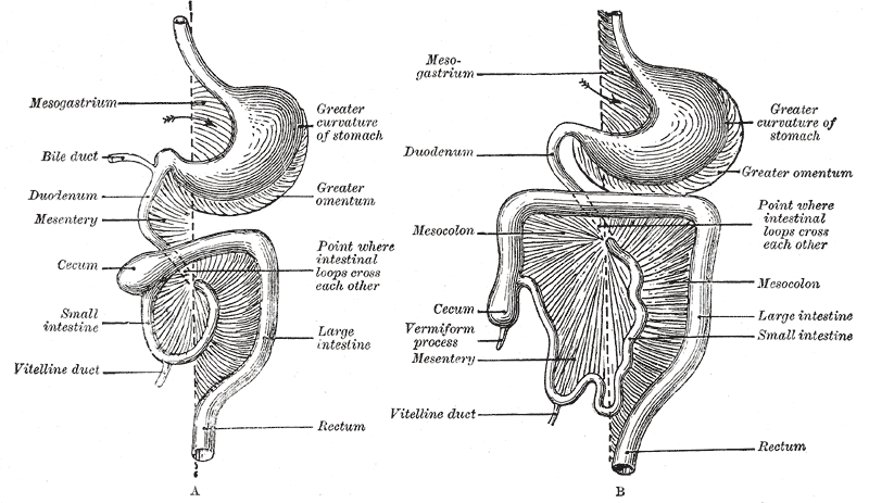
In human anatomy, the mesentery is an organ that attaches the intestines to the posterior abdominal wall, consisting of a double fold of the peritoneum. It helps (among other functions) in storing fat and allowing blood vessels, lymphatics, and nerves to supply the intestines. The (the part of the mesentery that attaches the colon to the abdominal wall) was formerly thought to be a fragmented structure, with all named parts—the ascending, transverse, descending, and sigmoid mesocolons, the mesoappendix, and the mesorectum—separately terminating their insertion into the posterior abdominal wall. However, in 2012, new microscopic and electron microscopic examinations showed the mesocolon to be a single structure derived from the duodenojejunal flexure and extending to the distal mesorectal layer. Thus the mesentery is an internal organ. Structure The mesentery of the small intestine arises from the root of the mesentery (or mesenteric root) and is the part connected ... [...More Info...] [...Related Items...] OR: [Wikipedia] [Google] [Baidu] |
Aortic Bifurcation
The aortic bifurcation is the point at which the abdominal aorta bifurcates (forks) into the left and right common iliac arteries. The aortic bifurcation is usually seen at the level of L4, just above the junction of the left and right common iliac veins. The right common iliac artery passes in front of the left common iliac vein. In some individuals, mainly women with lumbar lordosis, this vein can be compressed between the vertebra and the artery. This is the so-called Cockett syndrome or May–Thurner syndrome can cause a slower venous flow and the possibility of deep venous thrombosis in the left leg mainly in pregnancy. In surface anatomy, the bifurcation approximately corresponds to the umbilicus. Additional images Image:Gray531.png, The abdominal aorta and its branches. Image:Gray847.png, Abdominal portion of the sympathetic trunk, with the celiac and hypogastric plexuses. Image:Gray1121.png, Posterior abdominal wall, after removal of the peritoneum, showi ... [...More Info...] [...Related Items...] OR: [Wikipedia] [Google] [Baidu] |
Sacral Promontory
The sacrum (: sacra or sacrums), in human anatomy, is a triangular bone at the base of the spine that forms by the fusing of the sacral vertebrae (S1S5) between ages 18 and 30. The sacrum situates at the upper, back part of the pelvic cavity, between the two wings of the pelvis. It forms joints with four other bones. The two projections at the sides of the sacrum are called the alae (wings), and articulate with the ilium at the L-shaped sacroiliac joints. The upper part of the sacrum connects with the last lumbar vertebra (L5), and its lower part with the coccyx (tailbone) via the sacral and coccygeal cornua. The sacrum has three different surfaces which are shaped to accommodate surrounding pelvic structures. Overall, it is concave (curved upon itself). The base of the sacrum, the broadest and uppermost part, is tilted forward as the sacral promontory internally. The central part is curved outward toward the posterior, allowing greater room for the pelvic cavity. ... [...More Info...] [...Related Items...] OR: [Wikipedia] [Google] [Baidu] |
Lumbar Vertebrae
The lumbar vertebrae are located between the thoracic vertebrae and pelvis. They form the lower part of the back in humans, and the tail end of the back in quadrupeds. In humans, there are five lumbar vertebrae. The term is used to describe the anatomy of humans and quadrupeds, such as horses, pigs, or cattle. These bones are found in particular cuts of meat, including tenderloin or sirloin steak. Human anatomy In human anatomy, the five vertebrae are between the rib cage and the pelvis. They are the largest segments of the vertebral column and are characterized by the absence of the foramen transversarium within the transverse process (since it is only found in the cervical region) and by the absence of facets on the sides of the body (as found only in the thoracic region). They are designated L1 to L5, starting at the top. The lumbar vertebrae help support the weight of the body, and permit movement. General characteristics The adjacent figure depicts the general cha ... [...More Info...] [...Related Items...] OR: [Wikipedia] [Google] [Baidu] |
Renal Plexus
The renal plexus is a complex network of nerves formed by filaments from the celiac ganglia and plexus, aorticorenal ganglia, lower thoracic splanchnic nerves and first lumbar splanchnic nerve and aortic plexus. The nerves from these sources, fifteen or twenty in number, have a few ganglia developed upon them. It enters the kidneys on arterial branches to supply the vessels, renal glomerulus, and tubules with branches to the ureteric plexus. Some filaments are distributed to the spermatic plexus and, on the right side, to the inferior vena cava. The ovarian plexus arises from the renal plexus, and is one of two sympathetic supplies distributed to the ovary and fundus of the uterus The uterus (from Latin ''uterus'', : uteri or uteruses) or womb () is the hollow organ, organ in the reproductive system of most female mammals, including humans, that accommodates the embryonic development, embryonic and prenatal development, f .... Additional images File:Gray849.png ... [...More Info...] [...Related Items...] OR: [Wikipedia] [Google] [Baidu] |
Ureteric Plexus
The ureteric plexus (plexus: "braid") is a branching network of intersecting nerves (nerve plexus) covering and innervating the ureter. The plexus can be graduated into three parts, as the ureter itself can be divided: In the upper part of the ureter, the plexus gets its nerve fibers mainly from the renal plexus, but also from the abdominal aortic plexus. In the intermediate part the plexus receives nervous input from the superior hypogastric plexus and in the lower part from the inferior hypogastric plexus. The plexus contains both sympathetic and parasympathetic fibers, where the sympathetic components come from T11 to L2 levels of the spinal cord. Preganglionic vagal fibers (vagal fibers before passing through a ganglion) run through the celiac plexus The celiac plexus, also known as the solar plexus because of its radiating nerve fibers, is a nerve plexus, complex network of nerves located in the abdomen, near where the celiac trunk, superior mesenteric artery, and renal a ... [...More Info...] [...Related Items...] OR: [Wikipedia] [Google] [Baidu] |
Internal Iliac Artery
The internal iliac artery (formerly known as the hypogastric artery) is the main artery of the pelvis. Structure The internal iliac artery supplies the walls and viscera of the pelvis, the buttock, the reproductive organs, and the medial compartment of the thigh. The vesicular branches of the internal iliac arteries supply the bladder. It is a short, thick vessel, smaller than the external iliac artery, and about 3 to 4 cm in length. Course The internal iliac artery arises at the bifurcation of the common iliac artery, opposite the lumbosacral articulation, and, passing downward to the upper margin of the greater sciatic foramen, divides into two large trunks, an anterior and a posterior. It is posterior to the ureter, anterior to the internal iliac vein, anterior to the lumbosacral trunk, and anterior to the piriformis muscle. Near its origin, it is medial to the external iliac vein, which lies between it and the psoas major muscle. It is above the obturator ... [...More Info...] [...Related Items...] OR: [Wikipedia] [Google] [Baidu] |
Plexus
In anatomy, a plexus (from the Latin term for 'braid') is a branching network of blood vessels, lymphatic vessels, or nerves. The nerves are typically axons outside the central nervous system. The standard plural form in English is plexuses. Alternatively, the Latin plural plexūs may be used. Types Nerve plexuses The four primary nerve plexuses are the cervical plexus, brachial plexus, lumbar plexus, and the sacral plexus. Cardiac plexus Celiac plexus Renal plexus Venous plexus Choroid plexus The choroid plexus is a part of the central nervous system in the brain and consists of capillaries, brain ventricles, and ependymal cells. Invertebrates The plexus is the characteristic form of nervous system in the coelenterates and persists with modifications in the flatworms. The nerves of the radially symmetric echinoderms also take this form, where a plexus underlies the ectoderm of these animals and deeper in the body other nerve cells form plexuses of limited extent. ... [...More Info...] [...Related Items...] OR: [Wikipedia] [Google] [Baidu] |
Inferior Mesenteric Artery
In human anatomy, the inferior mesenteric artery (IMA) is the third main branch of the abdominal aorta and arises at the level of L3, supplying the large intestine from the distal transverse colon to the upper part of the anal canal. The regions supplied by the IMA are the descending colon, the sigmoid colon, and part of the rectum. Structure Origin The IMA arises from the anterior aspect of the abdominal aorta. Its origin is situated at the L3 vertebral level ( subcostal plane), below the origins of the two renal arteries, 3-4 cm above the aortic bifurcation, at the level of the umbilicus, and posterior to the inferior border of the horizontal (III) part of the duodenum. Branches Along its course, the IMA has the following branches: All these arterial branches further divide into arcades which then supply the colon at regular intervals. Relations The IMA is accompanied along its course by a similarly named vein, the inferior mesenteric vein, which drains into the ... [...More Info...] [...Related Items...] OR: [Wikipedia] [Google] [Baidu] |


