|
Strumal Carcinoid
The strumal carcinoid is a type of monodermal teratoma with histomorphologic features of (1) the thyroid gland and (2) a neuroendocrine tumour (carcinoid). See also *Struma ovarii *Teratoma A teratoma is a tumor made up of several different types of tissue, such as hair, muscle, teeth, or bone. Teratomata typically form in the ovary, testicle, or coccyx. Symptoms Symptoms may be minimal if the tumor is small. A testicular terato ... References External links Germ cell neoplasia {{oncology-stub ... [...More Info...] [...Related Items...] OR: [Wikipedia] [Google] [Baidu] |
Micrograph
A micrograph or photomicrograph is a photograph or digital image taken through a microscope or similar device to show a magnify, magnified image of an object. This is opposed to a macrograph or photomacrograph, an image which is also taken on a microscope but is only slightly magnified, usually less than 10 times. Micrography is the practice or art of using microscopes to make photographs. A micrograph contains extensive details of microstructure. A wealth of information can be obtained from a simple micrograph like behavior of the material under different conditions, the phases found in the system, failure analysis, grain size estimation, elemental analysis and so on. Micrographs are widely used in all fields of microscopy. Types Photomicrograph A light micrograph or photomicrograph is a micrograph prepared using an optical microscope, a process referred to as ''photomicroscopy''. At a basic level, photomicroscopy may be performed simply by connecting a camera to a micros ... [...More Info...] [...Related Items...] OR: [Wikipedia] [Google] [Baidu] |
H&E Stain
Hematoxylin and eosin stain ( or haematoxylin and eosin stain or hematoxylin-eosin stain; often abbreviated as H&E stain or HE stain) is one of the principal tissue stains used in histology. It is the most widely used stain in medical diagnosis and is often the gold standard. For example, when a pathologist looks at a biopsy of a suspected cancer, the histological section is likely to be stained with H&E. H&E is the combination of two histological stains: hematoxylin and eosin. The hematoxylin stains cell nuclei a purplish blue, and eosin stains the extracellular matrix and cytoplasm pink, with other structures taking on different shades, hues, and combinations of these colors. Hence a pathologist can easily differentiate between the nuclear and cytoplasmic parts of a cell, and additionally, the overall patterns of coloration from the stain show the general layout and distribution of cells and provides a general overview of a tissue sample's structure. Thus, pattern recogn ... [...More Info...] [...Related Items...] OR: [Wikipedia] [Google] [Baidu] |
Histomorphology
Histology, also known as microscopic anatomy or microanatomy, is the branch of biology which studies the microscopic anatomy of biological tissues. Histology is the microscopic counterpart to gross anatomy, which looks at larger structures visible without a microscope. Although one may divide microscopic anatomy into ''organology'', the study of organs, ''histology'', the study of tissues, and ''cytology'', the study of cells, modern usage places all of these topics under the field of histology. In medicine, histopathology is the branch of histology that includes the microscopic identification and study of diseased tissue. In the field of paleontology, the term paleohistology refers to the histology of fossil organisms. Biological tissues Animal tissue classification There are four basic types of animal tissues: muscle tissue, nervous tissue, connective tissue, and epithelial tissue. All animal tissues are considered to be subtypes of these four principal tissue types (fo ... [...More Info...] [...Related Items...] OR: [Wikipedia] [Google] [Baidu] |
Thyroid Gland
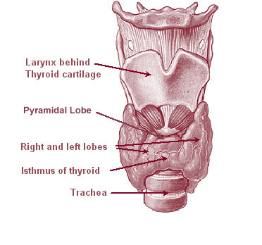
The thyroid, or thyroid gland, is an endocrine gland in vertebrates. In humans it is in the neck and consists of two connected lobes. The lower two thirds of the lobes are connected by a thin band of tissue called the thyroid isthmus. The thyroid is located at the front of the neck, below the Adam's apple. Microscopically, the functional unit of the thyroid gland is the spherical thyroid follicle, lined with follicular cells (thyrocytes), and occasional parafollicular cells that surround a lumen containing colloid. The thyroid gland secretes three hormones: the two thyroid hormones triiodothyronine (T3) and thyroxine (T4)and a peptide hormone, calcitonin. The thyroid hormones influence the metabolic rate and protein synthesis, and in children, growth and development. Calcitonin plays a role in calcium homeostasis. Secretion of the two thyroid hormones is regulated by thyroid-stimulating hormone (TSH), which is secreted from the anterior pituitary gland. TSH is regulated by ... [...More Info...] [...Related Items...] OR: [Wikipedia] [Google] [Baidu] |
Neuroendocrine Tumour
Neuroendocrine tumors (NETs) are neoplasms that arise from cells of the endocrine ( hormonal) and nervous systems. They most commonly occur in the intestine, where they are often called carcinoid tumors, but they are also found in the pancreas, lung, and the rest of the body. Although there are many kinds of NETs, they are treated as a group of tissue because the cells of these neoplasms share common features, such as looking similar, having special secretory granules, and often producing biogenic amines and polypeptide hormones. Classification WHO The World Health Organization (WHO) classification scheme places neuroendocrine tumors into three main categories, which emphasize the tumor grade rather than the anatomical origin: * well-differentiated neuroendocrine tumours, further subdivided into tumors with benign and those with uncertain behavior * well-differentiated (low grade) neuroendocrine carcinomas with low-grade malignant behavior * poorly differentiated (high grade) n ... [...More Info...] [...Related Items...] OR: [Wikipedia] [Google] [Baidu] |
Struma Ovarii
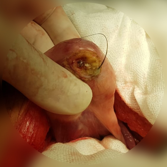
A struma ovarii (literally: goitre of the ovary) is a rare form of monodermal teratoma that contains mostly thyroid tissue, which may cause hyperthyroidism. Despite its name, struma ovarii is not restricted to the ovary. The vast majority of struma ovarii are benign tumours; however, malignant tumours of this type are found in a small percentage of cases. Radiologic findings The ultrasound (US) features of struma ovarii are nonspecific, but a heterogeneous, predominantly solid mass may be seen. US demonstrates a complex appearance with multiple cystic and solid areas, findings that reflect the gross pathologic appearance of the tumor. Magnetic resonance imaging findings may be more characteristic: The cystic spaces demonstrate both high and low signal intensity on T1- and T2-weighted images. Some of the cystic spaces may demonstrate low signal intensity on both T1- and T2-weighted images due to the thick, gelatinous colloid of the struma. No fat is evident in these lesions. ... [...More Info...] [...Related Items...] OR: [Wikipedia] [Google] [Baidu] |
Teratoma
A teratoma is a tumor made up of several different types of tissue, such as hair, muscle, teeth, or bone. Teratomata typically form in the ovary, testicle, or coccyx. Symptoms Symptoms may be minimal if the tumor is small. A testicular teratoma may present as a painless lump. Complications may include ovarian torsion, testicular torsion, or hydrops fetalis. They are a type of germ cell tumor (a tumor that begins in the cells that give rise to sperm or eggs). They are divided into two types: mature and immature. Mature teratomas include dermoid cysts and are generally benign. Immature teratomas may be cancerous. Most ovarian teratomas are mature. In adults, testicular teratomas are generally cancerous. Definitive diagnosis is based on a tissue biopsy. Treatment of coccyx, testicular, and ovarian teratomas is generally by surgery. Testicular and immature ovarian teratomas are also frequently treated with chemotherapy. Teratomas occur in the coccyx in about one in ... [...More Info...] [...Related Items...] OR: [Wikipedia] [Google] [Baidu] |


