|
Sphenoid Bone
The sphenoid bone is an unpaired bone of the neurocranium. It is situated in the middle of the skull towards the front, in front of the basilar part of occipital bone, basilar part of the occipital bone. The sphenoid bone is one of the seven bones that articulate to form the orbit (anatomy), orbit. Its shape somewhat resembles that of a butterfly, bat or wasp with its wings extended. The name presumably originates from this shape, since () means in Ancient Greek. Structure It is divided into the following parts: * a median portion, known as the body of sphenoid bone, containing the sella turcica, which houses the pituitary gland as well as the paired paranasal sinuses, the sphenoidal sinuses * two Greater wing of sphenoid bone, greater wings on the lateral side of the body and two Lesser wing of sphenoid bone, lesser wings from the anterior side. * Pterygoid processes of the sphenoides, directed downwards from the junction of the body and the greater wings. Two sphenoidal co ... [...More Info...] [...Related Items...] OR: [Wikipedia] [Google] [Baidu] |
Bone
A bone is a rigid organ that constitutes part of the skeleton in most vertebrate animals. Bones protect the various other organs of the body, produce red and white blood cells, store minerals, provide structure and support for the body, and enable mobility. Bones come in a variety of shapes and sizes and have complex internal and external structures. They are lightweight yet strong and hard and serve multiple functions. Bone tissue (osseous tissue), which is also called bone in the uncountable sense of that word, is hard tissue, a type of specialised connective tissue. It has a honeycomb-like matrix internally, which helps to give the bone rigidity. Bone tissue is made up of different types of bone cells. Osteoblasts and osteocytes are involved in the formation and mineralisation of bone; osteoclasts are involved in the resorption of bone tissue. Modified (flattened) osteoblasts become the lining cells that form a protective layer on the bone surface. The mine ... [...More Info...] [...Related Items...] OR: [Wikipedia] [Google] [Baidu] |
Sphenoidal Conchae
The sphenoidal conchae (sphenoidal turbinated processes) are two thin, curved plates, situated at the anterior and lower part of the body of the sphenoid. An aperture of variable size exists in the anterior wall of each, and through this the sphenoidal sinus opens into the nasal cavity. ''General Anatomy and Osteology of Head and Neck'' (I. K. International Pvt Ltd, 2009; by Mahdi Hasan)- Retrieved 2018-08-29 Each is irregular in form, and tapers to a point behind, being broader and thinner in front. Its upper surface is concave, and looks toward the cavity of the sinus; its under surface is convex, and forms part of the roof of the corresponding nasal cavity. Each bone articulates in front with the ethmoid, laterally with the Hard palate, palatine; its pointed poste ... [...More Info...] [...Related Items...] OR: [Wikipedia] [Google] [Baidu] |
Pterygoid Hamulus
The pterygoid hamulus is a hook-like process at the lower extremity of the medial pterygoid plate of the sphenoid bone of the skull. It is the superior origin of the pterygomandibular raphe, and the tensor veli palatini muscle courses around it before inserting into the palatine aponeurosis. Structure The pterygoid hamulus is part of the medial pterygoid plate of the sphenoid bone of the skull. Its tip is rounded off. It has an average length of 7.2 mm, an average depth of 1.4 mm, and an average width of 2.3 mm. The tendon of tensor veli palatini muscle glides around it. Function The pterygoid hamulus is the superior origin of the pterygomandibular raphe. It is also the origin of levator veli palatini muscle. Clinical significance Rarely, the pterygoid hamulus may be enlarged, which may cause mouth pain Pain is a distressing feeling often caused by intense or damaging Stimulus (physiology), stimuli. The International Association for the Study of Pain defines pai ... [...More Info...] [...Related Items...] OR: [Wikipedia] [Google] [Baidu] |
Scaphoid Fossa
In the pterygoid processes of the sphenoid, above the pterygoid fossa is a small, oval, shallow depression, the scaphoid fossa, which gives origin to the tensor veli palatini The tensor veli palatini muscle (tensor palati or tensor muscle of the velum palatinum) is a thin, triangular muscle of the head that tenses the soft palate and opens the Eustachian tube to equalise pressure in the middle ear. Structure The te .... It is not the same as and has to be distinguished from the scaphoid fossa of the external ear or pinna. References External links Diagram - look for #28(sourchere Bones of the head and neck {{musculoskeletal-stub ... [...More Info...] [...Related Items...] OR: [Wikipedia] [Google] [Baidu] |
Pterygoid Fossa
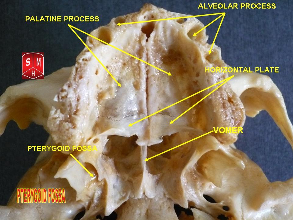
The pterygoid fossa is an anatomical term for the fossa formed by the divergence of the lateral pterygoid plate and the medial pterygoid plate of the sphenoid bone. Structure The lateral and medial pterygoid plates (of the pterygoid process of the sphenoid bone) diverge behind and enclose between them a V-shaped fossa, the pterygoid fossa. This fossa faces posteriorly, and contains the medial pterygoid muscle and the tensor veli palatini muscle. See also * Pterygoid fovea * Scaphoid fossa In the pterygoid processes of the sphenoid, above the pterygoid fossa is a small, oval, shallow depression, the scaphoid fossa, which gives origin to the tensor veli palatini The tensor veli palatini muscle (tensor palati or tensor muscle of t ... * Pterygoid process References Bones of the head and neck {{musculoskeletal-stub ... [...More Info...] [...Related Items...] OR: [Wikipedia] [Google] [Baidu] |
Pterygoid Notch
The Pterygoid notch (incisura pterygoidea) is a notch on the inferior portion of the pterygoid processes of the sphenoid bone The sphenoid bone is an unpaired bone of the neurocranium. It is situated in the middle of the skull towards the front, in front of the basilar part of occipital bone, basilar part of the occipital bone. The sphenoid bone is one of the seven bon ..., between the medial and lateral plates into which the pyramidal process of the palatine bone is fitted. Bones of the head and neck {{musculoskeletal-stub ... [...More Info...] [...Related Items...] OR: [Wikipedia] [Google] [Baidu] |
Pterygoalar Ligament
The pterygoalar ligament extends from the lamina of the lateral pterygoid to the undersurface of the greater wing of the sphenoid bone The sphenoid bone is an unpaired bone of the neurocranium. It is situated in the middle of the skull towards the front, in front of the basilar part of occipital bone, basilar part of the occipital bone. The sphenoid bone is one of the seven bon .... See also * Pterygospinous ligament References Joints {{ligament-stub ... [...More Info...] [...Related Items...] OR: [Wikipedia] [Google] [Baidu] |
Ossification
Ossification (also called osteogenesis or bone mineralization) in bone remodeling is the process of laying down new bone material by cells named osteoblasts. It is synonymous with bone tissue formation. There are two processes resulting in the formation of normal, healthy bone tissue: Intramembranous ossification is the direct laying down of bone into the primitive connective tissue ( mesenchyme), while endochondral ossification involves cartilage as a precursor. In fracture healing, endochondral osteogenesis is the most commonly occurring process, for example in fractures of long bones treated by plaster of Paris, whereas fractures treated by open reduction and internal fixation with metal plates, screws, pins, rods and nails may heal by intramembranous osteogenesis. Heterotopic ossification is a process resulting in the formation of bone tissue that is often atypical, at an extraskeletal location. Calcification is often confused with ossification. Calcificatio ... [...More Info...] [...Related Items...] OR: [Wikipedia] [Google] [Baidu] |
Middle Clinoid Process
The middle clinoid process is a small, bilaterally paired elevation on either side of the tuberculum sellae, at the anterior boundary of the sella turcica. A (larger) anterior clinoid process is situated lateral to each middle clinoid process. The diaphragma sellae (i.e. the dura forming the roof of the cavernous sinus) and the dura of the floor of the hypophyseal fossa (sella turcica) attach onto the middle clinoid processes. On each side of the body, the internal carotid artery passes between the anterior and middle clinoid processes. Etymology Clinoid likely comes from the Greek root ''klinein'' or the Latin Latin ( or ) is a classical language belonging to the Italic languages, Italic branch of the Indo-European languages. Latin was originally spoken by the Latins (Italic tribe), Latins in Latium (now known as Lazio), the lower Tiber area aroun ... ''clinare'', both meaning "sloped" as in "inclined." References Bones of the head and neck {{musculosk ... [...More Info...] [...Related Items...] OR: [Wikipedia] [Google] [Baidu] |
Posterior Clinoid Process
The posterior clinoid processes are the tubercles of the sphenoid bone situated at the superior angles of the dorsum sellae (one on each angle) which represents the posterior boundary of the sella turcica. They vary considerably in size and form. The posterior clinoid processes deepen the sella turcica, and give attachment to (the attached border of) the tentorium cerebelli, and the dura forming the floor of the hypophyseal fossa (sella turcica). The petroclinoid ligament The petroclinoid ligament is a fold of dura matter. It extends between the posterior clinoid process and anterior clinoid process and the petrosal part of the temporal bone The temporal bone is a paired bone situated at the sides and base of the skull, lateral to the temporal lobe of the cerebral cortex. The temporal bones are overlaid by the sides of the head known as the temples where four of the cranial bone ... of the skull. There are two separate bands of the ligament; named the anterior and pos ... [...More Info...] [...Related Items...] OR: [Wikipedia] [Google] [Baidu] |
Anterior Clinoid Process
The anterior clinoid process is a posterior projection of the sphenoid bone at the junction of the medial end of either lesser wing of sphenoid bone with the body of sphenoid bone. The bilateral processes flank the sella turcica anteriorly. The ACP is an important structure for cranial and endovascular surgical operations for several structures, including the pituitary gland and the internal carotid artery. Anatomy The anterior clinoid process is a pyramid-shaped bony projection of the lesser wing of the sphenoid bone and forms part of the lateral wall of the optic canal. Between each ACP lies the sella turcica, which holds the pituitary gland. Additionally, the ACP is part of the anterior roof of the cavernous sinus. The posterior and inferior portions of the ACP border the internal carotid artery. Attachments The free border of the tentorium cerebelli extends anteriorly on either side beyond the attached border of the same side (which ends anteriorly at the posterior clin ... [...More Info...] [...Related Items...] OR: [Wikipedia] [Google] [Baidu] |


