|
Renal Osteodystrophy
Renal osteodystrophy is defined as an alteration of bone in patients with chronic kidney disease (CKD). It is one measure of the skeletal component of the systemic disorder of chronic kidney disease-mineral and bone disorder (CKD-MBD). The term "renal osteodystrophy" was coined in 1943, 60 years after an association was identified between bone disease and kidney failure. The types of renal osteodystrophy have traditionally been defined on the basis of bone turnover and mineralization: 1) mild, slight increase in turnover and normal mineralization; 2) osteitis fibrosa, increased turnover and normal mineralization; 3) osteomalacia, decreased turnover and abnormal mineralization; 4) adynamic, decreased turnover and acellularity; and, 5) mixed, increased turnover with abnormal mineralization.A Kidney Disease: Improving Global Outcomes (KDIGO) report has suggested that bone biopsies in patients with CKD should be characterized by determining bone turnover, mineralization, and volume ( ... [...More Info...] [...Related Items...] OR: [Wikipedia] [Google] [Baidu] |
Chronic Kidney Disease
Chronic kidney disease (CKD) is a type of long-term kidney disease, defined by the sustained presence of abnormal kidney function and/or abnormal kidney structure. To meet criteria for CKD, the abnormalities must be present for at least three months. Early in the course of CKD, patients are usually asymptomatic, but later symptoms may include pedal edema, leg swelling, feeling tired, vomiting, loss of appetite, and confusion. Complications can relate to hormonal dysfunction of the kidneys and include (in chronological order) Hypertension, high blood pressure (often related to activation of the renin–angiotensin system), renal osteodystrophy, bone disease, and anemia. Additionally CKD patients have markedly increased Cardiovascular disease, cardiovascular complications with increased risks of death and hospitalization. CKD can lead to kidney failure, end-stage kidney failure requiring kidney dialysis or kidney transplantation. Causes of chronic kidney disease include diabetic ne ... [...More Info...] [...Related Items...] OR: [Wikipedia] [Google] [Baidu] |
Calcium Carbonate
Calcium carbonate is a chemical compound with the chemical formula . It is a common substance found in Rock (geology), rocks as the minerals calcite and aragonite, most notably in chalk and limestone, eggshells, gastropod shells, shellfish skeletons and pearls. Materials containing much calcium carbonate or resembling it are described as calcareous. Calcium carbonate is the active ingredient in agricultural lime and is produced when calcium ions in hard water react with carbonate ions to form limescale. It has medical use as a calcium supplement or as an antacid, but excessive consumption can be hazardous and cause hypercalcemia and digestive issues. Chemistry Calcium carbonate shares the typical properties of other carbonates. Notably, it: *reacts with acids, releasing carbonic acid which quickly disintegrates into carbon dioxide and water: : *releases carbon dioxide upon heating, called a thermal decomposition reaction, or calcination (to above 840 °C in the case of ), t ... [...More Info...] [...Related Items...] OR: [Wikipedia] [Google] [Baidu] |
Phosphate Binders
Phosphate binders are medications used to reduce the absorption of dietary phosphate; they are taken along with meals and snacks. They are frequently used in people with chronic kidney failure (CKF), who are less able to excrete phosphate, resulting in an elevated serum phosphate. Mechanism of action These agents work by binding to phosphate in the GI tract, thereby making it unavailable to the body for absorption. Hence, these drugs are usually taken with meals to bind any phosphate that may be present in the ingested food. Phosphate binders may be simple molecular entities (such as magnesium, aluminium, calcium, or lanthanum salts) that react with phosphate and form an insoluble compound. Calcium carbonate Calcium-based phosphate binders, such as calcium carbonate, directly decrease phosphate levels by creating insoluble calcium–phosphate complexes which gets eliminated in the feces. Lanthanum carbonate Non-calcium-based phosphate binders, including lanthanum carbonate, ... [...More Info...] [...Related Items...] OR: [Wikipedia] [Google] [Baidu] |
Vitamin D
Vitamin D is a group of structurally related, fat-soluble compounds responsible for increasing intestinal absorption of calcium, magnesium, and phosphate, along with numerous other biological functions. In humans, the most important compounds within this group are vitamin D3 ( cholecalciferol) and vitamin D2 ( ergocalciferol). Unlike the other twelve vitamins, vitamin D is only conditionally essential, as with adequate skin exposure to the ultraviolet B (UVB) radiation component of sunlight there is synthesis of cholecalciferol in the lower layers of the skin's epidermis. For most people, skin synthesis contributes more than diet sources. Vitamin D can also be obtained through diet, food fortification and dietary supplements. In the U.S., cow's milk and plant-based milk substitutes are fortified with vitamin D3, as are many breakfast cereals. Government dietary recommendations typically assume that all of a person's vitamin D is taken by mouth, given the potential for ... [...More Info...] [...Related Items...] OR: [Wikipedia] [Google] [Baidu] |
Brown Tumor
The brown tumor is a bone lesion that arises in settings of excess osteoclast activity, such as hyperparathyroidism. They are a form of osteitis fibrosa cystica. It is not a neoplasm, but rather simply a mass. It most commonly affects the maxilla and mandible, though any bone may be affected. Brown tumours are radiolucent on x-ray. Pathology Brown tumours consist of fibrous tissue, woven bone and supporting vasculature, but no matrix. The osteoclasts consume the trabecular bone that osteoblasts lay down and this front of reparative bone deposition followed by additional resorption can expand beyond the usual shape of the bone, involving the periosteum thus causing bone pain. The characteristic brown coloration results from hemosiderin deposition into the osteolytic cysts. Hemosiderin deposition is not a distinctive feature of brown tumors; it may also be seen in giant cell tumors of the bone. Brown tumors may be rarely associated with ectopic parathyroid adenomas or end stag ... [...More Info...] [...Related Items...] OR: [Wikipedia] [Google] [Baidu] |
Osteoporosis
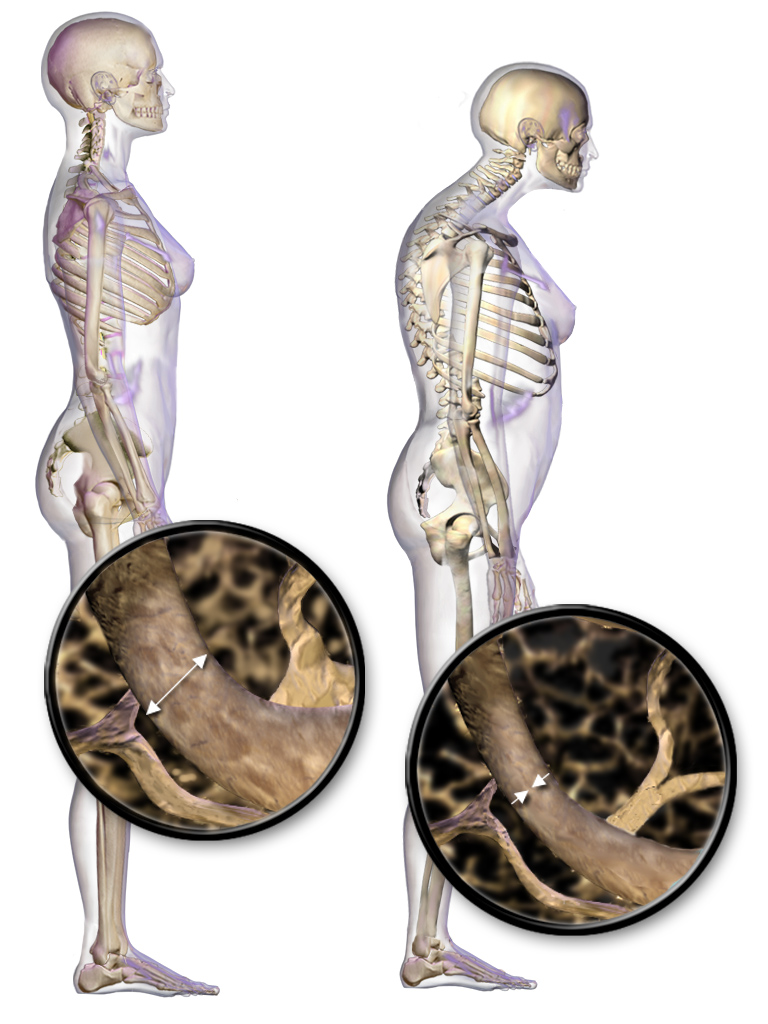
Osteoporosis is a systemic skeletal disorder characterized by low bone mass, micro-architectural deterioration of bone tissue leading to more porous bone, and consequent increase in Bone fracture, fracture risk. It is the most common reason for a broken bone among the Old age, elderly. Bones that commonly break include the vertebrae in the Vertebral column, spine, the bones of the forearm, the wrist, and the hip. Until a broken bone occurs there are typically no symptoms. Bones may weaken to such a degree that a break may occur with minor stress or spontaneously. After the broken bone heals, some people may have chronic pain and a decreased ability to carry out normal activities. Osteoporosis may be due to lower-than-normal peak bone mass, maximum bone mass and greater-than-normal bone loss. Bone loss increases after menopause in women due to lower levels of estrogen, and after andropause in older men due to lower levels of testosterone. Osteoporosis may also occur due to a ... [...More Info...] [...Related Items...] OR: [Wikipedia] [Google] [Baidu] |
Sacroiliac Joint
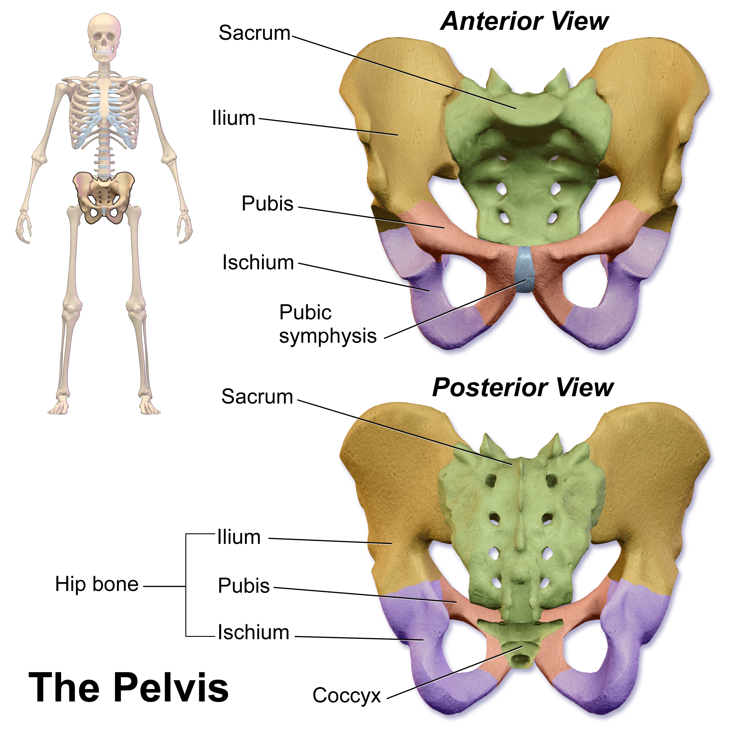
The sacroiliac joint or SI joint (SIJ) is the joint between the sacrum and the ilium bones of the pelvis, which are connected by strong ligaments. In humans, the sacrum supports the spine and is supported in turn by an ilium on each side. The joint is strong, supporting the entire weight of the upper body. It is a synovial plane joint with irregular elevations and depressions that produce interlocking of the two bones. The human body has two sacroiliac joints, one on the left and one on the right, that often match each other but are highly variable from person to person. Structure Sacroiliac joints are paired C-shaped or L-shaped joints capable of a small amount of movement (2–18 degrees, which is debatable at this time) that are formed between the auricular surfaces of the sacrum and the ilium bones. However, mostBogduk, Nicolai "Clinical and Radiological Anatomy of the Lumbar Spine" Elsevier Health Sciences, 2022, p. 172. agree that only slight movements occur on thes ... [...More Info...] [...Related Items...] OR: [Wikipedia] [Google] [Baidu] |
Periosteal Reaction
A periosteal reaction is the formation of new bone in response to injury or other stimuli of the periosteum surrounding the bone. It is most often identified on X-ray films of the bones. Cause A periosteal reaction can result from a large number of causes, including injury and chronic irritation due to a medical condition such as hypertrophic osteopathy, bone healing in response to fracture, chronic stress injuries, subperiosteal hematomas, osteomyelitis, and cancer of the bone. It may also occur as part of thyroid acropachy, a severe sign of the autoimmune thyroid disorder Graves' disease. Other causes include Menkes disease and hypervitaminosis A. It can take about three weeks to appear. Diagnosis The morphological appearance can be helpful in determining the cause of a periosteal reaction (for example, if other features of periostitis are present), but is usually not enough to be definitive. Diagnosis can be helped by establishing if bone formation is localized to a sp ... [...More Info...] [...Related Items...] OR: [Wikipedia] [Google] [Baidu] |
Projectional Radiography
Projectional radiography, also known as conventional radiography, is a form of radiography and medical imaging that produces two-dimensional images by X-ray radiation. The image acquisition is generally performed by radiographers, and the images are often examined by radiologists. Both the procedure and any resultant images are often simply called 'X-ray'. Plain radiography or roentgenography generally refers to projectional radiography (without the use of more advanced techniques such as CT scan, computed tomography that can generate 3D-images). ''Plain radiography'' can also refer to radiography without a radiocontrast agent or radiography that generates single static images, as contrasted to fluoroscopy, which are technically also projectional. Equipment X-ray generator Projectional radiographs generally use X-rays created by X-ray generators, which generate X-rays from X-ray tubes. Grid An anti-scatter grid may be placed between the patient and the detector to reduce the q ... [...More Info...] [...Related Items...] OR: [Wikipedia] [Google] [Baidu] |
Osteopenia
Osteopenia, known as "low bone mass" or "low bone density", is a condition in which bone mineral density is low. Because their bones are weaker, people with osteopenia may have a higher risk of fractures, and some people may go on to develop osteoporosis. In 2010, 43 million older adults in the US had osteopenia. Unlike osteoporosis, osteopenia does not usually cause symptoms, and losing bone density in itself does not cause pain. There is no single cause for osteopenia, although there are several risk factors, including modifiable (behavioral, including dietary and use of certain drugs) and non-modifiable (for instance, loss of bone mass with age). For people with risk factors, screening via a DXA scanner may help to detect the development and progression of low bone density. Prevention of low bone density may begin early in life and includes a healthy diet and weight-bearing exercise, as well as avoidance of tobacco and alcohol. The treatment of osteopenia is controversial ... [...More Info...] [...Related Items...] OR: [Wikipedia] [Google] [Baidu] |
Pubic Symphysis
The pubic symphysis (: symphyses) is a secondary cartilaginous joint between the left and right superior rami of the pubis of the hip bones. It is in front of and below the urinary bladder. In males, the suspensory ligament of the penis attaches to the pubic symphysis. In females, the pubic symphysis is attached to the suspensory ligament of the clitoris. In most adults, it can be moved roughly 2 mm and with 1 degree rotation. This increases for women at the time of childbirth. The name comes from the Greek word ''symphysis'', meaning 'growing together'. Structure The pubic symphysis is a nonsynovial amphiarthrodial joint. The width of the pubic symphysis at the front is 3–5 mm greater than its width at the back. This joint is connected by fibrocartilage and may contain a fluid-filled cavity; the center is avascular, possibly due to the nature of the compressive forces passing through this joint, which may lead to harmful vascular disease. The ends of both pubi ... [...More Info...] [...Related Items...] OR: [Wikipedia] [Google] [Baidu] |


