|
Red Reflex
The red reflex (also called the fundal reflex) refers to the reddish-orange reflection of light from the back of the eye, or fundus, observed when using an ophthalmoscope or retinoscope. The red reflex may be absent or poorly visible in people with dark eyes, and may appear yellow in Asians or green/blue in Africans. The reflex relies on the transparency of optical media (tear film, cornea, aqueous humor, crystalline lens, vitreous humor) and reflects off the fundus back through media into the aperture of the ophthalmoscope. The red reflex is considered abnormal if there is any asymmetry between the eyes, dark spots, or white reflex ( Leukocoria). Generally, it is a physical exam done on neonates and children by healthcare providers but occasionally occurs in flash photography seen when the pupil does not have enough time to constrict and reflects the fundus known as the red-eye effect. This is a recommended screening by the American Academy of Pediatrics and American A ... [...More Info...] [...Related Items...] OR: [Wikipedia] [Google] [Baidu] |
Fundus (eye)
The fundus of the eye is the interior surface of the eye opposite the lens and includes the retina, optic disc, macula, fovea, and posterior pole.Cassin, B. and Solomon, S. ''Dictionary of Eye Terminology''. Gainesville, Florida: Triad Publishing Company, 1990. The fundus can be examined by ophthalmoscopy and/or fundus photography. Variation The color of the fundus varies both between and within species. In one study of primates the retina is blue, green, yellow, orange, and red; only the human fundus (from a lightly pigmented blond person) is red. The major differences noted among the "higher" primate species were size and regularity of the border of macular area, size and shape of the optic disc, apparent 'texturing' of retina, and pigmentation of retina. Clinical significance Medical signs that can be detected from observation of eye fundus (generally by funduscopy) include hemorrhages, exudates, cotton wool spots, blood vessel abnormalities ( tortuosity, puls ... [...More Info...] [...Related Items...] OR: [Wikipedia] [Google] [Baidu] |
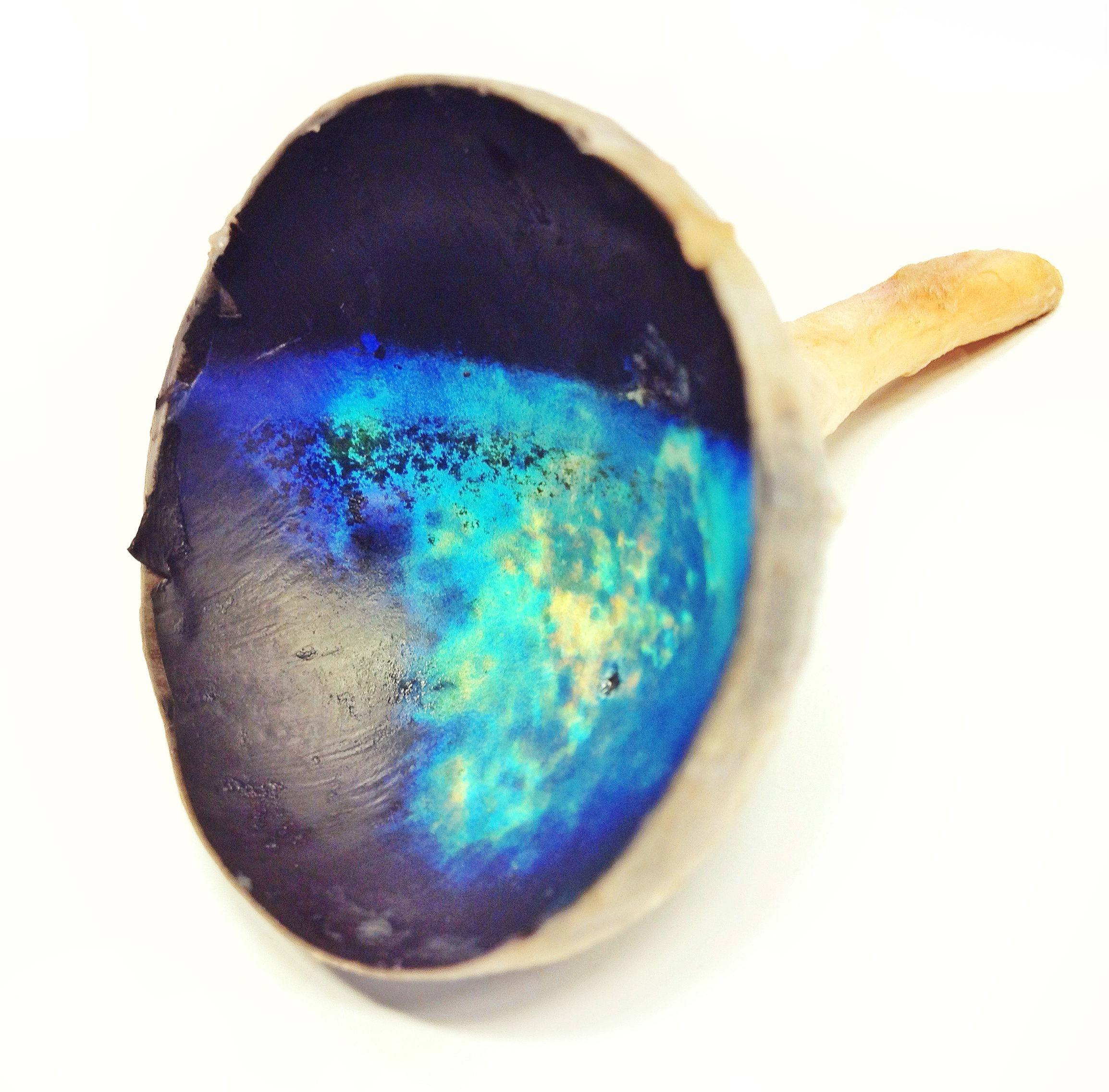
Tapetum Lucidum
The ; ; : tapeta lucida) is a layer of tissue in the eye of many vertebrates and some other animals. Lying immediately behind the retina, it is a retroreflector. It Reflection (physics), reflects visible light back through the retina, increasing the light available to the Photoreceptor cell, photoreceptors (although slightly blurring the image). The tapetum lucidum contributes to the superior night vision of some animals. Many of these animals are nocturnality, nocturnal, especially carnivores, while others are Deep-sea community, deep-sea animals. Similar adaptations occur in some species of spiders. Haplorhini, Haplorhine primates, including humans, are Diurnality, diurnal and lack a tapetum lucidum. Function and mechanism The presence of a tapetum lucidum enables animals to see in dimmer light than would otherwise be possible. The tapetum lucidum, which is iridescent, reflects light roughly on the Interference (wave propagation), interference principles of thin-film opti ... [...More Info...] [...Related Items...] OR: [Wikipedia] [Google] [Baidu] |
Red-eye Effect
The red-eye effect in photography is the common appearance of red pupils in color photographs of eyes. It occurs when using a photographic flash at low lighting or at night. When a flash passes through the eyes and rebounds at the back of the eye, it causes a red reflex in an image, turning the subject's eyes red. The hue is mostly caused by a high concentration of blood in the choroid. The effect can also be influenced by the near proximity of the flash and camera lens. In children, a different hue red reflex, such as white or yellow, may indicate an illness. In animals, a similar effect could cause their eyes to change colors in photographs. The effect can be avoided physically by instructing the subject to look away from the lens, increasing the brightness of the photographic location, or moving the flash further away from the lens, or digitally by using the red-eye correction option on digital cameras or by removing the effect in editing software. Scholars have developed ... [...More Info...] [...Related Items...] OR: [Wikipedia] [Google] [Baidu] |
Ophthalmologist
Ophthalmology (, ) is the branch of medicine that deals with the diagnosis, treatment, and surgery of eye diseases and disorders. An ophthalmologist is a physician who undergoes subspecialty training in medical and surgical eye care. Following a medical degree, a doctor specialising in ophthalmology must pursue additional postgraduate residency training specific to that field. In the United States, following graduation from medical school, one must complete a four-year residency in ophthalmology to become an ophthalmologist. Following residency, additional specialty training (or fellowship) may be sought in a particular aspect of eye pathology. Ophthalmologists prescribe medications to treat ailments, such as eye diseases, implement laser therapy, and perform surgery when needed. Ophthalmologists provide both primary and specialty eye care—medical and surgical. Most ophthalmologists participate in academic research on eye diseases at some point in their training and many incl ... [...More Info...] [...Related Items...] OR: [Wikipedia] [Google] [Baidu] |
Familial Exudative Vitreoretinopathy
Familial exudative vitreoretinopathy (FEVR, pronounced as fever) is a genetic disorder affecting the growth and development of blood vessels in the retina of the eye. This disease can lead to visual impairment and sometimes complete blindness in one or both eyes. FEVR is characterized by incomplete vascularization of the peripheral retina. This can lead to the growth of new blood vessels which are prone to Exudative, leakage and hemorrhage and can cause retinal folds, tears, and detachments. Treatment involves Laser coagulation, laser photocoagulation of the avascular portions of the retina to reduce new blood vessel growth and risk of complications including leakage of retinal blood vessels and retinal detachments. Pathophysiology FEVR is caused by genetic defects involving the regulation of blood vessel growth in developing eyes. As a result, there is poor blood vessel growth to the periphery of the retina. The lack of blood supply to the peripheral retina triggers the release o ... [...More Info...] [...Related Items...] OR: [Wikipedia] [Google] [Baidu] |
Pupillary Membranes
Persistent pupillary membrane (PPM) is a condition of the eye involving remnants of a fetal membrane that persist as strands of tissue crossing the pupil. The pupillary membrane in mammals exists in the fetus as a source of blood supply for the lens. It normally atrophies from the time of birth to the age of four to eight weeks. PPM occurs when this atrophy is incomplete. It generally does not cause any symptoms. The strands can connect to the cornea or lens, but most commonly to other parts of the iris. Attachment to the cornea can cause small corneal opacities, while attachment to the lens can cause small cataracts. Using topical atropine to dilate the pupil may help break down PPMs. In dogs, PPM is inherited in the Basenji but can occur in other breeds such as the Pembroke Welsh Corgi, Chow Chow, Mastiff, and English Cocker Spaniel. It can also be observed in cats, horses, and cattle Cattle (''Bos taurus'') are large, domesticated, bovid ungulates widely kept a ... [...More Info...] [...Related Items...] OR: [Wikipedia] [Google] [Baidu] |
Retinal Detachment
Retinal detachment is a condition where the retina pulls away from the tissue underneath it. It may start in a small area, but without quick treatment, it can spread across the entire retina, leading to serious vision loss and possibly blindness. Retinal detachment is a medical emergency that requires surgery. The retina is a thin layer at the back of the eye that processes visual information and sends it to the brain. When the retina detaches, common symptoms include seeing floaters, flashing lights, a dark shadow in vision, and sudden blurry vision. The most common type of retinal detachment is rhegmatogenous, which occurs when a tear or hole in the retina lets fluid from the center of the eye get behind it, causing the retina to pull away. Rhegmatogenous retinal detachment is most commonly caused by posterior vitreous detachment, a condition where the gel inside the eye breaks down and pulls on the retina. Risk factors include older age, nearsightedness ( myopia), eye injury, ... [...More Info...] [...Related Items...] OR: [Wikipedia] [Google] [Baidu] |
Strabismus
Strabismus is an eye disorder in which the eyes do not properly align with each other when looking at an object. The eye that is pointed at an object can alternate. The condition may be present occasionally or constantly. If present during a large part of childhood, it may result in amblyopia, or lazy eyes, and loss of depth perception. If onset is during adulthood, it is more likely to result in diplopia, double vision. Strabismus can occur out of muscle dysfunction (e.g., myasthenia gravis), farsightedness, problems in the brain, trauma, or infections. Risk factors include premature birth, cerebral palsy, and a family history of the condition. Types include esotropia, where the eyes are crossed ("cross eyed"); exotropia, where the eyes diverge ("lazy eyed" or "wall eyed"); and hypertropia or hypotropia, where they are vertically misaligned. They can also be classified by whether the problem is present in all directions a person looks (comitant) or varies by direction (inco ... [...More Info...] [...Related Items...] OR: [Wikipedia] [Google] [Baidu] |
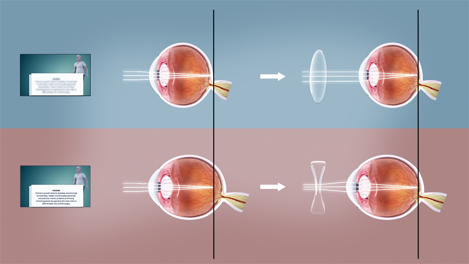
Refractive Error
Refractive error is a problem with focus (optics), focusing light accurately on the retina due to the shape of the eye and/or cornea. The most common types of refractive error are myopia, near-sightedness, hyperopia, far-sightedness, astigmatism, and presbyopia. Near-sightedness results in far away objects being blurred vision, blurry, far-sightedness and presbyopia result in close objects being blurry, and astigmatism causes objects to appear stretched out or blurry. Other symptoms may include double vision, headaches, and eye strain. Near-sightedness is due to the length of the eyeball being too long; far-sightedness the eyeball too short; astigmatism the cornea being the wrong shape, while presbyopia results from aging of the lens of the eye such that it cannot change shape sufficiently. Some refractive errors occur more often among those whose parents are affected. Diagnosis is by eye examination. Refractive errors are corrected with eyeglasses, contact lenses, or surgery ... [...More Info...] [...Related Items...] OR: [Wikipedia] [Google] [Baidu] |
Congenital Cataract
Congenital cataracts are a lens opacity that is present at birth. Congenital cataracts occur in a broad range of severity. Some lens opacities do not progress and are visually insignificant, others can produce profound visual impairment. Congenital cataracts may be unilateral or bilateral. They can be classified by morphology, presumed or defined genetic cause, presence of specific metabolic disorders, or associated ocular anomalies or systemic findings. Treatment options depend on the severity of the condition. For children under the age of two years old whose vision is affected by the cataracts in both eyes, surgical options include intraocular lens implantation or a lensectomy. Congenital cataracts are considered to be a significant cause of childhood blindness. This condition is considered 'treatable' with early intervention and compared to other types of childhood visual loss problems, however, in parts of the world where treatment options are not available such as som ... [...More Info...] [...Related Items...] OR: [Wikipedia] [Google] [Baidu] |
Antimuscarinic
A muscarinic acetylcholine receptor antagonist, also simply known as a muscarinic antagonist or as an antimuscarinic agent, is a type of anticholinergic drug that blocks the activity of the muscarinic acetylcholine receptors (mAChRs). The muscarinic receptors are proteins involved in the transmission of signals through certain parts of the nervous system, and muscarinic receptor antagonists work to prevent this transmission from occurring. Notably, muscarinic antagonists reduce the activation of the parasympathetic nervous system. The normal function of the parasympathetic system is often summarised as "rest-and-digest", and includes slowing of the heart, an increased rate of digestion, Bronchoconstriction, narrowing of the airways, promotion of urination, and sexual arousal. Muscarinic antagonists counter this parasympathetic "rest-and-digest" response, and also work elsewhere in both the Central nervous system, central and peripheral nervous systems. Drugs with muscarinic antagon ... [...More Info...] [...Related Items...] OR: [Wikipedia] [Google] [Baidu] |


