|
Recognition Sequence
A recognition sequence is a DNA sequence to which a structural motif of a DNA-binding domain exhibits binding specificity. Recognition sequences are palindromes. The transcription factor Sp1 for example, binds the sequences 5'-(G/T)GGGCGG(G/A)(G/A)(C/T)-3', where (G/T) indicates that the domain will bind a guanine or thymine at this position. The restriction endonuclease PstI recognizes, binds, and cleaves the sequence 5'-CTGCAG-3'. A recognition sequence is different from a recognition site. A given recognition sequence can occur one or more times, or not at all, on a specific DNA fragment. A recognition site is specified by the position of the site. For example, there are two PstI recognition sites in the following DNA sequence fragment, starting at base 9 and 31 respectively. A recognition sequence is a specific sequence, usually very short (less than 10 bases). Depending on the degree of specificity of the protein, a DNA-binding protein can bind to more than one specific ... [...More Info...] [...Related Items...] OR: [Wikipedia] [Google] [Baidu] |
|
|
DNA Sequence
A nucleic acid sequence is a succession of bases within the nucleotides forming alleles within a DNA (using GACT) or RNA (GACU) molecule. This succession is denoted by a series of a set of five different letters that indicate the order of the nucleotides. By convention, sequences are usually presented from the 5' end to the 3' end. For DNA, with its double helix, there are two possible directions for the notated sequence; of these two, the sense strand is used. Because nucleic acids are normally linear (unbranched) polymers, specifying the sequence is equivalent to defining the covalent structure of the entire molecule. For this reason, the nucleic acid sequence is also termed the primary structure. The sequence represents genetic information. Biological deoxyribonucleic acid represents the information which directs the functions of an organism. Nucleic acids also have a secondary structure and tertiary structure. Primary structure is sometimes mistakenly referred to as "prim ... [...More Info...] [...Related Items...] OR: [Wikipedia] [Google] [Baidu] |
|
|
Structural Motif
In a chain-like biological molecule, such as a protein or nucleic acid, a structural motif is a common three-dimensional structure that appears in a variety of different, evolutionarily unrelated molecules. A structural motif does not have to be associated with a sequence motif; it can be represented by different and completely unrelated sequences in different proteins or RNA. In nucleic acids Depending upon the sequence and other conditions, nucleic acids can form a variety of structural motifs which is thought to have biological significance. ;Stem-loop: Stem-loop intramolecular base pairing is a pattern that can occur in single-stranded DNA or, more commonly, in RNA. The structure is also known as a hairpin or hairpin loop. It occurs when two regions of the same strand, usually complementary in nucleotide sequence when read in opposite directions, base-pair to form a double helix that ends in an unpaired loop. The resulting structure is a key building block of many ... [...More Info...] [...Related Items...] OR: [Wikipedia] [Google] [Baidu] |
|
 |
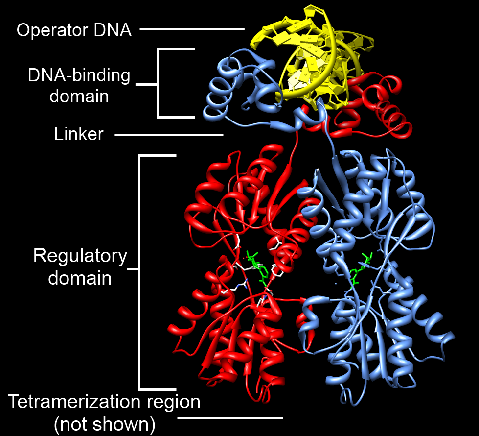
DNA-binding Domain
A DNA-binding domain (DBD) is an independently folded protein domain that contains at least one structural motif that recognizes double- or single-stranded DNA. A DBD can recognize a specific DNA sequence (a recognition sequence) or have a general affinity to DNA. Some DNA-binding domains may also include nucleic acids in their folded structure. Function One or more DNA-binding domains are often part of a larger protein consisting of further protein domains with differing function. The extra domains often regulate the activity of the DNA-binding domain. The function of DNA binding is either structural or involves transcription regulation, with the two roles sometimes overlapping. DNA-binding domains with functions involving DNA structure have biological roles in DNA replication, repair, storage, and modification, such as methylation. Many proteins involved in the regulation of gene expression contain DNA-binding domains. For example, proteins that regulate transcription ... [...More Info...] [...Related Items...] OR: [Wikipedia] [Google] [Baidu] |
 |
Chemical Specificity
Chemical specificity is the ability of binding site of a macromolecule (such as a protein) to bind specific ligands. The fewer ligands a protein can bind, the greater its specificity. Specificity describes the strength of binding between a given protein and ligand. This relationship can be described by a dissociation constant, which characterizes the balance between bound and unbound states for the protein-ligand system. In the context of a single enzyme and a pair of binding molecules, the two ligands can be compared as stronger or weaker ligands (for the enzyme) on the basis of their dissociation constants. (A lower value corresponds to a stronger binding.) Specificity for a set of ligands is unrelated to the ability of an enzyme to catalyze a given reaction, with the ligand as a substrate. If a given enzyme has a high chemical specificity, this means that the set of ligands to which it binds is limited, such that neither binding events nor catalysis can occur at an apprecia ... [...More Info...] [...Related Items...] OR: [Wikipedia] [Google] [Baidu] |
|
Palindromic Sequence
A palindromic sequence is a nucleic acid sequence in a double-stranded DNA or RNA molecule whereby reading in a certain direction (e.g. 5' to 3') on one strand is identical to the sequence in the same direction (e.g. 5' to 3') on the complementary strand. This definition of palindrome thus depends on complementary strands being palindromic of each other. The meaning of palindrome in the context of genetics is slightly different from the definition used for words and sentences. Since a double helix is formed by two paired antiparallel strands of nucleotides that run in opposite directions, and the nucleotides always pair in the same way (adenine (A) with thymine (T) in DNA or uracil (U) in RNA; cytosine (C) with guanine (G)), a (single-stranded) nucleotide sequence is said to be a palindrome if it is equal to its reverse complement. For example, the DNA sequence ACCTAGGT is palindromic with its nucleotide-by-nucleotide complement TGGATCCA because reversing the order of the n ... [...More Info...] [...Related Items...] OR: [Wikipedia] [Google] [Baidu] |
|
|
Transcription Factor
In molecular biology, a transcription factor (TF) (or sequence-specific DNA-binding factor) is a protein that controls the rate of transcription (genetics), transcription of genetics, genetic information from DNA to messenger RNA, by binding to a specific DNA sequence. The function of TFs is to regulate—turn on and off—genes in order to make sure that they are Gene expression, expressed in the desired Cell (biology), cells at the right time and in the right amount throughout the life of the cell and the organism. Groups of TFs function in a coordinated fashion to direct cell division, cell growth, and cell death throughout life; cell migration and organization (body plan) during embryonic development; and intermittently in response to signals from outside the cell, such as a hormone. There are approximately 1600 TFs in the human genome. Transcription factors are members of the proteome as well as regulome. TFs work alone or with other proteins in a complex, by promoting (a ... [...More Info...] [...Related Items...] OR: [Wikipedia] [Google] [Baidu] |
|
|
Transcription Factor Sp1
Transcription factor Sp1, also known as specificity protein 1* is a protein that in humans is encoded by the ''SP1'' gene. Function The protein encoded by this gene is a zinc finger transcription factor that binds to GC-rich motifs of many promoters. The encoded protein is involved in many cellular processes, including cell differentiation, cell growth, apoptosis, immune responses, response to DNA damage, and chromatin remodeling. post-translational modifications such as phosphorylation, acetylation, ''O''-GlcNAcylation, and proteolytic processing significantly affect the activity of this protein, which can be an activator or a repressor. In the SV40 virus, Sp1 binds to the GC boxes in the regulatory sequence of the genome. Structure SP1 belongs to the Sp/KLF family of transcription factors. The protein is 785 amino acids long, with a molecular weight of 81 kDa. The SP1 transcription factor contains two glutamine-rich activation domains at its N-terminus that are belie ... [...More Info...] [...Related Items...] OR: [Wikipedia] [Google] [Baidu] |
|
|
Guanine
Guanine () (symbol G or Gua) is one of the four main nucleotide bases found in the nucleic acids DNA and RNA, the others being adenine, cytosine, and thymine ( uracil in RNA). In DNA, guanine is paired with cytosine. The guanine nucleoside is called guanosine. With the formula C5H5N5O, guanine is a derivative of purine, consisting of a fused pyrimidine- imidazole ring system with conjugated double bonds. This unsaturated arrangement means the bicyclic molecule is planar. Properties Guanine, along with adenine and cytosine, is present in both DNA and RNA, whereas thymine is usually seen only in DNA, and uracil only in RNA. Guanine has multiple tautomeric forms. For the imidazole ring, the proton can reside on either nitrogen. For the pyrimidine ring, the ring N-H can center can reside on either of the ring nitrogens. The latter tautomer does not apply to nucleoside or nucleotide versions of guanine. It binds to cytosine through three hydrogen bonds. In cytosine, t ... [...More Info...] [...Related Items...] OR: [Wikipedia] [Google] [Baidu] |
|
|
Thymine
Thymine () (symbol T or Thy) is one of the four nucleotide bases in the nucleic acid of DNA that are represented by the letters G–C–A–T. The others are adenine, guanine, and cytosine. Thymine is also known as 5-methyluracil, a pyrimidine nucleobase. In RNA, thymine is replaced by the nucleobase uracil. Thymine was first isolated in 1893 by Albrecht Kossel and Albert Neumann from calf thymus glands, hence its name. Derivation As its alternate name (5-methyluracil) suggests, thymine may be derived by methylation of uracil at the 5th carbon. In RNA, thymine is replaced with uracil in most cases. In DNA, thymine (T) binds to adenine (A) via two hydrogen bonds, thereby stabilizing the nucleic acid structures. Thymine combined with deoxyribose creates the nucleoside deoxythymidine, which is synonymous with the term thymidine. Thymidine can be phosphorylated with up to three phosphoric acid groups, producing dTMP (deoxythymidine monophosphate), dTDP, or dTTP (for the ... [...More Info...] [...Related Items...] OR: [Wikipedia] [Google] [Baidu] |
|
 |
Restriction Endonuclease
A restriction enzyme, restriction endonuclease, REase, ENase or'' restrictase '' is an enzyme that cleaves DNA into fragments at or near specific recognition sites within molecules known as restriction sites. Restriction enzymes are one class of the broader endonuclease group of enzymes. Restriction enzymes are commonly classified into five types, which differ in their structure and whether they cut their DNA enzyme substrate (biology), substrate at their recognition site, or if the recognition and cleavage sites are separate from one another. To cut DNA, all restriction enzymes make two incisions, once through each backbone chain, sugar-phosphate backbone (i.e. each strand) of the DNA double helix. These enzymes are found in bacteria and archaea and provide a defense mechanism against invading viruses. Inside a prokaryote, the restriction enzymes selectively cut up ''foreign'' DNA in a process called ''restriction digestion''; meanwhile, host DNA is protected by a modification ... [...More Info...] [...Related Items...] OR: [Wikipedia] [Google] [Baidu] |
|
PstI
PstI is a type II restriction endonuclease isolated from the Gram negative species, ''Providencia stuartii''. Function PstI cleaves DNA at the recognition sequence 5′-CTGCA/G-3′ generating fragments with 3′-cohesive termini. This cleavage yields sticky ends 4 base pairs long. PstI is catalytically active as a dimer. The two subunits are related by a 2-fold symmetry axis which in the complex with the substrate coincides with the dyad axis of the recognition sequence. It has a molecular weight of 69,500 and contains 54 positive and 41 negatively charged residues. The PstI restriction/modification (R/M) system has two components: a restriction enzyme that cleaves foreign DNA, and a methyltransferase which protect native DNA strands by methylation of the adenine base inside the recognition sequence. The combination of both provide is a defense mechanism against invading viruses. The methyltransferase and endonuclease are encoded as two separate proteins and act independentl ... [...More Info...] [...Related Items...] OR: [Wikipedia] [Google] [Baidu] |
|
|
Recognition Site
In molecular biology, restriction sites, or restriction recognition sites, are regions of a DNA molecule containing specific (4-8 base pairs in length) sequences of nucleotides; these are recognized by restriction enzymes, which cleave the DNA at or near the site. These are generally palindromic sequences (because restriction enzymes usually bind as homodimers), and a particular restriction enzyme may cut the sequence between two nucleotides within its recognition site, or somewhere nearby. Function For example, the common restriction enzyme EcoRI recognizes the palindromic sequence GAATTC and cuts between the G and the A on both the top and bottom strands. This leaves an overhang (an end-portion of a DNA strand with no attached complement) known as a sticky end on each end of AATT. The overhang can then be used to ligate in (see DNA ligase) a piece of DNA with a complementary overhang (another EcoRI-cut piece, for example). Some restriction enzymes cut DNA at a restriction sit ... [...More Info...] [...Related Items...] OR: [Wikipedia] [Google] [Baidu] |