|
Ramariopsis Corcea
''Ramariopsis'' is a genus of coral fungi in the family Clavariaceae. The genus has a collectively widespread distribution and contains about 40 species. The name means 'having the appearance of ''Ramaria. Taxonomy ''Ramariopsis'' was originally defined as a subgenus of ''Clavaria'' by Dutch mycologist Marinus Anton Donk in 1933. Several European species similar to the type, '' Clavaria kunzei'', were included: '' Clavaria subtilis'', '' Clavaria pyxidata'', '' Clavaria angulispora'', and '' Clavaria pulchella''. In Donk's concept, defining characteristics of the group included small, branching, fruitbodies with a stipe, and an almost cartilaginous consistency to the flesh. Spores are small and hyaline (translucent), spherical to ellipsoid, and have a surface ornamentation ranging from echinulate (spiny) to verruculose (covered with small warts). E.J.H. Corner promoted the subgenus to generic status in his 1950 world monograph of clavarioid fungi. Ron Petersen emended the genu ... [...More Info...] [...Related Items...] OR: [Wikipedia] [Google] [Baidu] |
Ramariopsis Kunzei
''Ramariopsis kunzei'' is an edible species of coral fungi in the family Clavariaceae, and the type species of the genus ''Ramariopsis''. It is commonly known as white coral because of the branched structure of the fruit bodies that resemble marine coral. The fruit bodies are up to tall by wide, with numerous branches originating from a short rudimentary stem. The branches are one to two millimeters thick, smooth, and white, sometimes with yellowish tips in age. ''Ramariopsis kunzei'' has a widespread distribution, and is found in North America, Eurasia, and Australia. Taxonomy The species was first described as ''Clavaria kunzei'' by pioneer mycologist Elias Magnus Fries in 1821. E. J. H. Corner transferred the species to ''Ramariopsis'' in 1950, and made it the type species. In general, coral fungi often have extensive taxonomic histories, as mycologists have not agreed on the best way to classify them. In addition to ''Clavaria'' and ''Ramariopsis'', the ''R. ... [...More Info...] [...Related Items...] OR: [Wikipedia] [Google] [Baidu] |
Cartilage
Cartilage is a resilient and smooth type of connective tissue. Semi-transparent and non-porous, it is usually covered by a tough and fibrous membrane called perichondrium. In tetrapods, it covers and protects the ends of long bones at the joints as articular cartilage, and is a structural component of many body parts including the rib cage, the neck and the bronchial tubes, and the intervertebral discs. In other taxa, such as chondrichthyans and cyclostomes, it constitutes a much greater proportion of the skeleton. It is not as hard and rigid as bone, but it is much stiffer and much less flexible than muscle. The matrix of cartilage is made up of glycosaminoglycans, proteoglycans, collagen fibers and, sometimes, elastin. It usually grows quicker than bone. Because of its rigidity, cartilage often serves the purpose of holding tubes open in the body. Examples include the rings of the trachea, such as the cricoid cartilage and carina. Cartilage is composed of specialized c ... [...More Info...] [...Related Items...] OR: [Wikipedia] [Google] [Baidu] |
Basidia
A basidium (: basidia) is a microscopic spore-producing structure found on the hymenophore of reproductive bodies of basidiomycete fungi. The presence of basidia is one of the main characteristic features of the group. These bodies are also called tertiary mycelia, which are highly coiled versions of secondary mycelia. A basidium usually bears four sexual spores called basidiospores. Occasionally the number may be two or even eight. Each reproductive spore is produced at the tip of a narrow prong or horn called a sterigma (), and is forcefully expelled at full growth. The word ''basidium'' literally means "little pedestal". This is the way the basidium supports the spores. However, some biologists suggest that the structure looks more like a club. A partially grown basidium is known as a basidiole. Structure Most basidiomycota have single celled basidia (holobasidia), but some have ones with many cells (a phragmobasidia). For instance, rust fungi in the order ''Puccinal ... [...More Info...] [...Related Items...] OR: [Wikipedia] [Google] [Baidu] |
Clamp Connection
A clamp connection is a hook-like structure formed by growing hyphal cells of certain fungi. It is a characteristic feature of basidiomycete fungi. It is created to ensure that each cell, or segment of hypha separated by septa (cross walls), receives a set of differing nuclei, which are obtained through mating of hyphae of differing sexual types. It is used to maintain genetic variation within the hypha much like the mechanisms found in croziers (hooks) during the sexual reproduction of ascomycetes. Formation Clamp connections are formed by the terminal hypha during elongation. Before the clamp connection is formed this terminal segment contains two nuclei. Once the terminal segment is long enough it begins to form the clamp connection. At the same time, each nucleus undergoes mitotic division to produce two daughter nuclei. As the clamp continues to develop it uptakes one of the daughter (green circle) nuclei and separates it from its sister nucleus. While this is occurring t ... [...More Info...] [...Related Items...] OR: [Wikipedia] [Google] [Baidu] |
Hypha
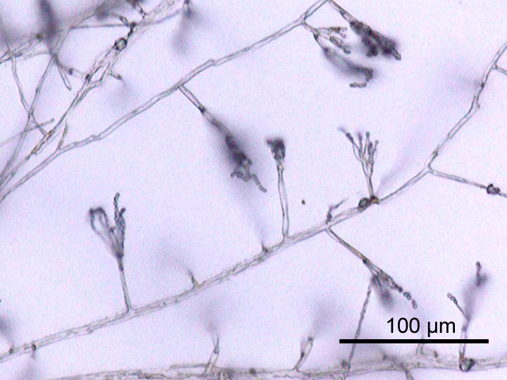
A hypha (; ) is a long, branching, filamentous structure of a fungus, oomycete, or actinobacterium. In most fungi, hyphae are the main mode of vegetative growth, and are collectively called a mycelium. Structure A hypha consists of one or more cells surrounded by a tubular cell wall. In most fungi, hyphae are divided into cells by internal cross-walls called "septa" (singular septum). Septa are usually perforated by pores large enough for ribosomes, mitochondria, and sometimes nuclei to flow between cells. The major structural polymer in fungal cell walls is typically chitin, in contrast to plants and oomycetes that have cellulosic cell walls. Some fungi have aseptate hyphae, meaning their hyphae are not partitioned by septa. Hyphae have an average diameter of 4–6 μm. Growth Hyphae grow at their tips. During tip growth, cell walls are extended by the external assembly and polymerization of cell wall components, and the internal production of new cell membrane. ... [...More Info...] [...Related Items...] OR: [Wikipedia] [Google] [Baidu] |
Basidiocarp
In fungi, a basidiocarp, basidiome, or basidioma () is the sporocarp of a basidiomycete, the multicellular structure on which the spore-producing hymenium is borne. Basidiocarps are characteristic of the hymenomycetes; rusts and smuts do not produce such structures. As with other sporocarps, epigeous (above-ground) basidiocarps that are visible to the naked eye (especially those with a more or less agaricoid morphology) are commonly referred to as mushrooms, while hypogeous (underground) basidiocarps are usually called false truffles. Structure All basidiocarps serve as the structure on which the hymenium is produced. Basidia are found on the surface of the hymenium, and the basidia ultimately produce spores. In its simplest form, a basidiocarp consists of an undifferentiated fruiting structure with a hymenium on the surface; such a structure is characteristic of many simple jelly and club fungi. In more complex basidiocarps, there is differentiation into a stipe, a p ... [...More Info...] [...Related Items...] OR: [Wikipedia] [Google] [Baidu] |
Ultrastructure
Ultrastructure (or ultra-structure) is the architecture of cells and biomaterials that is visible at higher magnifications than found on a standard optical light microscope. This traditionally meant the resolution and magnification range of a conventional transmission electron microscope (TEM) when viewing biological specimens such as cells, tissue, or organs. Ultrastructure can also be viewed with scanning electron microscopy and super-resolution microscopy, although TEM is a standard histology technique for viewing ultrastructure. Such cellular structures as organelles, which allow the cell to function properly within its specified environment, can be examined at the ultrastructural level. Ultrastructure, along with molecular phylogeny, is a reliable phylogenetic way of classifying organisms. Features of ultrastructure are used industrially to control material properties and promote biocompatibility. History In 1931, German engineers Max Knoll and Ernst Ruska invente ... [...More Info...] [...Related Items...] OR: [Wikipedia] [Google] [Baidu] |
Electron Microscopy
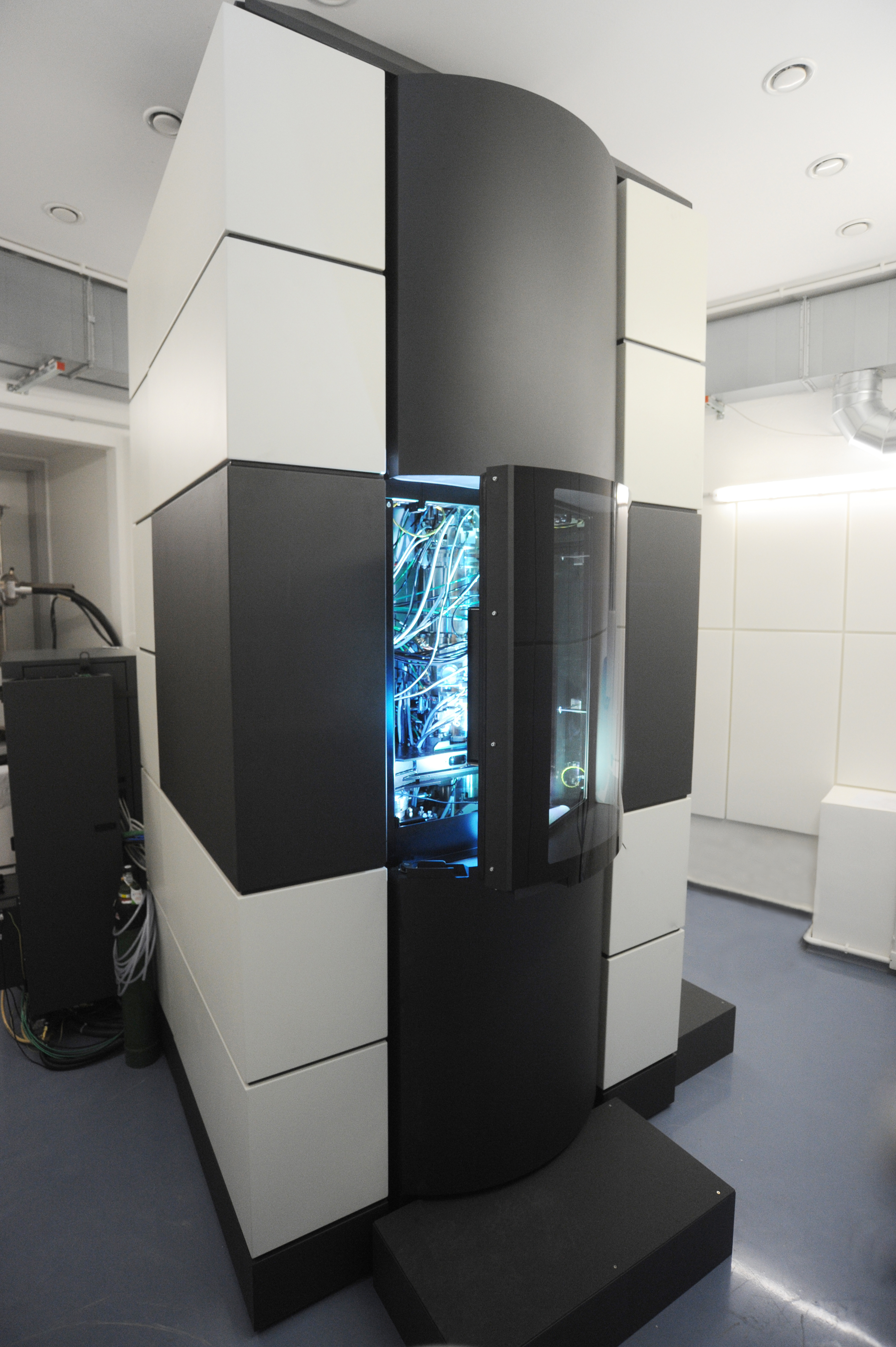
An electron microscope is a microscope that uses a beam of electrons as a source of illumination. It uses electron optics that are analogous to the glass lenses of an optical light microscope to control the electron beam, for instance focusing it to produce magnified images or electron diffraction patterns. As the wavelength of an electron can be up to 100,000 times smaller than that of visible light, electron microscopes have a much higher resolution of about 0.1 nm, which compares to about 200 nm for light microscopes. ''Electron microscope'' may refer to: * Transmission electron microscope (TEM) where swift electrons go through a thin sample * Scanning transmission electron microscope (STEM) which is similar to TEM with a scanned electron probe * Scanning electron microscope (SEM) which is similar to STEM, but with thick samples * Electron microprobe similar to a SEM, but more for chemical analysis * Low-energy electron microscope (LEEM), used to image surfaces * ... [...More Info...] [...Related Items...] OR: [Wikipedia] [Google] [Baidu] |
Ramariopsis Minutula
''Ramariopsis'' is a genus of coral fungi in the family Clavariaceae. The genus has a collectively widespread distribution and contains about 40 species. The name means 'having the appearance of ''Ramaria. Taxonomy ''Ramariopsis'' was originally defined as a subgenus of ''Clavaria'' by Dutch mycologist Marinus Anton Donk in 1933. Several European species similar to the type species, type, ''Clavaria kunzei'', were included: ''Ramariopsis subtilis, Clavaria subtilis'', ''Artomyces pyxidatus, Clavaria pyxidata'', ''Scytinopogon angulisporus, Clavaria angulispora'', and ''Ramariopsis pulchella, Clavaria pulchella''. In Donk's concept, defining characteristics of the group included small, branching, fruitbodies with a stipe (mycology), stipe, and an almost cartilage, cartilaginous consistency to the trama (mycology), flesh. Basidiospore, Spores are small and hyaline (translucent), spherical to ellipsoid, and have a surface ornamentation ranging from echinulate (spiny) to verruculose ... [...More Info...] [...Related Items...] OR: [Wikipedia] [Google] [Baidu] |
Ron Petersen
Ronald H. Petersen, more commonly known as Ron Petersen, born in 1934, is a mycologist and professor emeritus at the University of Tennessee The University of Tennessee, Knoxville (or The University of Tennessee; UT; UT Knoxville; or colloquially UTK or Tennessee) is a Public university, public Land-grant university, land-grant research university in Knoxville, Tennessee, United St ... known for his work on chanterelle mushrooms and the genus Flammulina. He was the editor-in-chief of the journal '' Mycologia'' from 1986 to 1990. See also * :Taxa named by Ron Petersen References 1934 births Living people American mycologists University of Tennessee faculty Place of birth missing (living people) {{Mycologist-stub ... [...More Info...] [...Related Items...] OR: [Wikipedia] [Google] [Baidu] |
Monograph
A monograph is generally a long-form work on one (usually scholarly) subject, or one aspect of a subject, typically created by a single author or artist (or, sometimes, by two or more authors). Traditionally it is in written form and published as a book, but it may be an artwork, audiovisual work, or exhibition made up of visual artworks. In library cataloguing, the word has a specific and broader meaning, while in the United States, the Food and Drug Administration uses the term to mean a set of published standards. Written works Academic works The English term ''monograph'' is derived from modern Latin , which has its root in Greek. In the English word, ''mono-'' means and ''-graph'' means . Unlike a textbook, which surveys the state of knowledge in a field, the main purpose of a monograph is to present primary research and original scholarship. This research is presented at length, distinguishing a monograph from an article. For these reasons, publication of a monograph ... [...More Info...] [...Related Items...] OR: [Wikipedia] [Google] [Baidu] |
Ellipsoid
An ellipsoid is a surface that can be obtained from a sphere by deforming it by means of directional Scaling (geometry), scalings, or more generally, of an affine transformation. An ellipsoid is a quadric surface; that is, a Surface (mathematics), surface that may be defined as the zero set of a polynomial of degree two in three variables. Among quadric surfaces, an ellipsoid is characterized by either of the two following properties. Every planar Cross section (geometry), cross section is either an ellipse, or is empty, or is reduced to a single point (this explains the name, meaning "ellipse-like"). It is Bounded set, bounded, which means that it may be enclosed in a sufficiently large sphere. An ellipsoid has three pairwise perpendicular Rotational symmetry, axes of symmetry which intersect at a Central symmetry, center of symmetry, called the center of the ellipsoid. The line segments that are delimited on the axes of symmetry by the ellipsoid are called the ''principal ax ... [...More Info...] [...Related Items...] OR: [Wikipedia] [Google] [Baidu] |


