|
Portosystemic Shunts In Animals
Congenital portosystemic shunts (PSS) is a hereditary condition in dogs and cats, its frequency varying depending on the breed. The shunts found mainly in small dog breeds such as Shih Tzus, Tibetan Spaniels, Miniature Schnauzers and Yorkshire Terriers, and in cats such as Persians, British Shorthairs, Himalayans, and mixed breeds are usually extrahepatic (outside the liver), while the shunts found in large dog breeds such as Irish Wolfhounds and Labrador Retrievers tend to be intrahepatic (inside the liver). Acquired PSS is uncommon and is found in dogs and cats with liver disease such as cirrhosis causing portal hypertension, which is high blood pressure in the portal vein. Symptoms Symptoms of congenital PSS usually appear by six months of age and include failure to gain weight, vomiting, and signs of hepatic encephalopathy (a condition where toxins normally removed by the liver accumulate in the blood and impair the function of brain cells) such as seizures, depression, tre ... [...More Info...] [...Related Items...] OR: [Wikipedia] [Google] [Baidu] |
Portosystemic Shunt
A portosystemic shunt or portasystemic shunt (medical subject heading term; PSS), also known as a liver shunt, is a bypass of the liver by the body's circulatory system. It can be either a congenital (present at birth) or acquired condition and occurs in humans as well as in other species of animals. Congenital PSS are extremely rare in humans but are relatively common in dogs. Thus a large part of medical and scientific literature on the subject is grounded in veterinary medicine. Background Blood leaving the digestive tract is rich in nutrients, as well as in toxins, which under normal conditions undergo processing and detoxification in the liver. The liver's position downstream to the intestines in the body's circulatory system - the hepatic portal vein conveys blood from the intestines to the liver - allows it to filter this nutrient rich blood before it passes to the rest of the body. The presence of a shunt, a bypass of the liver, causes blood to flow directly to the hear ... [...More Info...] [...Related Items...] OR: [Wikipedia] [Google] [Baidu] |
Uric Acid
Uric acid is a heterocyclic compound of carbon, nitrogen, oxygen, and hydrogen with the formula C5H4N4O3. It forms ions and salts known as urates and acid urates, such as ammonium acid urate. Uric acid is a product of the metabolic breakdown of purine nucleotides, and it is a normal component of urine. High blood concentrations of uric acid can lead to gout and are associated with other medical conditions, including diabetes and the formation of ammonium acid urate kidney stones. Chemistry Uric acid was first isolated from kidney stones in 1776 by Swedish chemist Carl Wilhelm Scheele. In 1882, the Ukrainian chemist Ivan Horbaczewski first synthesized uric acid by melting urea with glycine. Uric acid displays lactam–lactim tautomerism (also often described as keto–enol tautomerism). Although the lactim form is expected to possess some degree of aromaticity, uric acid crystallizes in the lactam form, with computational chemistry also indicating that tautomer to ... [...More Info...] [...Related Items...] OR: [Wikipedia] [Google] [Baidu] |
Specificity (tests)
In medicine and statistics, sensitivity and specificity mathematically describe the accuracy of a test that reports the presence or absence of a medical condition. If individuals who have the condition are considered "positive" and those who do not are considered "negative", then sensitivity is a measure of how well a test can identify true positives and specificity is a measure of how well a test can identify true negatives: * Sensitivity (true positive rate) is the probability of a positive test result, conditioned on the individual truly being positive. * Specificity (true negative rate) is the probability of a negative test result, conditioned on the individual truly being negative. If the true status of the condition cannot be known, sensitivity and specificity can be defined relative to a " gold standard test" which is assumed correct. For all testing, both diagnoses and screening, there is usually a trade-off between sensitivity and specificity, such that higher sensit ... [...More Info...] [...Related Items...] OR: [Wikipedia] [Google] [Baidu] |
Bile Acid
Bile acids are steroid acids found predominantly in the bile of mammals and other vertebrates. Diverse bile acids are synthesized in the liver. Bile acids are conjugated with taurine or glycine residues to give anions called bile salts. Primary bile acids are those synthesized by the liver. Secondary bile acids result from bacterial actions in the colon. In humans, taurocholic acid and glycocholic acid (derivatives of cholic acid) and taurochenodeoxycholic acid and glycochenodeoxycholic acid (derivatives of chenodeoxycholic acid) are the major bile salts. They are roughly equal in concentration. The salts of their 7-alpha-dehydroxylated derivatives, deoxycholic acid and lithocholic acid, are also found, with derivatives of cholic, chenodeoxycholic and deoxycholic acids accounting for over 90% of human biliary bile acids. Bile acids comprise about 80% of the organic compounds in bile (others are phospholipids and cholesterol). An increased secretion of bile acids produ ... [...More Info...] [...Related Items...] OR: [Wikipedia] [Google] [Baidu] |
Hepatitis
Hepatitis is inflammation of the liver parenchyma, liver tissue. Some people or animals with hepatitis have no symptoms, whereas others develop yellow discoloration of the skin and whites of the eyes (jaundice), Anorexia (symptom), poor appetite, vomiting, fatigue (medicine), tiredness, abdominal pain, and diarrhea. Hepatitis is ''acute (medicine), acute'' if it resolves within six months, and ''chronic condition, chronic'' if it lasts longer than six months. Acute hepatitis can self-limiting (biology), resolve on its own, progress to chronic hepatitis, or (rarely) result in acute liver failure. Chronic hepatitis may progress to scarring of the liver (cirrhosis), liver failure, and liver cancer. Hepatitis is most commonly caused by the virus ''hepatovirus A'', ''hepatitis B virus, B'', ''hepatitis C virus, C'', ''hepatitis D virus, D'', and ''hepatitis E virus, E''. Other Viral hepatitis, viruses can also cause liver inflammation, including cytomegalovirus, Epstein–Barr virus, ... [...More Info...] [...Related Items...] OR: [Wikipedia] [Google] [Baidu] |
Fibrosis
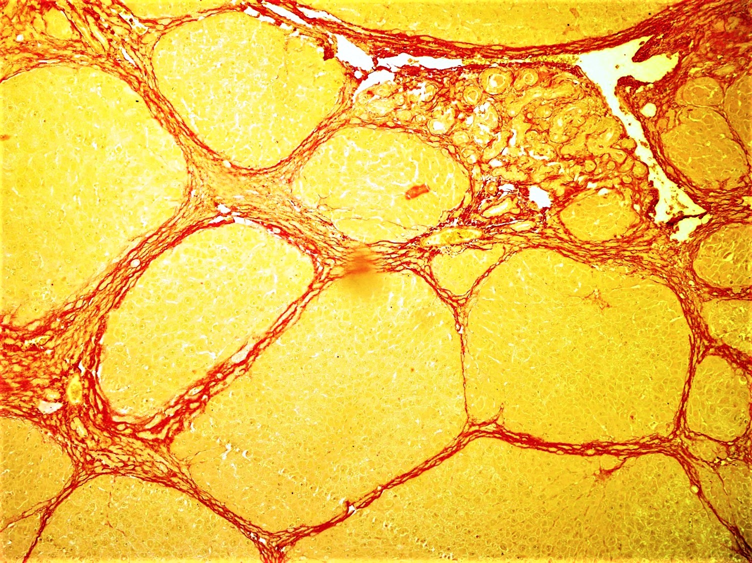
Fibrosis, also known as fibrotic scarring, is a pathological wound healing in which connective tissue replaces normal parenchymal tissue to the extent that it goes unchecked, leading to considerable tissue remodelling and the formation of permanent scar tissue. Repeated injuries, chronic inflammation and repair are susceptible to fibrosis where an accidental excessive accumulation of extracellular matrix components, such as the collagen is produced by fibroblasts, leading to the formation of a permanent fibrotic scar. In response to injury, this is called scarring, and if fibrosis arises from a single cell line, this is called a fibroma. Physiologically, fibrosis acts to deposit connective tissue, which can interfere with or totally inhibit the normal architecture and function of the underlying organ or tissue. Fibrosis can be used to describe the pathological state of excess deposition of fibrous tissue, as well as the process of connective tissue deposition in healing. Define ... [...More Info...] [...Related Items...] OR: [Wikipedia] [Google] [Baidu] |
Left Gastric Vein
The left gastric vein (or coronary vein) is a vein that derives from tributaries draining the lesser curvature of the stomach. Structure The left gastric vein runs from right to left along the lesser curvature of the stomach. It passes to the esophageal opening of the stomach, where it receives some esophageal veins. It then turns backward and passes from left to right behind the omental bursa. It drains into the portal vein near the superior border of the pancreas. Function The left gastric vein drains deoxygenated blood from the lesser curvature of the stomach. It also acts as collaterals between the portal vein and the systemic venous system of the lower esophagus (azygous vein). Clinical significance Esophageal and paraesophageal varices are supplied primarily by the left gastric vein (due to flow reversal) and typically drain into the azygos/hemiazygos venous system.Siegelman, E.: "Body MRI", page 47. Saunders, 2004 See also * Right gastric vein The right g ... [...More Info...] [...Related Items...] OR: [Wikipedia] [Google] [Baidu] |
Ammonia
Ammonia is an inorganic compound of nitrogen and hydrogen with the formula . A stable binary hydride, and the simplest pnictogen hydride, ammonia is a colourless gas with a distinct pungent smell. Biologically, it is a common nitrogenous waste, particularly among aquatic organisms, and it contributes significantly to the nutritional needs of terrestrial organisms by serving as a precursor to 45% of the world's food and fertilizers. Around 70% of ammonia is used to make fertilisers in various forms and composition, such as urea and Diammonium phosphate. Ammonia in pure form is also applied directly into the soil. Ammonia, either directly or indirectly, is also a building block for the synthesis of many pharmaceutical products and is used in many commercial cleaning products. It is mainly collected by downward displacement of both air and water. Although common in nature—both terrestrially and in the outer planets of the Solar System—and in wide use, ammonia i ... [...More Info...] [...Related Items...] OR: [Wikipedia] [Google] [Baidu] |
Journal Of The American Animal Hospital Association
The American Animal Hospital Association (AAHA) is a non-profit organization for companion animal veterinary hospitals. Established in 1933, the association is the only accrediting body for small animal hospitals in the U.S. and Canada. The association develops standards for veterinary business practices, publications, and educational programs. Any veterinary hospital can join AAHA as a member, but must then pass an evaluation in order to receive AAHA accreditation. AAHA Accreditation Unlike human hospitals, veterinary hospitals are not required to be accredited. Accredited hospitals are the only hospitals in the U.S. and Canada that choose to be evaluated on approximately 900 quality standards that go above and beyond basic state regulations, ranging from patient care and pain management to staff training and advanced diagnostic services. To become AAHA-accredited, practices undergo a rigorous evaluation process to ensure they meet the AAHA Standards of Accreditation, which inc ... [...More Info...] [...Related Items...] OR: [Wikipedia] [Google] [Baidu] |
Inferior Vena Cava
The inferior vena cava is a large vein that carries the deoxygenated blood from the lower and middle body into the right atrium of the heart. It is formed by the joining of the right and the left common iliac veins, usually at the level of the fifth lumbar vertebra. The inferior vena cava is the lower ("inferior") of the two venae cavae, the two large veins that carry deoxygenated blood from the body to the right atrium of the heart: the inferior vena cava carries blood from the lower half of the body whilst the superior vena cava carries blood from the upper half of the body. Together, the venae cavae (in addition to the coronary sinus, which carries blood from the muscle of the heart itself) form the venous counterparts of the aorta. It is a large retroperitoneal vein that lies posterior to the abdominal cavity and runs along the right side of the vertebral column. It enters the right auricle at the lower right, back side of the heart. The name derives from la, ven ... [...More Info...] [...Related Items...] OR: [Wikipedia] [Google] [Baidu] |
Vitelline Veins
The vitelline veins are veins that drain blood from the yolk sac and the gut tube during gestation. Path They run upward at first in front, and subsequently on either side of the intestinal canal. They unite on the ventral aspect of the canal. Beyond this, they are connected to one another by two anastomotic branches, one on the dorsal, and the other on the ventral aspect of the duodenal portion of the intestine. This is encircled by two venous rings; into the middle or dorsal anastomosis the superior mesenteric vein opens. The portions of the veins above the upper ring become interrupted by the developing liver and broken up by it into a plexus of small capillary-like vessels termed sinusoids. Derivatives The vitelline veins give rise to: * Hepatic veins * Inferior portion of Inferior vena cava * Portal vein * Superior mesenteric vein * Inferior mesenteric vein The branches conveying the blood to the plexus are named the venae advehentes, and become the branches of the po ... [...More Info...] [...Related Items...] OR: [Wikipedia] [Google] [Baidu] |


-3D-balls.png)

