|
Piriform Aperture
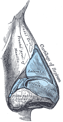
The piriform aperture, pyriform aperture, or anterior nasal aperture is a pear-shaped opening in the human skull. Its long axis is vertical, and narrow end upward; in the recent state it is much contracted by the lateral nasal cartilage and the greater and lesser alar cartilages of the nose. It is bounded above by the inferior borders of the nasal bones; laterally by the thin, sharp margins which separate the anterior from the nasal surfaces of the maxilla; and below by the same borders, where they curve medialward to join each other at the anterior nasal spine The anterior nasal spine, or anterior nasal spine of maxilla, is a bony projection in the skull that serves as a cephalometric landmark. The anterior nasal spine is the projection formed by the fusion of the two maxillary bones at the intermaxill .... References Nose {{musculoskeletal-stub ... [...More Info...] [...Related Items...] OR: [Wikipedia] [Google] [Baidu] |
Michiel Sweerts
Michiel Sweerts or Michael Sweerts (29 September 1618 – 1 June 1664) was a Flemish painter and printmaker of the Baroque period, who is known for his allegorical and genre paintings, portraits and tronies. The artist led an itinerant life and worked in Rome, Brussels, Amsterdam, Persia and India (Goa). While in Rome Sweerts became linked to the group of Dutch and Flemish painters of low-life scenes known as the ''Bamboccianti''. Sweerts' contributions to the Bamboccianti genre display generally greater stylistic mastery and social-philosophical sensitivity than the other artists working in this manner. While he was successful during his lifetime, Sweerts and his work fell into obscurity until he was rediscovered in the 20th century as one of the most intriguing and enigmatic artists of his time. [...More Info...] [...Related Items...] OR: [Wikipedia] [Google] [Baidu] |
Pear
Pears are fruits produced and consumed around the world, growing on a tree and harvested in late summer into mid-autumn. The pear tree and shrub are a species of genus ''Pyrus'' , in the Family (biology), family Rosaceae, bearing the Pome, pomaceous fruit of the same name. Several species of pears are valued for their edible fruit and juices, while others are cultivated as trees. The tree is medium-sized and native to coastal and mildly temperate regions of Europe, North Africa, and Asia. Pear wood is one of the preferred materials in the manufacture of high-quality woodwind instruments and furniture. About 3,000 known varieties of pears are grown worldwide, which vary in both shape and taste. The fruit is consumed fresh, canning, canned, as juice, Dried fruit, dried, or fermented as perry. Etymology The word ''pear'' is probably from Germanic ''pera'' as a loanword of Vulgar Latin ''pira'', the plural of ''pirum'', akin to Greek ''apios'' (from Mycenaean ''ápisos''), of ... [...More Info...] [...Related Items...] OR: [Wikipedia] [Google] [Baidu] |
Human Skull
The skull, or cranium, is typically a bony enclosure around the brain of a vertebrate. In some fish, and amphibians, the skull is of cartilage. The skull is at the head end of the vertebrate. In the human, the skull comprises two prominent parts: the neurocranium and the facial skeleton, which evolved from the first pharyngeal arch. The skull forms the frontmost portion of the axial skeleton and is a product of cephalization and vesicular enlargement of the brain, with several special senses structures such as the eyes, ears, nose, tongue and, in fish, specialized tactile organs such as barbels near the mouth. The skull is composed of three types of bone: cranial bones, facial bones and ossicles, which is made up of a number of fused flat and irregular bones. The cranial bones are joined at firm fibrous junctions called sutures and contains many foramina, fossae, processes, and sinuses. In zoology, the openings in the skull are called fenestrae, the ... [...More Info...] [...Related Items...] OR: [Wikipedia] [Google] [Baidu] |
Lateral Nasal Cartilage
The lateral nasal cartilage (upper lateral cartilage, lateral process of septal nasal cartilage) is situated below the inferior margin of the nasal bone, and is flattened, and triangular in shape. Its anterior margin is thicker than the posterior, and is continuous above with the septal nasal cartilage, but separated from it below by a narrow fissure; its superior margin is attached to the nasal bone and the frontal process of the maxilla; its inferior margin is connected by fibrous tissue with the greater alar cartilage. Where the lateral cartilage meets the greater alar cartilage, the lateral cartilage often curls up, to join with an inward curl of the greater alar cartilage. That curl of the inferior portion of the lateral cartilage is called its "scroll." ''Thieme Atlas of Anatomy ... [...More Info...] [...Related Items...] OR: [Wikipedia] [Google] [Baidu] |
Greater Alar Cartilage
The major alar cartilage (greater alar cartilage) (lower lateral cartilage) is a thin, flexible plate, situated immediately below the lateral nasal cartilage, and bent upon itself in such a manner as to form the Nasal septum, medial wall and lateral wall of the nostril of its own side. The portion which forms the Nasal septum, medial wall (crus mediale) is loosely connected with the corresponding portion of the opposite cartilage, the two forming, together with the thickened integument and subjacent tissue, the nasal septum. The part which forms the lateral wall (crus laterale) is curved to correspond with the Human nose#External nose, ala of the nose; it is oval and flattened, narrow behind, where it is connected with the frontal process of the maxilla by a tough fibrous membrane, in which are found three or four small cartilaginous plates, the lesser alar cartilages (cartilagines alares minores; sesamoid cartilages). Above, it is connected by fibrous tissue to the lateral carti ... [...More Info...] [...Related Items...] OR: [Wikipedia] [Google] [Baidu] |
Lesser Alar Cartilages
In human anatomy, the part of the nose which forms the lateral wall is curved to correspond with the ala of the nose; it is oval and flattened, narrow behind, where it is connected with the frontal process of the maxilla In vertebrates, the maxilla (: maxillae ) is the upper fixed (not fixed in Neopterygii) bone of the jaw formed from the fusion of two maxillary bones. In humans, the upper jaw includes the hard palate in the front of the mouth. The two maxil ... by a tough fibrous membrane, in which are found three or four small nasal cartilages the minor alar cartilages, also referred to as lesser alar or sesamoid cartilages or accessory cartilages. ''An atlas of human anatomy for students and physicians'' (Rebman, 1919; by Carl Toldt, Alois Dalla Rosa, Eden Paul, p. 942-944)- Re ... [...More Info...] [...Related Items...] OR: [Wikipedia] [Google] [Baidu] |
Human Nose
The human nose is the first organ of the respiratory system. It is also the principal organ in the olfactory system. The shape of the nose is determined by the nasal bones and the nasal cartilages, including the nasal septum, which separates the nostrils and divides the nasal cavity into two. The nose has an important function in breathing. The nasal mucosa lining the nasal cavity and the paranasal sinuses carries out the necessary conditioning of inhaled air by warming and moistening it. Nasal conchae, shell-like bones in the walls of the cavities, play a major part in this process. Filtering of the air by nasal hair in the nostrils prevents large particles from entering the lungs. Sneezing is a reflex to expel unwanted particles from the nose that irritate the mucosal lining. Sneezing can Transmission (medicine), transmit infections, because aerosols are created in which the Respiratory droplets, droplets can harbour pathogens. Another major function of the nose is olfactio ... [...More Info...] [...Related Items...] OR: [Wikipedia] [Google] [Baidu] |
Nasal Bones
The nasal bones are two small oblong bones, varying in size and form in different individuals; they are placed side by side at the middle and upper part of the face and by their junction, form the bridge of the upper one third of the nose. Each has two surfaces and four borders. Structure There is heavy variation in the structure of the nasal bones, accounting for the differences in sizes and shapes of the nose seen across different people. Angles, shapes, and configurations of both the bone and cartilage are heavily varied between individuals. Broadly, most nasal bones can be categorized as "V-shaped" or "S-shaped" but these are not scientific or medical categorizations. When viewing anatomical drawings of these bones, consider that they are unlikely to be accurate for a majority of people. The two nasal bones are joined at the midline internasal suture and make up the bridge of the nose. Surfaces The ''outer surface'' is concavo-convex from above downward, convex from ... [...More Info...] [...Related Items...] OR: [Wikipedia] [Google] [Baidu] |
Maxilla
In vertebrates, the maxilla (: maxillae ) is the upper fixed (not fixed in Neopterygii) bone of the jaw formed from the fusion of two maxillary bones. In humans, the upper jaw includes the hard palate in the front of the mouth. The two maxillary bones are fused at the intermaxillary suture, forming the anterior nasal spine. This is similar to the mandible (lower jaw), which is also a fusion of two mandibular bones at the mandibular symphysis. The mandible is the movable part of the jaw. Anatomy Structure The maxilla is a paired bone - the two maxillae unite with each other at the intermaxillary suture. The maxilla consists of: * The body of the maxilla: pyramid-shaped; has an orbital, a nasal, an infratemporal, and a facial surface; contains the maxillary sinus. * Four processes: ** the zygomatic process ** the frontal process ** the alveolar process ** the palatine process It has three surfaces: * the anterior, posterior, medial Features of the maxilla include: * t ... [...More Info...] [...Related Items...] OR: [Wikipedia] [Google] [Baidu] |
Anterior Nasal Spine
The anterior nasal spine, or anterior nasal spine of maxilla, is a bony projection in the skull that serves as a cephalometric landmark. The anterior nasal spine is the projection formed by the fusion of the two maxillary bones at the intermaxillary suture. It is placed at the level of the nostrils, at the uppermost part of the philtrum. It rarely fractures. Additional images File:Anterior nasal spine of maxilla - animation02.gif, Animation. Anterior nasal spine shown in red. File:Anterior nasal spine of maxilla - animation00.gif, Left maxilla. Anterior nasal spine shown in red. File:Anterior nasal spine of maxilla - skull - anterior view.png, Skull. Anterior view. Anterior nasal spine shown in red. File:Slide12hhhh.JPG, Right maxilla. Anterior nasal spine labeled at center left. See also * Posterior nasal spine The posterior nasal spine is part of the horizontal plate of the palatine bone of the skull. It is found at the medial end of its posterior border. It is paired ... [...More Info...] [...Related Items...] OR: [Wikipedia] [Google] [Baidu] |


