|
Periptychus
''Periptychus'' is an extinct genus of mammal belonging to the family Periptychidae. It lived from the Early to Late Paleocene and its fossil remains have been found in North America. Description This animal was of medium size and could exceed one meter in overall length; ''Periptychus'' is supposed to have weighed about 23 kilograms. ''Periptychus'' was an unusual mammal that combined a number of rather specialized dental, cranial, and postcranial features with a relatively generalized skeletal structure. Skull The shape of the skull of ''Periptychus'' was almost identical to that of early Eutheria, although it was more robust. The snout of ''Periptychus'' was moderately elongated and tall, and tapered anteriorly without a rostral constriction. The morphology of the rostrum of this animal was very similar to that found in related genera such as '' Carsioptychus'' or ''Ectoconus''; the snout was not elongated as in other condylarths such as ''Arctocyon'', but it was long ... [...More Info...] [...Related Items...] OR: [Wikipedia] [Google] [Baidu] |
Periptychus Teeth
''Periptychus'' is an extinct genus of mammal belonging to the family Periptychidae. It lived from the Early to Late Paleocene and its fossil remains have been found in North America. Description This animal was of medium size and could exceed one meter in overall length; ''Periptychus'' is supposed to have weighed about 23 kilograms. ''Periptychus'' was an unusual mammal that combined a number of rather specialized dental, cranial, and postcranial features with a relatively generalized skeletal structure. Skull The shape of the skull of ''Periptychus'' was almost identical to that of early Eutheria, although it was more robust. The snout of ''Periptychus'' was moderately elongated and tall, and tapered anteriorly without a rostral constriction. The morphology of the rostrum of this animal was very similar to that found in related genera such as '' Carsioptychus'' or ''Ectoconus''; the snout was not elongated as in other condylarths such as '' Arctocyon'', but it was long ... [...More Info...] [...Related Items...] OR: [Wikipedia] [Google] [Baidu] |
Periptychidae
Periptychidae is a family of Paleocene placental mammals, known definitively only from North America. The family is part of a radiation of early herbivorous and omnivorous mammals formerly classified in the extinct order "Condylarthra", which may be related to some or all living ungulates (hoofed mammals). Periptychids are distinguished from other "condylarths" by their teeth, which have swollen premolar The premolars, also called premolar teeth, or bicuspids, are transitional teeth located between the canine and molar teeth. In humans, there are two premolars per quadrant in the permanent set of teeth, making eight premolars total in the mouth ...s and unusual vertical enamel ridges. The family includes both large and small genera, with the larger forms having robust skeletons. Known skeletons of periptychids suggest generalized terrestrial habits. References *McKenna, Malcolm C., and Bell, Susan K. 1997. ''Classification of Mammals Above the Species Level.'' Columbia Univ ... [...More Info...] [...Related Items...] OR: [Wikipedia] [Google] [Baidu] |
Paleocene
The Paleocene, ( ) or Palaeocene, is a geological epoch that lasted from about 66 to 56 million years ago (mya). It is the first epoch of the Paleogene Period in the modern Cenozoic Era. The name is a combination of the Ancient Greek ''palaiós'' meaning "old" and the Eocene Epoch (which succeeds the Paleocene), translating to "the old part of the Eocene". The epoch is bracketed by two major events in Earth's history. The K–Pg extinction event, brought on by an asteroid impact and possibly volcanism, marked the beginning of the Paleocene and killed off 75% of living species, most famously the non-avian dinosaurs. The end of the epoch was marked by the Paleocene–Eocene Thermal Maximum (PETM), which was a major climatic event wherein about 2,500–4,500 gigatons of carbon were released into the atmosphere and ocean systems, causing a spike in global temperatures and ocean acidification. In the Paleocene, the continents of the Northern Hemisphere were still connected v ... [...More Info...] [...Related Items...] OR: [Wikipedia] [Google] [Baidu] |
Mandible
In anatomy, the mandible, lower jaw or jawbone is the largest, strongest and lowest bone in the human facial skeleton. It forms the lower jaw and holds the lower teeth in place. The mandible sits beneath the maxilla. It is the only movable bone of the skull (discounting the ossicles of the middle ear). It is connected to the temporal bones by the temporomandibular joints. The bone is formed in the fetus from a fusion of the left and right mandibular prominences, and the point where these sides join, the mandibular symphysis, is still visible as a faint ridge in the midline. Like other symphyses in the body, this is a midline articulation where the bones are joined by fibrocartilage, but this articulation fuses together in early childhood.Illustrated Anatomy of the Head and Neck, Fehrenbach and Herring, Elsevier, 2012, p. 59 The word "mandible" derives from the Latin word ''mandibula'', "jawbone" (literally "one used for chewing"), from '' mandere'' "to chew" and ''-bula'' ... [...More Info...] [...Related Items...] OR: [Wikipedia] [Google] [Baidu] |
Diaphysis
The diaphysis is the main or midsection (shaft) of a long bone. It is made up of cortical bone and usually contains bone marrow and adipose tissue (fat). It is a middle tubular part composed of compact bone which surrounds a central marrow cavity which contains red or yellow marrow. In diaphysis, primary ossification occurs. Ewing sarcoma Ewing sarcoma is a type of cancer that forms in bone or soft tissue. Symptoms may include swelling and pain at the site of the tumor, fever, and a bone fracture. The most common areas where it begins are the legs, pelvis, and chest wall. In abou ... tends to occur at the diaphysis.Physical Medicine and Rehabilitation Board Review, Cuccurullo Additional images Illu long bone.jpg File:EpiMetaDiaphyse.jpg, Long bone See also * Epiphysis * Metaphysis References Skeletal system Long bones {{musculoskeletal-stub ... [...More Info...] [...Related Items...] OR: [Wikipedia] [Google] [Baidu] |
Radius (bone)
The radius or radial bone is one of the two large bones of the forearm, the other being the ulna. It extends from the lateral side of the elbow to the thumb side of the wrist and runs parallel to the ulna. The ulna is usually slightly longer than the radius, but the radius is thicker. Therefore the radius is considered to be the larger of the two. It is a long bone, prism-shaped and slightly curved longitudinally. The radius is part of two joints: the elbow and the wrist. At the elbow, it joins with the capitulum of the humerus, and in a separate region, with the ulna at the radial notch. At the wrist, the radius forms a joint with the ulna bone. The corresponding bone in the lower leg is the fibula. Structure The long narrow medullary cavity is enclosed in a strong wall of compact bone. It is thickest along the interosseous border and thinnest at the extremities, same over the cup-shaped articular surface (fovea) of the head. The trabeculae of the spongy t ... [...More Info...] [...Related Items...] OR: [Wikipedia] [Google] [Baidu] |
Ulna
The ulna (''pl''. ulnae or ulnas) is a long bone found in the forearm that stretches from the elbow to the smallest finger, and when in anatomical position, is found on the medial side of the forearm. That is, the ulna is on the same side of the forearm as the little finger. It runs parallel to the radius, the other long bone in the forearm. The ulna is usually slightly longer than the radius, but the radius is thicker. Therefore, the radius is considered to be the larger of the two. Structure The ulna is a long bone found in the forearm that stretches from the elbow to the smallest finger, and when in anatomical position, is found on the medial side of the forearm. It is broader close to the elbow, and narrows as it approaches the wrist. Close to the elbow, the ulna has a bony process, the olecranon process, a hook-like structure that fits into the olecranon fossa of the humerus. This prevents hyperextension and forms a hinge joint with the trochlea of the humerus. There ... [...More Info...] [...Related Items...] OR: [Wikipedia] [Google] [Baidu] |
Humerus
The humerus (; ) is a long bone in the arm that runs from the shoulder to the elbow. It connects the scapula and the two bones of the lower arm, the radius and ulna, and consists of three sections. The humeral upper extremity consists of a rounded head, a narrow neck, and two short processes (tubercles, sometimes called tuberosities). The body is cylindrical in its upper portion, and more prismatic below. The lower extremity consists of 2 epicondyles, 2 processes (trochlea & capitulum), and 3 fossae (radial fossa, coronoid fossa, and olecranon fossa). As well as its true anatomical neck, the constriction below the greater and lesser tubercles of the humerus is referred to as its surgical neck due to its tendency to fracture, thus often becoming the focus of surgeons. Etymology The word "humerus" is derived from la, humerus, umerus meaning upper arm, shoulder, and is linguistically related to Gothic ''ams'' shoulder and Greek ''ōmos''. Structure Upper extremity The upper or pr ... [...More Info...] [...Related Items...] OR: [Wikipedia] [Google] [Baidu] |
Lumbar Vertebrae
The lumbar vertebrae are, in human anatomy, the five vertebrae between the rib cage and the pelvis. They are the largest segments of the vertebral column and are characterized by the absence of the foramen transversarium within the transverse process (since it is only found in the cervical region) and by the absence of facets on the sides of the body (as found only in the thoracic region). They are designated L1 to L5, starting at the top. The lumbar vertebrae help support the weight of the body, and permit movement. Human anatomy General characteristics The adjacent figure depicts the general characteristics of the first through fourth lumbar vertebrae. The fifth vertebra contains certain peculiarities, which are detailed below. As with other vertebrae, each lumbar vertebra consists of a ''vertebral body'' and a ''vertebral arch''. The vertebral arch, consisting of a pair of ''pedicles'' and a pair of ''laminae'', encloses the ''vertebral foramen'' (opening) and s ... [...More Info...] [...Related Items...] OR: [Wikipedia] [Google] [Baidu] |
Plantigrade
151px, Portion of a human skeleton, showing plantigrade habit In terrestrial animals, plantigrade locomotion means walking with the toes and metatarsals flat on the ground. It is one of three forms of locomotion adopted by terrestrial mammals. The other options are digitigrade, walking on the toes with the heel and wrist permanently raised, and unguligrade, walking on the nail or nails of the toes (the hoof) with the heel/wrist and the digits permanently raised. The leg of a plantigrade mammal includes the bones of the upper leg (femur/humerus) and lower leg (tibia and fibula/radius and ulna). The leg of a digitigrade mammal also includes the metatarsals/metacarpals, the bones that in a human compose the arch of the foot and the palm of the hand. The leg of an unguligrade mammal also includes the phalanges, the finger and toe bones. Among extinct animals, most early mammals such as pantodonts were plantigrade. A plantigrade foot is the primitive condition for mammals; di ... [...More Info...] [...Related Items...] OR: [Wikipedia] [Google] [Baidu] |
Canine Tooth
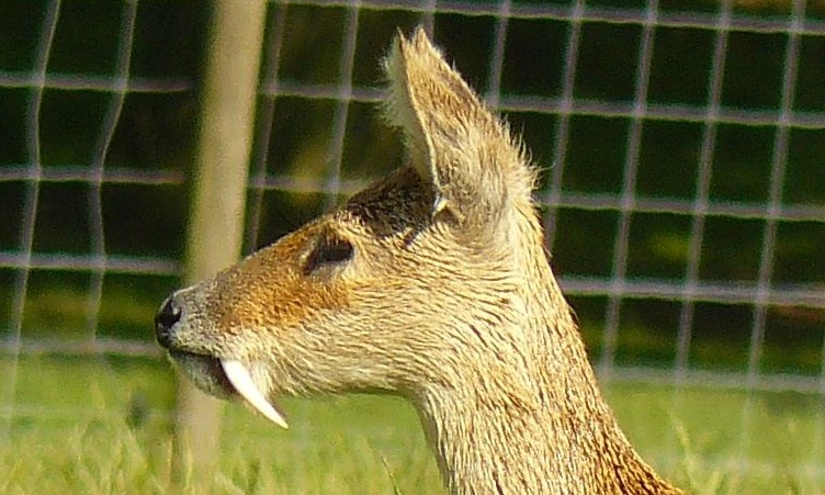
In mammalian oral anatomy, the canine teeth, also called cuspids, dog teeth, or (in the context of the upper jaw) fangs, eye teeth, vampire teeth, or vampire fangs, are the relatively long, pointed teeth. They can appear more flattened however, causing them to resemble incisors and leading them to be called ''incisiform''. They developed and are used primarily for firmly holding food in order to tear it apart, and occasionally as weapons. They are often the largest teeth in a mammal's mouth. Individuals of most species that develop them normally have four, two in the upper jaw and two in the lower, separated within each jaw by incisors; humans and dogs are examples. In most species, canines are the anterior-most teeth in the maxillary bone. The four canines in humans are the two maxillary canines and the two mandibular canines. Details There are generally four canine teeth: two in the upper (maxillary) and two in the lower (mandibular) arch. A canine is placed laterall ... [...More Info...] [...Related Items...] OR: [Wikipedia] [Google] [Baidu] |
Tooth Enamel
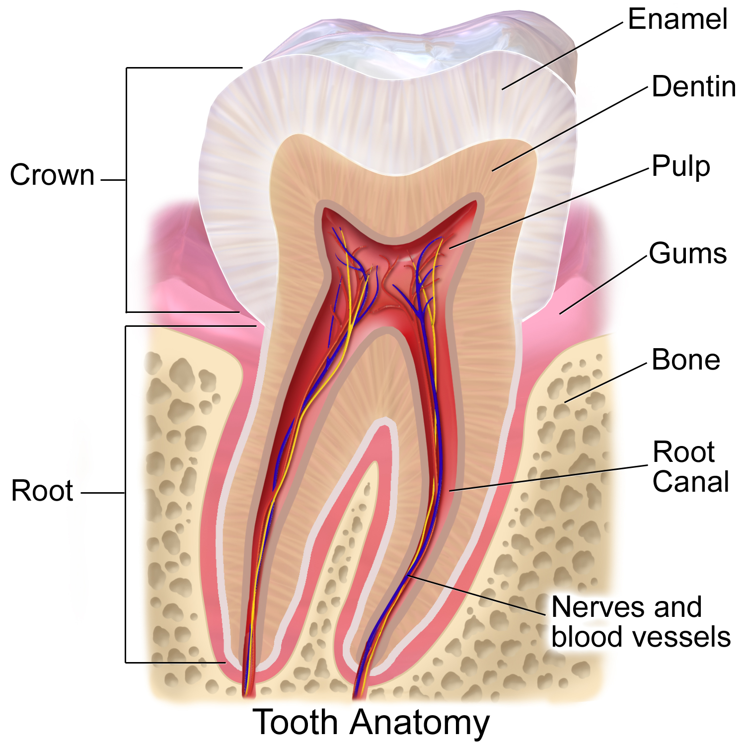
Tooth enamel is one of the four major tissues that make up the tooth in humans and many other animals, including some species of fish. It makes up the normally visible part of the tooth, covering the crown. The other major tissues are dentin, cementum, and dental pulp. It is a very hard, white to off-white, highly mineralised substance that acts as a barrier to protect the tooth but can become susceptible to degradation, especially by acids from food and drink. Calcium hardens the tooth enamel. In rare circumstances enamel fails to form, leaving the underlying dentin exposed on the surface. Features Enamel is the hardest substance in the human body and contains the highest percentage of minerals (at 96%),Ross ''et al.'', p. 485 with water and organic material composing the rest.Ten Cate's Oral Histology, Nancy, Elsevier, pp. 70–94 The primary mineral is hydroxyapatite, which is a crystalline calcium phosphate. Enamel is formed on the tooth while the tooth develops wit ... [...More Info...] [...Related Items...] OR: [Wikipedia] [Google] [Baidu] |


