|
Patella Aspera
The patella (: patellae or patellas), also known as the kneecap, is a flat, rounded triangular bone which articulates with the femur (thigh bone) and covers and protects the anterior articular surface of the knee joint. The patella is found in many tetrapods, such as mice, cats, birds, and dogs, but not in whales, or most reptiles. In humans, the patella is the largest sesamoid bone (i.e., embedded within a tendon or a muscle) in the body. Babies are born with a patella of soft cartilage which begins to ossify into bone at about four years of age. Structure The patella is a sesamoid bone roughly triangular in shape, with the apex of the patella facing downwards. The apex is the most inferior (lowest) part of the patella. It is pointed in shape, and gives attachment to the patellar ligament. The front and back surfaces are joined by a thin margin and towards centre by a thicker margin. The tendon of the quadriceps femoris muscle attaches to the base of the patella., with the ... [...More Info...] [...Related Items...] OR: [Wikipedia] [Google] [Baidu] |
Bone
A bone is a rigid organ that constitutes part of the skeleton in most vertebrate animals. Bones protect the various other organs of the body, produce red and white blood cells, store minerals, provide structure and support for the body, and enable mobility. Bones come in a variety of shapes and sizes and have complex internal and external structures. They are lightweight yet strong and hard and serve multiple functions. Bone tissue (osseous tissue), which is also called bone in the uncountable sense of that word, is hard tissue, a type of specialised connective tissue. It has a honeycomb-like matrix internally, which helps to give the bone rigidity. Bone tissue is made up of different types of bone cells. Osteoblasts and osteocytes are involved in the formation and mineralisation of bone; osteoclasts are involved in the resorption of bone tissue. Modified (flattened) osteoblasts become the lining cells that form a protective layer on the bone surface. The mine ... [...More Info...] [...Related Items...] OR: [Wikipedia] [Google] [Baidu] |
Vastus Medialis
The vastus medialis (vastus internus or teardrop muscle) is an extensor muscle located medially in the thigh that extends the knee. The vastus medialis is part of the quadriceps muscle group. Structure The vastus medialis is a muscle present in the anterior compartment of thigh, and is one of the four muscles that make up the quadriceps muscle. The others are the vastus lateralis, vastus intermedius and rectus femoris. It is the most medial of the "vastus" group of muscles. The vastus medialis arises medially along the entire length of the femur, and attaches with the other muscles of the quadriceps in the quadriceps tendon. The vastus medialis muscle originates from a continuous line of attachment on the femur, which begins on the front and middle side (anteromedially) on the intertrochanteric line of the femur. It continues down and back (posteroinferiorly) along the pectineal line and then descends along the inner (medial) lip of the linea aspera and onto the medi ... [...More Info...] [...Related Items...] OR: [Wikipedia] [Google] [Baidu] |
Medial Patellofemoral Ligament
The medial patellofemoral ligament (MPFL) is one of several ligaments on the medial aspect of the knee. It originates in the superomedial aspect of the patella and inserts in the space between the adductor tubercle and the medial femoral epicondyle. The ligament itself extends from the femur to the superomedial patella, and its shape is similar to a trapezoid. It keeps the patella in place, but its main function is to prevent lateral displacement of the patella. Structure The MPFL is located in the second soft tissue layer in the knee; this layer also includes the medial collateral ligament. The middle layer has the most consequential role in the patella's stabilization. The MPFL's origin is on the femur between the medial femoral epicondyle and the adductor tubercle, while being superior to the superficial medial collateral ligament. From the origin, it moves anteriorly, and combines with the deep portion of the vastus medialus oblique, inserting to the superomedial side of t ... [...More Info...] [...Related Items...] OR: [Wikipedia] [Google] [Baidu] |
Lateral Condyle Of Femur
The lateral condyle is one of the two projections on the lower extremity of the femur. The other one is the medial condyle. The lateral condyle is the more prominent and is broader both in its front-to-back and transverse diameters. Clinical significance The most common injury to the lateral femoral condyle is an osteochondral fracture combined with a patellar dislocation. The osteochondral fracture occurs on the weight-bearing portion of the lateral condyle. Typically, the condyle will fracture (and the patella may dislocate) as a result of severe impaction from activities such as downhill skiing and parachuting. Open reduction and internal fixation surgery is typically used to repair an osteochondral fracture. For a AO Type B1 partial articular fracture of the lateral condyle, interfragmentary lag screws are used to secure the bone back together. Supplementation of buttress screws or a buttress plate is used if the fracture extends to the proximal metaphysis or distal diaphysi ... [...More Info...] [...Related Items...] OR: [Wikipedia] [Google] [Baidu] |
Quadriceps Tendon
In human anatomy Human anatomy (gr. ἀνατομία, "dissection", from ἀνά, "up", and τέμνειν, "cut") is primarily the scientific study of the morphology of the human body. Anatomy is subdivided into gross anatomy and microscopic anatomy. Gross ..., the quadriceps tendon works with the quadriceps muscle to extend the leg. All four parts of the quadriceps muscle attach to the shin via the patella (knee cap), where the quadriceps tendon becomes the patellar ligament. It attaches the quadriceps to the top of the patella, which in turn is connected to the shin from its bottom by the patellar ligament. A tendon connects muscle to bone, while a ligament connects bone to bone.Saladin, Kenneth S. Anatomy & Physiology: The Unity of Form and Function. 6th ed. New York: McGraw-Hill, 2012. Print. Injuries are common to this tendon, with tears, either partial or complete, being the most common. If the quadriceps tendon is completely torn, surgery will be required to ... [...More Info...] [...Related Items...] OR: [Wikipedia] [Google] [Baidu] |
Lever
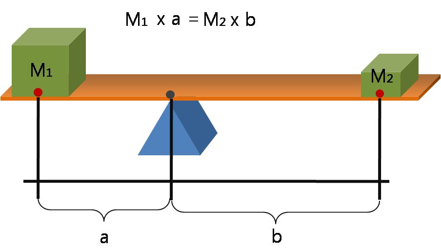
A lever is a simple machine consisting of a beam (structure), beam or rigid rod pivoted at a fixed hinge, or '':wikt:fulcrum, fulcrum''. A lever is a rigid body capable of rotating on a point on itself. On the basis of the locations of fulcrum, load, and effort, the lever is divided into Lever#Types of levers, three types. It is one of the six simple machines identified by Renaissance scientists. A lever amplifies an input force to provide a greater output force, which is said to provide leverage, which is mechanical advantage gained in the system, equal to the ratio of the output force to the input force. As such, the lever is a mechanical advantage device, trading off force against movement. Etymology The word "lever" entered English language, English around 1300 from . This sprang from the stem of the verb ''lever'', meaning "to raise". The verb, in turn, goes back to , itself from the adjective ''levis'', meaning "light" (as in "not heavy"). The word's primary origin is the ... [...More Info...] [...Related Items...] OR: [Wikipedia] [Google] [Baidu] |
Ossification
Ossification (also called osteogenesis or bone mineralization) in bone remodeling is the process of laying down new bone material by cells named osteoblasts. It is synonymous with bone tissue formation. There are two processes resulting in the formation of normal, healthy bone tissue: Intramembranous ossification is the direct laying down of bone into the primitive connective tissue ( mesenchyme), while endochondral ossification involves cartilage as a precursor. In fracture healing, endochondral osteogenesis is the most commonly occurring process, for example in fractures of long bones treated by plaster of Paris, whereas fractures treated by open reduction and internal fixation with metal plates, screws, pins, rods and nails may heal by intramembranous osteogenesis. Heterotopic ossification is a process resulting in the formation of bone tissue that is often atypical, at an extraskeletal location. Calcification is often confused with ossification. Calcificatio ... [...More Info...] [...Related Items...] OR: [Wikipedia] [Google] [Baidu] |
Hypoplastic
Hypoplasia (; adjective form ''hypoplastic'') is underdevelopment or incomplete development of a Tissue (biology), tissue or Organ (biology), organ. Dictionary of Cell and Molecular Biology (11 March 2008) Although the term is not always used precisely, it properly refers to an inadequate or below-normal number of cells.Hypoplasia Stedman's Medical Dictionary. lww.com Hypoplasia is similar to aplasia, but less severe. It is technically ''not'' the opposite of hyperplasia (too many cells). Hypoplasia is a congenital condition, while hyperpla ... [...More Info...] [...Related Items...] OR: [Wikipedia] [Google] [Baidu] |
Bipartite Patella
Bipartite patella is a condition where the patella, or kneecap, is composed of two separate bone, bones. Instead of ossification, fusing together as normally Patella#development, occurs in early childhood, the bones of the patella remain separated. The condition occurs in approximately 12% of the population and is no more likely to occur in male, males than female, females. It is often asymptomatic and most commonly diagnosed as an incidental findings, incidental finding, with about 2% of cases becoming symptom, symptomatic. Saupe introduced a classification system for Bipartite Patella back in 1921. Type 1: Fragment is located at the bottom of the kneecap (5% of cases) Type 2: Fragment is located on the lateral side of the kneecap (20% of cases) Type 3: Fragment is located on the upper lateral border of the kneecap (75% of cases) References External links Congenital disorders of musculoskeletal system Knee injuries and disorders Patella {{musculoskeletal-stub ... [...More Info...] [...Related Items...] OR: [Wikipedia] [Google] [Baidu] |
Patella Bipartita
Bipartite patella is a condition where the patella, or kneecap, is composed of two separate bones. Instead of fusing together as normally occurs in early childhood, the bones of the patella remain separated. The condition occurs in approximately 12% of the population and is no more likely to occur in males than females. It is often asymptomatic and most commonly diagnosed as an incidental finding, with about 2% of cases becoming symptomatic Signs and symptoms are diagnostic indications of an illness, injury, or condition. Signs are objective and externally observable; symptoms are a person's reported subjective experiences. A sign for example may be a higher or lower temperature .... Saupe introduced a classification system for Bipartite Patella back in 1921. Type 1: Fragment is located at the bottom of the kneecap (5% of cases) Type 2: Fragment is located on the lateral side of the kneecap (20% of cases) Type 3: Fragment is located on the upper lateral border of the kn ... [...More Info...] [...Related Items...] OR: [Wikipedia] [Google] [Baidu] |
Infrapatellar Fat Pad
The infrapatellar fat pad (Hoffa's fat pad) is a cylindrical piece of fat that is situated inferior and posterior to the patella bone within the knee, intervening between the patellar ligament and synovial fold of the knee joint. Clinical significance The fat pad is a normal structure but it can sometimes become a problem: * It can become damaged and painful * It can be deliberately removed at arthroscopic surgery Arthroscopy (also called arthroscopic or keyhole surgery) is a minimally invasive surgery, surgical procedure on a joint in which an examination and sometimes treatment of damage is performed using an arthroscope, an endoscope that is inserted in ... to make it easier for the surgeon to see what they are doing - but this can also lead to scarring and pain. * It can become hypertrophic and may become impinged between the patella and the femoral condyle, causing sharp pain when the leg is extended. This is called infrapatellar fat pad syndrome or Hoffa syndrome. * It c ... [...More Info...] [...Related Items...] OR: [Wikipedia] [Google] [Baidu] |
Joint
A joint or articulation (or articular surface) is the connection made between bones, ossicles, or other hard structures in the body which link an animal's skeletal system into a functional whole.Saladin, Ken. Anatomy & Physiology. 7th ed. McGraw-Hill Connect. Webp.274/ref> They are constructed to allow for different degrees and types of movement. Some joints, such as the knee, elbow, and shoulder, are self-lubricating, almost frictionless, and are able to withstand compression and maintain heavy loads while still executing smooth and precise movements. Other joints such as suture (joint), sutures between the bones of the skull permit very little movement (only during birth) in order to protect the brain and the sense organs. The connection between a tooth and the jawbone is also called a joint, and is described as a fibrous joint known as a gomphosis. Joints are classified both structurally and functionally. Joints play a vital role in the human body, contributing to movement, sta ... [...More Info...] [...Related Items...] OR: [Wikipedia] [Google] [Baidu] |


