|
Pander's Islands
Blood islands are structures around the developing embryo which lead to many different parts of the circulatory system. Blood islands arise external to the developing embryo on the umbilical vesicle, allantois, connecting stalk and chorion. They are also known as Pander's islands or Wolff's islands, after Heinz Christian Pander or Caspar Friedrich Wolff. Development In humans, the formation of extraembryonic blood vessels starts at the beginning of the third week after fertilization. Vasculogenesis begins as mesodermal cells differentiate into hemangioblasts, which in turn differentiate into angioblasts. Clusters of angioblasts make up the blood islands. Within the blood islands, lumens begin to appear by the growth of intercellular clefts. The flattened cells at the periphery form the endothelium. Mesenchymal cells exterior to this form the muscular and connective tissue components of blood vessels. Roughly 3 weeks after fertilization, red blood cells, still with a ... [...More Info...] [...Related Items...] OR: [Wikipedia] [Google] [Baidu] |
Bird
Birds are a group of warm-blooded vertebrates constituting the class Aves (), characterised by feathers, toothless beaked jaws, the laying of hard-shelled eggs, a high metabolic rate, a four-chambered heart, and a strong yet lightweight skeleton. Birds live worldwide and range in size from the bee hummingbird to the ostrich. There are about ten thousand living species, more than half of which are passerine, or "perching" birds. Birds have whose development varies according to species; the only known groups without wings are the extinct moa and elephant birds. Wings, which are modified forelimbs, gave birds the ability to fly, although further evolution has led to the loss of flight in some birds, including ratites, penguins, and diverse endemic island species. The digestive and respiratory systems of birds are also uniquely adapted for flight. Some bird species of aquatic environments, particularly seabirds and some waterbirds, have further evolved for swimm ... [...More Info...] [...Related Items...] OR: [Wikipedia] [Google] [Baidu] |
Lumen (anatomy)
In biology, a lumen (plural lumina) is the inside space of a tubular structure, such as an artery or intestine. It comes . It can refer to: *The interior of a vessel, such as the central space in an artery, vein or capillary through which blood flows. *The interior of the gastrointestinal tract *The pathways of the bronchi in the lungs *The interior of renal tubules and urinary collecting ducts *The pathways of the female genital tract, starting with a single pathway of the vagina, splitting up in two lumina in the uterus, both of which continue through the Fallopian tubes In cell biology, a lumen is a membrane-defined space that is found inside several organelles, cellular components, or structures: *thylakoid, endoplasmic reticulum, Golgi apparatus, lysosome, mitochondrion, or microtubule Transluminal procedures ''Transluminal procedures'' are procedures occurring through lumina, including: * Natural orifice transluminal endoscopic surgery in the lumina of, for example, ... [...More Info...] [...Related Items...] OR: [Wikipedia] [Google] [Baidu] |
Dorsal Aorta
The dorsal aortae are paired (left and right) embryological vessels which progress to form the descending aorta. The paired dorsal aortae arise from aortic arches that in turn arise from the aortic sac. The primary dorsal aorta is located deep to the lateral plate of mesoderm and move from lateral to medial position with development and eventually will fuse with the other dorsal aorta to form the descending aorta. Each primitive aorta anteriorly receives the vitelline vein from the yolk-sac, and is prolonged backward on the lateral aspect of the notochord under the name of the dorsal aorta. The dorsal aortae give branches to the yolk-sac, and are continued backward through the body-stalk as the umbilical arteries to the villi of the chorion The chorion is the outermost fetal membrane around the embryo in mammals, birds and reptiles ( amniotes). It develops from an outer fold on the surface of the yolk sac, which lies outside the zona pellucida (in mammals), known a ... [...More Info...] [...Related Items...] OR: [Wikipedia] [Google] [Baidu] |
Vitelline Arteries
The vitelline arteries are the arterial counterpart to the vitelline veins. Like the veins, they play an important role in the vitelline circulation Vitelline circulation refers to the system of blood flowing from the embryo to the yolk sac and back again. The yolk-sac is situated on the ventral aspect of the embryo; it is lined by endoderm, outside of which is a layer of mesoderm. It is fill ... of blood to and from the yolk sac of a fetus. They are a branch of the dorsal aorta. They give rise to the celiac artery, superior mesenteric artery, and inferior mesenteric artery. References External links * * https://web.archive.org/web/20070623132305/http://isc.temple.edu/marino/embryology/Heart98/heart_text.htm * * https://web.archive.org/web/20070915072304/http://www.ana.ed.ac.uk/database/humat/notes/extraemb/yolksac/vitart.htm * http://www.med.umich.edu/lrc/coursepages/M1/embryology/embryo/13cardiovascular_system.htm * https://web.archive.org/web/20070812190309/http:// ... [...More Info...] [...Related Items...] OR: [Wikipedia] [Google] [Baidu] |
Vitelline Veins
The vitelline veins are veins that drain blood from the yolk sac and the gut tube during gestation. Path They run upward at first in front, and subsequently on either side of the intestinal canal. They unite on the ventral aspect of the canal. Beyond this, they are connected to one another by two anastomotic branches, one on the dorsal, and the other on the ventral aspect of the duodenal portion of the intestine. This is encircled by two venous rings; into the middle or dorsal anastomosis the superior mesenteric vein opens. The portions of the veins above the upper ring become interrupted by the developing liver and broken up by it into a plexus of small capillary-like vessels termed sinusoids. Derivatives The vitelline veins give rise to: * Hepatic veins * Inferior portion of Inferior vena cava * Portal vein * Superior mesenteric vein * Inferior mesenteric vein The branches conveying the blood to the plexus are named the venae advehentes, and become the branches of the po ... [...More Info...] [...Related Items...] OR: [Wikipedia] [Google] [Baidu] |
Thymus
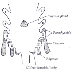
The thymus is a specialized primary lymphoid organ of the immune system. Within the thymus, thymus cell lymphocytes or ''T cells'' mature. T cells are critical to the adaptive immune system, where the body adapts to specific foreign invaders. The thymus is located in the upper front part of the chest, in the anterior superior mediastinum, behind the sternum, and in front of the heart. It is made up of two lobes, each consisting of a central medulla and an outer cortex, surrounded by a capsule. The thymus is made up of immature T cells called thymocytes, as well as lining cells called epithelial cells which help the thymocytes develop. T cells that successfully develop react appropriately with MHC immune receptors of the body (called ''positive selection'') and not against proteins of the body (called ''negative selection''). The thymus is largest and most active during the neonatal and pre-adolescent periods. By the early teens, the thymus begins to decrease in size and ... [...More Info...] [...Related Items...] OR: [Wikipedia] [Google] [Baidu] |
Red Bone Marrow
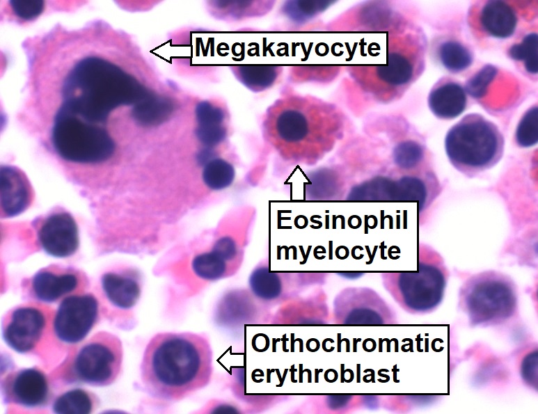
Bone marrow is a semi-solid tissue found within the spongy (also known as cancellous) portions of bones. In birds and mammals, bone marrow is the primary site of new blood cell production (or haematopoiesis). It is composed of hematopoietic cells, marrow adipose tissue, and supportive stromal cells. In adult humans, bone marrow is primarily located in the ribs, vertebrae, sternum, and bones of the pelvis. Bone marrow comprises approximately 5% of total body mass in healthy adult humans, such that a man weighing 73 kg (161 lbs) will have around 3.7 kg (8 lbs) of bone marrow. Human marrow produces approximately 500 billion blood cells per day, which join the systemic circulation via permeable vasculature sinusoids within the medullary cavity. All types of hematopoietic cells, including both myeloid and lymphoid lineages, are created in bone marrow; however, lymphoid cells must migrate to other lymphoid organs (e.g. thymus) in order to complete matu ... [...More Info...] [...Related Items...] OR: [Wikipedia] [Google] [Baidu] |
Spleen
The spleen is an organ found in almost all vertebrates. Similar in structure to a large lymph node, it acts primarily as a blood filter. The word spleen comes .σπλήν Henry George Liddell, Robert Scott, ''A Greek-English Lexicon'', on Perseus Digital Library The spleen plays very important roles in regard to red blood cells (erythrocytes) and the . It removes old red blood cells and holds a reserve of blood, which can be valuable in case of [...More Info...] [...Related Items...] OR: [Wikipedia] [Google] [Baidu] |
Liver
The liver is a major organ only found in vertebrates which performs many essential biological functions such as detoxification of the organism, and the synthesis of proteins and biochemicals necessary for digestion and growth. In humans, it is located in the right upper quadrant of the abdomen, below the diaphragm. Its other roles in metabolism include the regulation of glycogen storage, decomposition of red blood cells, and the production of hormones. The liver is an accessory digestive organ that produces bile, an alkaline fluid containing cholesterol and bile acids, which helps the breakdown of fat. The gallbladder, a small pouch that sits just under the liver, stores bile produced by the liver which is later moved to the small intestine to complete digestion. The liver's highly specialized tissue, consisting mostly of hepatocytes, regulates a wide variety of high-volume biochemical reactions, including the synthesis and breakdown of small and complex molecu ... [...More Info...] [...Related Items...] OR: [Wikipedia] [Google] [Baidu] |
Plexus
In neuroanatomy, a plexus (from the Latin term for "braid") is a branching network of vessels or nerves. The vessels may be blood vessels (veins, capillaries) or lymphatic vessels. The nerves are typically axons outside the central nervous system. The standard plural form in English is plexuses. Alternatively, the Latin plural plexūs may be used. Types Nerve plexuses The four primary nerve plexuses are the cervical plexus, brachial plexus, lumbar plexus, and the sacral plexus. Cardiac plexus Celiac plexus Renal plexus Venous plexus Choroid plexus The choroid plexus is a part of the central nervous system in the brain and consists of capillaries, brain ventricles, and ependymal cells. Invertebrates The plexus is the characteristic form of nervous system in the coelenterates and persists with modifications in the flatworms. The nerves of the radially symmetric echinoderm An echinoderm () is any member of the phylum Echinodermata (). The adults are re ... [...More Info...] [...Related Items...] OR: [Wikipedia] [Google] [Baidu] |
Blood Plasma
Blood plasma is a light amber-colored liquid component of blood in which blood cells are absent, but contains proteins and other constituents of whole blood in suspension. It makes up about 55% of the body's total blood volume. It is the intravascular part of extracellular fluid (all body fluid outside cells). It is mostly water (up to 95% by volume), and contains important dissolved proteins (6–8%; e.g., serum albumins, globulins, and fibrinogen), glucose, clotting factors, electrolytes (, , , , , etc.), hormones, carbon dioxide (plasma being the main medium for excretory product transportation), and oxygen. It plays a vital role in an intravascular osmotic effect that keeps electrolyte concentration balanced and protects the body from infection and other blood-related disorders. Blood plasma is separated from the blood by spinning a vessel of fresh blood containing an anticoagulant in a centrifuge until the blood cells fall to the bottom of the tube. The bloo ... [...More Info...] [...Related Items...] OR: [Wikipedia] [Google] [Baidu] |
Cell Nucleus
The cell nucleus (pl. nuclei; from Latin or , meaning ''kernel'' or ''seed'') is a membrane-bound organelle found in eukaryotic cells. Eukaryotic cells usually have a single nucleus, but a few cell types, such as mammalian red blood cells, have no nuclei, and a few others including osteoclasts have many. The main structures making up the nucleus are the nuclear envelope, a double membrane that encloses the entire organelle and isolates its contents from the cellular cytoplasm; and the nuclear matrix, a network within the nucleus that adds mechanical support. The cell nucleus contains nearly all of the cell's genome. Nuclear DNA is often organized into multiple chromosomes – long stands of DNA dotted with various proteins, such as histones, that protect and organize the DNA. The genes within these chromosomes are structured in such a way to promote cell function. The nucleus maintains the integrity of genes and controls the activities of the cell by regulating g ... [...More Info...] [...Related Items...] OR: [Wikipedia] [Google] [Baidu] |
.jpg)


