|
Outline Of Brain Mapping
The following outline is provided as an overview of and topical guide to brain mapping: Brain mapping – set of neuroscience techniques predicated on the mapping of (biological) quantities or properties onto spatial representations of the (human or non-human) brain resulting in maps. Brain mapping is further defined as the study of the anatomy and function of the brain and spinal cord through the use of imaging (including intra-operative, microscopic, endoscopic and multi-modality imaging), immunohistochemistry, molecular and optogenetics, stem cell and cellular biology, engineering (material, electrical and biomedical), neurophysiology and nanotechnology. Broad scope * History of neuroscience * History of neurology * Brain mapping * Human brain * Neuroscience * Nervous system. The neuron doctrine * Neuron doctrine – A set of carefully constructed elementary set of observations regarding neurons. ''For more granularity, more current, and more advanced topics, see the ce ... [...More Info...] [...Related Items...] OR: [Wikipedia] [Google] [Baidu] |
Myelin
Myelin Sheath ( ) is a lipid-rich material that in most vertebrates surrounds the axons of neurons to insulate them and increase the rate at which electrical impulses (called action potentials) pass along the axon. The myelinated axon can be likened to an electrical wire (the axon) with insulating material (myelin) around it. However, unlike the plastic covering on an electrical wire, myelin does not form a single long sheath over the entire length of the axon. Myelin ensheaths part of an axon known as an internodal segment, in multiple myelin layers of a tightly regulated internodal length. The ensheathed segments are separated at regular short unmyelinated intervals, called nodes of Ranvier. Each node of Ranvier is around one micrometre long. Nodes of Ranvier enable a much faster rate of conduction known as saltatory conduction where the action potential recharges at each node to jump over to the next node, and so on till it reaches the axon terminal. At the terminal the ... [...More Info...] [...Related Items...] OR: [Wikipedia] [Google] [Baidu] |
Blue Brain Project
The Blue Brain Project was a Swiss brain research initiative that aimed to create a digital reconstruction of the mouse brain. The project was founded in May 2005 by the Brain Mind Institute of ''École Polytechnique Fédérale de Lausanne'' (EPFL) in Switzerland. The project ended in December 2024. Its mission was to use biologically-detailed digital reconstructions and simulations of the mammalian brain to identify the fundamental principles of brain structure and function. The project was headed by the founding director Henry Markram—who also launched the European Human Brain Project—and was co-directed by Felix Schürmann, Adriana Salvatore and Sean Hill. Using a Blue Gene supercomputer running Michael Hines's NEURON, the simulation involved a biologically realistic model of neurons and an empirically reconstructed model connectome. There were a number of collaborations, including the Cajal Blue Brain, which is coordinated by the Supercomputing and Visualization Center ... [...More Info...] [...Related Items...] OR: [Wikipedia] [Google] [Baidu] |
Medical Image Computing
Medical image computing (MIC) is an interdisciplinary field at the intersection of computer science Computer science is the study of computation, information, and automation. Computer science spans Theoretical computer science, theoretical disciplines (such as algorithms, theory of computation, and information theory) to Applied science, ..., information engineering, electrical engineering, physics, mathematics and medicine. This field develops computational and mathematical methods for solving problems pertaining to medical images and their use for biomedical research and clinical care. The main goal of MIC is to extract clinically relevant information or knowledge from medical images. While closely related to the field of medical imaging, MIC focuses on the computational analysis of the images, not their acquisition. The methods can be grouped into several broad categories: image segmentation, image registration, image-based physiological modeling, and others. Data f ... [...More Info...] [...Related Items...] OR: [Wikipedia] [Google] [Baidu] |
Jean Talairach
Jean Talairach (January 15, 1911 – March 15, 2007) was a psychiatrist and neurosurgeon who practiced at the Sainte-Anne Hospital Center in Paris, and who is noted for the Talairach coordinates, which are relevant in stereotactic neurosurgery. Early life Talairach was the son of a pianist, and learned the cello to a professional level. However, instead of pursuing a musical career, he then developed a passion for geometry and architecture, and was particularly interested in the lecture halls in the medieval medical college in Montpellier. This interest, in turn, led him to become interested in medicine, especially psychiatry. In 1938 he traveled to Paris to study medicine. He completed his doctoral studies at Ste. Anne's Hospital, one of the oldest and most renowned hospitals in France. World War II During the German occupation of France in the second World War, Talairach joined the French resistance. He created a detailed map of the tunnels under Paris, which he gave to t ... [...More Info...] [...Related Items...] OR: [Wikipedia] [Google] [Baidu] |
Decade Of The Brain
A decade (from , , ) is a period of 10 years. Decades may describe any 10-year period, such as those of a person's life, or refer to specific groupings of calendar years. Usage Any period of ten years is a "decade". For example, the statement that "during his last decade, Mozart explored chromatic harmony to a degree rare at the time" refers to the last 10 years of Wolfgang Amadeus Mozart's life without regard to which calendar years are encompassed. Also, 'the first decade' of a person's life begins on the day of their birth and ends at the end of their 10th year of life when they have their 10th birthday; the second decade of life starts with their 11th year of life (during which one is typically still referred to as being "10") and ends at the end of their 20th year of life, on their 20th birthday; similarly, the third decade of life, when one is in one's twenties or 20s, starts with the 21st year of life, and so on, with subsequent decades of life similarly described by refe ... [...More Info...] [...Related Items...] OR: [Wikipedia] [Google] [Baidu] |
NeuroNames
''NeuroNames'' is an integrated nomenclature for structures in the brain and spinal cord of the four species most studied by neuroscientists: human, macaque, rat and mouse. It offers a standard, controlled vocabulary of common names for structures, which is suitable for unambiguous neuroanatomical indexing of information in digital databases. Terms in the standard vocabulary have been selected for ease of pronunciation, mnemonic value, and frequency of use in recent neuroscientific publications. Structures and their relations to each other are defined in terms of the standard vocabulary. Currently NeuroNames contains standard names, synonyms and definitions of some 2,500 neuroanatomical entities. The nomenclature is maintained by the University of Washington and is the core component of a tool called "BrainInfo". BrainInfo helps one identify structures in the brain. One can either search by a structure name or locate the structure in a brain atlas and get information such as it ... [...More Info...] [...Related Items...] OR: [Wikipedia] [Google] [Baidu] |
BrainMaps
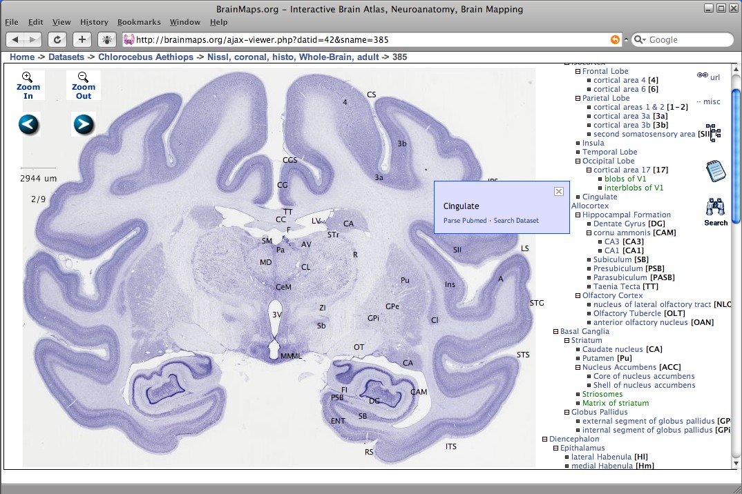
BrainMaps is an NIH-funded interactive zoomable high-resolution digital brain atlas and virtual microscope that is based on more than 140 million megapixels (140 terabytes) of scanned images of serial sections of both primate and non-primate brains and that is integrated with a high-speed database for querying and retrieving data about brain structure and function over the internet. Currently featured are complete brain atlas datasets for 16 species; a few of which are: ''Macaca mulatta'', '' Chlorocebus aethiops'', '' Felis silvestris catus'', ''Mus musculus'', ''Rattus norvegicus'', and ''Tyto alba''. The project's principal investigator was UC Davis neuroscientist Ted Jones from 2005 through 2011, after which the role was taken by W. Martin Usrey. Description BrainMaps uses multiresolution image formats for representing massive brain images, and a dHTML/Javascript front-end user interface for image navigation, both similar to the way that Google Maps works for geospatial ... [...More Info...] [...Related Items...] OR: [Wikipedia] [Google] [Baidu] |
Allen Brain Atlas
The Allen Mouse and Human Brain Atlases are projects within the Allen Institute for Brain Science which seek to combine genomics with neuroanatomy by creating gene expression maps for the mouse and human brain. They were initiated in September 2003 with a $100 million donation from Paul G. Allen and the first atlas went public in September 2006. , seven brain atlases have been published: Mouse Brain Atlas, Human Brain Atlas, Developing Mouse Brain Atlas, Developing Human Brain Atlas, Mouse Connectivity Atlas, Non-Human Primate Atlas, and Mouse Spinal Cord Atlas. There are also three related projects with data banks: Glioblastoma, Mouse Diversity, and Sleep. It is the hope of the Allen Institute that their findings will help advance various fields of science, especially those surrounding the understanding of neurobiological diseases. The atlases are free and available for public use online. History In 2001, Paul Allen gathered a group of scientists, including James Watson and St ... [...More Info...] [...Related Items...] OR: [Wikipedia] [Google] [Baidu] |
Connectome
A connectome () is a comprehensive map of neural connections in the brain, and may be thought of as its " wiring diagram". These maps are available in varying levels of detail. A functional connectome shows connections between various brain regions, but not individual neurons. These are available for large animals, including mice and humans, are normally obtained by techniques such as MRI, and have a scale of millimeters. At the other extreme are neural connectomes, which show individual neurons and their interconnections. These are usually obtained by electron microscopy (EM) and have a scale of nanometers. They are only available for small creatures such as the worm ''C. Elegans'' and the fruit fly ''Drosophila melanogaster'', and small regions of mammal brains. Finally there are chemical connectomes, showing which neurons emit, and are sensitive to, a wide variety of neuromodulators. The significance of the connectome stems from the realization that the structure an ... [...More Info...] [...Related Items...] OR: [Wikipedia] [Google] [Baidu] |
Human Connectome Project
The Human Connectome Project (HCP) was a five-year project (later extended to 10 years) sponsored by sixteen components of the National Institutes of Health, split between two consortia of research institutions. The project was launched in July 2009 as the first of three Grand Challenges of the NIH's Blueprint for Neuroscience Research. On September 15, 2010, the NIH announced that it would award two grants: $30 million over five years to a consortium led by Washington University in St. Louis and the University of Minnesota, with strong contributions from University of Oxford (FMRIB) and $8.5 million over three years to a consortium led by Harvard University, Massachusetts General Hospital and the University of California Los Angeles. The goal of the Human Connectome Project was to build a "network map" (connectome) that sheds light on the anatomical and functional connectivity within the healthy human brain, as well as to produce a body of data that will facilitate research into ... [...More Info...] [...Related Items...] OR: [Wikipedia] [Google] [Baidu] |
BigBrain
BigBrain is a freely accessible high-resolution 3D digital atlas of the human brain, released in June 2013 by a team of researchers at the Montreal Neurological Institute and the German Forschungszentrum Jülich and is part of the European Human Brain Project.The isotropic 3D spatial resolution of the BigBrain atlas is 20 μm, much finer than the typical 1 mm resolution of other existing 3D models of the human brain such as the Allen Brain Atlas. In 2014, BigBrain was cited in the top 10 MIT Technology Review. Acquisition The atlas was created from the brain of an unidentified 65-year-old man (it was "65-year-old female", according to "BigBrain: An Ultrahigh-Resolution3D Human Brain Model", page 1472, Amunts K et al., SCIENCE, 21 JUNE 2013 VOL 340) who died with no known brain pathology. His brain, after being removed from the skull, was first scanned using an MRI machine, then embedded in paraffin and sliced into 7,404 20 μm thick sections using a large-scale micr ... [...More Info...] [...Related Items...] OR: [Wikipedia] [Google] [Baidu] |


