|
Ophthalmic Nerve
The ophthalmic nerve (CN V1) is a sensory nerve of the head. It is one of three divisions of the trigeminal nerve (CN V), a cranial nerve. It has three major branches which provide sensory innervation to the eye, and the skin of the upper face and anterior scalp, as well as other structures of the head. Structure Origin The ophthalmic nerve is the first branch of the trigeminal nerve (CN V), the first and smallest of its three divisions. It arises from the superior part of the trigeminal ganglion. Course It passes anterior-ward along the lateral wall of the cavernous sinus inferior to the oculomotor nerve (CN III) and trochlear nerve (N IV). It exits the skull into the orbit through the superior orbital fissure. Branches Within the skull, the ophthalmic nerve produces: * meningeal branch (tentorial nerve) The ophthalmic nerve divides into three major branches which pass through the superior orbital fissure: * frontal nerve ** supraorbital nerve ** supratrochlea ... [...More Info...] [...Related Items...] OR: [Wikipedia] [Google] [Baidu] |
Cavernous Sinus
The cavernous sinus within the human head is one of the dural venous sinuses creating a cavity called the lateral sellar compartment bordered by the temporal bone of the skull and the sphenoid bone, lateral to the sella turcica. Structure The cavernous sinus is one of the dural venous sinuses of the head. It is a network of veins that sit in a cavity. It sits on both sides of the sphenoidal bone and pituitary gland, approximately 1 × 2 cm in size in an adult. The carotid siphon of the internal carotid artery, and cranial nerves III, IV, V (branches V1 and V2) and VI all pass through this blood filled space. Both sides of cavernous sinus are connected to each other via intercavernous sinuses. The cavernous sinus lies in between the inner and outer layers of dura mater. Nearby structures * Above: optic tract, optic chiasma, internal carotid artery. * Inferiorly: foramen lacerum, and the junction of the body and greater wing of sphenoid bone. * Medially: p ... [...More Info...] [...Related Items...] OR: [Wikipedia] [Google] [Baidu] |
Posterior Ethmoidal Nerve
The posterior ethmoidal nerve is a nerve of the head. It is a branch of the nasociliary nerve (itself a branch of the Ophthalmic nerve, ophthalmic nerve (CN V1)). It provides sensory innervation to the sphenoid sinus and ethmoid sinus, and part of the dura mater in the anterior cranial fossa. Structure Origin The posterior ethmoidal nerve is a branch of the nasociliary nerve. Course It passes through the posterior ethmoidal foramen alongside the posterior ethmoidal artery. Branches Within the anterior cranial fossa, it issues a branch to which innervates part of the dura mater. It gives branches to the sphenoid sinus and the ethmoid sinus. Variation The posterior ethmoidal nerve is absent in a significant proportion of people. This may be around 30%. Function The posterior ethmoidal nerve supplies sensation to the sphenoid sinus and the ethmoid sinus. It also supplies sensation to part of the dura mater in the anterior cranial fossa. Other animals The posterio ... [...More Info...] [...Related Items...] OR: [Wikipedia] [Google] [Baidu] |
Eyelids
An eyelid ( ) is a thin fold of skin that covers and protects an eye. The levator palpebrae superioris muscle retracts the eyelid, exposing the cornea to the outside, giving vision. This can be either voluntarily or involuntarily. "Palpebral" (and "blepharal") means relating to the eyelids. Its key function is to regularly spread the tears and other secretions on the eye surface to keep it moist, since the cornea must be continuously moist. They keep the eyes from drying out when asleep. Moreover, the blink reflex protects the eye from foreign bodies. A set of specialized hairs known as lashes grow from the upper and lower eyelid margins to further protect the eye from dust and debris. The appearance of the human upper eyelid often varies between different populations. The prevalence of an epicanthic fold covering the inner corner of the eye account for the majority of East Asian and Southeast Asian populations, and is also found in varying degrees among other populatio ... [...More Info...] [...Related Items...] OR: [Wikipedia] [Google] [Baidu] |
Nasal Cavity
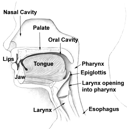
The nasal cavity is a large, air-filled space above and behind the nose in the middle of the face. The nasal septum divides the cavity into two cavities, also known as fossae. Each cavity is the continuation of one of the two nostrils. The nasal cavity is the uppermost part of the respiratory system and provides the nasal passage for inhaled air from the nostrils to the nasopharynx and rest of the respiratory tract. The paranasal sinuses surround and drain into the nasal cavity. Structure The term "nasal cavity" can refer to each of the two cavities of the nose, or to the two sides combined. The lateral wall of each nasal cavity mainly consists of the maxilla. However, there is a deficiency that is compensated for by the perpendicular plate of the palatine bone, the medial pterygoid plate, the labyrinth of ethmoid and the inferior concha. The paranasal sinuses are connected to the nasal cavity through small orifices called ostia. Most of these ostia communicat ... [...More Info...] [...Related Items...] OR: [Wikipedia] [Google] [Baidu] |
Mucous Membrane
A mucous membrane or mucosa is a membrane that lines various cavities in the body of an organism and covers the surface of internal organs. It consists of one or more layers of epithelial cells overlying a layer of loose connective tissue. It is mostly of endodermal origin and is continuous with the skin at body openings such as the eyes, eyelids, ears, inside the nose, inside the mouth, lips, the genital areas, the urethral opening and the anus. Some mucous membranes secrete mucus, a thick protective fluid. The function of the membrane is to stop pathogens and dirt from entering the body and to prevent bodily tissues from becoming dehydrated. Structure The mucosa is composed of one or more layers of epithelial cells that secrete mucus, and an underlying lamina propria of loose connective tissue. The type of cells and type of mucus secreted vary from organ to organ and each can differ along a given tract. Mucous membranes line the digestive, respiratory and rep ... [...More Info...] [...Related Items...] OR: [Wikipedia] [Google] [Baidu] |
Conjunctiva
In the anatomy of the eye, the conjunctiva (: conjunctivae) is a thin mucous membrane that lines the inside of the eyelids and covers the sclera (the white of the eye). It is composed of non-keratinized, stratified squamous epithelium with goblet cells, stratified columnar epithelium and stratified cuboidal epithelium (depending on the zone). The conjunctiva is highly Angiogenesis, vascularised, with many microvessels easily accessible for imaging studies. Structure The conjunctiva is typically divided into three parts: Blood supply Blood to the bulbar conjunctiva is primarily derived from the ophthalmic artery. The blood supply to the palpebral conjunctiva (the eyelid) is derived from the external carotid artery. However, the circulations of the bulbar conjunctiva and palpebral conjunctiva are linked, so both bulbar conjunctival and palpebral conjunctival vessels are supplied by both the ophthalmic artery and the external carotid artery, to varying extents. Nerve supply Se ... [...More Info...] [...Related Items...] OR: [Wikipedia] [Google] [Baidu] |
Lacrimal Gland
The lacrimal glands are paired exocrine glands, one for each eye, found in most terrestrial vertebrates and some marine mammals, that secrete the aqueous layer of the tear film. In humans, they are situated in the upper lateral region of each orbit, in the lacrimal fossa of the orbit formed by the frontal bone. Inflammation of the lacrimal glands is called dacryoadenitis. The lacrimal gland produces tears which are secreted by the lacrimal ducts, and flow over the ocular surface, and then into canals that connect to the lacrimal sac. From that sac, the tears drain through the lacrimal duct into the nose. Anatomists divide the gland into two sections, a palpebral lobe, or portion, and an orbital lobe or portion. The smaller ''palpebral lobe'' lies close to the eye, along the inner surface of the eyelid; if the upper eyelid is everted, the palpebral portion can be seen. The orbital lobe of the gland, contains fine interlobular ducts that connect the orbital lobe and the palpe ... [...More Info...] [...Related Items...] OR: [Wikipedia] [Google] [Baidu] |
Iris (anatomy)
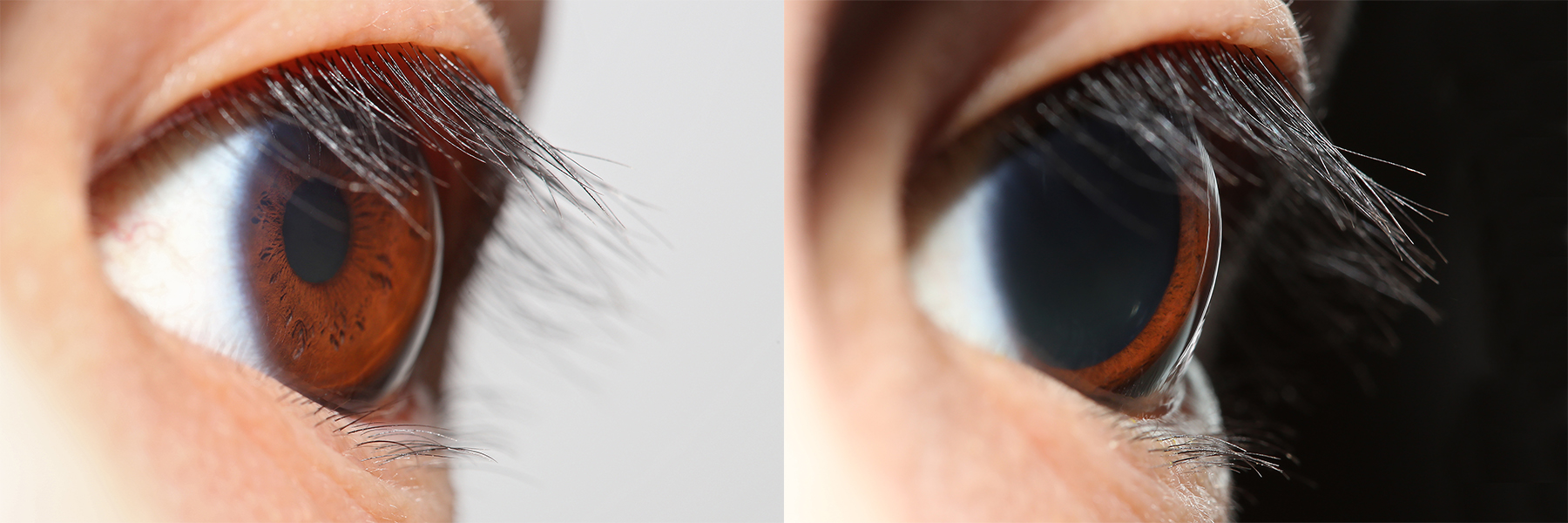
The iris (: irides or irises) is a thin, annular structure in the eye in most mammals and birds that is responsible for controlling the diameter and size of the pupil, and thus the amount of light reaching the retina. In optical terms, the pupil is the eye's aperture, while the iris is the diaphragm (optics), diaphragm. Eye color is defined by the iris. Etymology The word "iris" is derived from the Greek word for "rainbow", also Iris (mythology), its goddess plus messenger of the gods in the ''Iliad'', because of the many eye color, colours of this eye part. Structure The iris consists of two layers: the front pigmented Wikt:fibrovascular, fibrovascular layer known as a stroma of iris, stroma and, behind the stroma, pigmented epithelial cells. The stroma is connected to a sphincter muscle (sphincter pupillae), which contracts the pupil in a circular motion, and a set of dilator muscles (dilator pupillae), which pull the iris radially to enlarge the pupil, pulling it in folds. ... [...More Info...] [...Related Items...] OR: [Wikipedia] [Google] [Baidu] |
Ciliary Body
The ciliary body is a part of the eye that includes the ciliary muscle, which controls the shape of the lens, and the ciliary epithelium, which produces the aqueous humor. The aqueous humor is produced in the non-pigmented portion of the ciliary body. The ciliary body is part of the uvea, the layer of tissue that delivers oxygen and nutrients to the eye tissues. The ciliary body joins the ora serrata of the choroid to the root of the iris.Cassin, B. and Solomon, S. ''Dictionary of Eye Terminology''. Gainesville, Florida: Triad Publishing Company, 1990. Structure The ciliary body is a ring-shaped thickening of tissue inside the eye that divides the posterior chamber from the vitreous body. It contains the ciliary muscle, vessels, and fibrous connective tissue. Folds on the inner ciliary epithelium are called ciliary processes, and these secrete aqueous humor into the posterior chamber. The aqueous humor then flows through the iris into the anterior chamber. The ciliary bo ... [...More Info...] [...Related Items...] OR: [Wikipedia] [Google] [Baidu] |
Cornea
The cornea is the transparency (optics), transparent front part of the eyeball which covers the Iris (anatomy), iris, pupil, and Anterior chamber of eyeball, anterior chamber. Along with the anterior chamber and Lens (anatomy), lens, the cornea Refraction, refracts light, accounting for approximately two-thirds of the eye's total optical power. In humans, the refractive power of the cornea is approximately 43 dioptres. The cornea can be reshaped by surgical procedures such as LASIK. While the cornea contributes most of the eye's focusing power, its Focus (optics), focus is fixed. Accommodation (eye), Accommodation (the refocusing of light to better view near objects) is accomplished by changing the geometry of the lens. Medical terms related to the cornea often start with the prefix "''wikt:kerat-, kerat-''" from the Ancient Greek, Greek word κέρας, ''horn''. Structure The cornea has myelinated, unmyelinated nerve endings sensitive to touch, temperature and chemicals; a to ... [...More Info...] [...Related Items...] OR: [Wikipedia] [Google] [Baidu] |
Long Root Of Ciliary Ganglion
The ciliary ganglion is a parasympathetic ganglion located just behind the eye in the posterior orbit. Three types of axons enter the ciliary ganglion but only the preganglionic parasympathetic axons synapse there. The entering axons are arranged into three roots of the ciliary ganglion, which join enter the posterior surface of the ganglion. Sympathetic root The sympathetic root of ciliary ganglion is one of three roots of the ciliary ganglion. It contains ''postganglionic'' sympathetic fibers whose cell bodies are located in the superior cervical ganglion. Their axons ascend with the internal carotid artery as a plexus of nerves, the internal carotid plexus. Sympathetic fibers supplying the eye separate from the carotid plexus within the cavernous sinus. They run forward through the superior orbital fissure and merge with the long ciliary nerves (branches of the nasociliary nerve) and the short ciliary nerves (from the ciliary ganglion). Sympathetic fibers in the short c ... [...More Info...] [...Related Items...] OR: [Wikipedia] [Google] [Baidu] |
Infratrochlear Nerve
The infratrochlear nerve is a branch of the nasociliary nerve (itself a branch of the ophthalmic nerve (CN V1)) in the orbit In celestial mechanics, an orbit (also known as orbital revolution) is the curved trajectory of an object such as the trajectory of a planet around a star, or of a natural satellite around a planet, or of an artificial satellite around an .... It exits the orbit inferior to the trochlea of superior oblique. It provides sensory innervation to structures of the orbit and skin of adjacent structures. Structure The nasociliary nerve terminates by bifurcating into the infratrochlear and the anterior ethmoidal nerves. The infratrochlear nerve travels anteriorly in the orbit along the upper border of the medial rectus muscle and underneath the trochlea of the superior oblique muscle. It exits the orbit medially and divides into small sensory branches. Distribution The infratrochlear nerve provides sensory innervation to the skin of the eyelids ... [...More Info...] [...Related Items...] OR: [Wikipedia] [Google] [Baidu] |


