|
Omental Infarction
Omental infarction, or omental torsion, is an acute vascular disorder which compromises tissue of the greater omentum—the largest peritoneal fold in the abdomen. Signs and symptoms Patients will present with a sudden onset of cramps/abdominal pain or a 'stitch'. The pain localises in the area of the umbilicus and can radiate to the lumbar and surrounding regions. Causes Omental infarction is a rare cause of acute abdomen pain with reported incidence being less than 4 per 1000 cases of appendicitis. Omental infarction usually presents as right-sided abdominal pain although seldom causing left-sided abdominal pain and even epigastric pain. The dominion of right-sided abdominal pain in omental infarction has been attributed to right segmental infarction as a result of the tenuous blood vessels in this part of the omentum as well as its longer size and higher mobility in comparison to the left side which subjects it to torsion. Obesity is a known risk factor for omental infarction ... [...More Info...] [...Related Items...] OR: [Wikipedia] [Google] [Baidu] |
Infarction
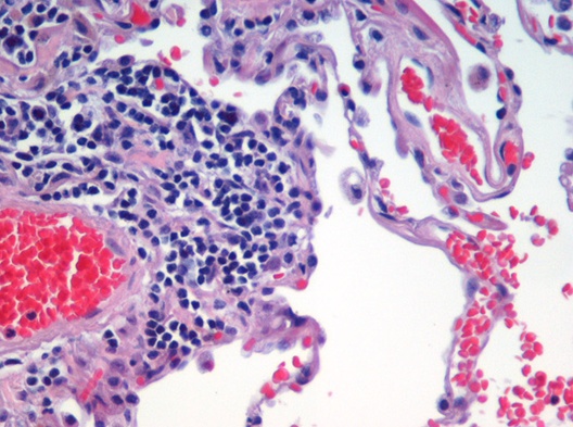
Infarction is tissue death (necrosis) due to inadequate blood supply to the affected area. It may be caused by artery blockages, rupture, mechanical compression, or vasoconstriction. The resulting lesion is referred to as an infarct (from the Latin ''infarctus'', "stuffed into"). Causes Infarction occurs as a result of prolonged ischemia, which is the insufficient supply of oxygen and nutrition to an area of tissue due to a disruption in blood supply. The blood vessel supplying the affected area of tissue may be blocked due to an obstruction in the vessel (e.g., an arterial embolus, thrombus, or atherosclerotic plaque), compressed by something outside of the vessel causing it to narrow (e.g., tumor, volvulus, or hernia), ruptured by trauma causing a loss of blood pressure downstream of the rupture, or vasoconstricted, which is the narrowing of the blood vessel by contraction of the muscle wall rather than an external force (e.g., cocaine vasoconstriction le ... [...More Info...] [...Related Items...] OR: [Wikipedia] [Google] [Baidu] |
Vascular System
The blood circulatory system is a system of organs that includes the heart, blood vessels, and blood which is circulated throughout the entire body of a human or other vertebrate. It includes the cardiovascular system, or vascular system, that consists of the heart and blood vessels (from Greek ''kardia'' meaning ''heart'', and from Latin ''vascula'' meaning ''vessels''). The circulatory system has two divisions, a systemic circulation or circuit, and a pulmonary circulation or circuit. Some sources use the terms ''cardiovascular system'' and ''vascular system'' interchangeably with the ''circulatory system''. The network of blood vessels are the great vessels of the heart including large elastic arteries, and large veins; other arteries, smaller arterioles, capillaries that join with venules (small veins), and other veins. The circulatory system is closed in vertebrates, which means that the blood never leaves the network of blood vessels. Some invertebrates such as arthro ... [...More Info...] [...Related Items...] OR: [Wikipedia] [Google] [Baidu] |
Tissue (biology)
In biology, tissue is a biological organizational level between cells and a complete organ. A tissue is an ensemble of similar cells and their extracellular matrix from the same origin that together carry out a specific function. Organs are then formed by the functional grouping together of multiple tissues. The English word "tissue" derives from the French word "tissu", the past participle of the verb tisser, "to weave". The study of tissues is known as histology or, in connection with disease, as histopathology. Xavier Bichat is considered as the "Father of Histology". Plant histology is studied in both plant anatomy and physiology. The classical tools for studying tissues are the paraffin block in which tissue is embedded and then sectioned, the histological stain, and the optical microscope. Developments in electron microscopy, immunofluorescence, and the use of frozen tissue-sections have enhanced the detail that can be observed in tissues. With these tools, th ... [...More Info...] [...Related Items...] OR: [Wikipedia] [Google] [Baidu] |
Greater Omentum
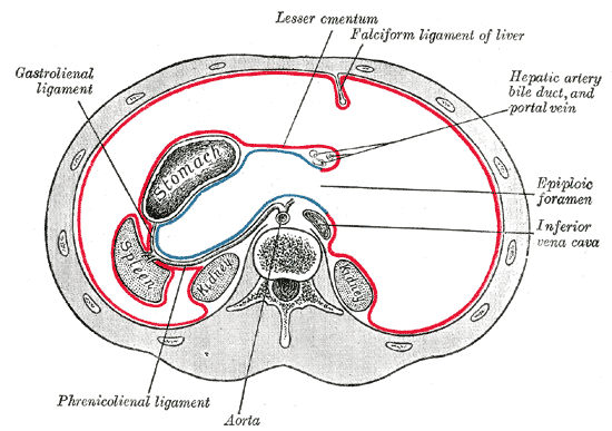
The greater omentum (also the great omentum, omentum majus, gastrocolic omentum, epiploon, or, especially in animals, caul) is a large apron-like fold of visceral peritoneum that hangs down from the stomach. It extends from the greater curvature of the stomach, passing in front of the small intestines and doubles back to ascend to the transverse colon before reaching to the posterior abdominal wall. The greater omentum is larger than the lesser omentum, which hangs down from the liver to the lesser curvature. The common anatomical term "epiploic" derives from "epiploon", from the Greek ''epipleein'', meaning to float or sail on, since the greater omentum appears to float on the surface of the intestines. It is the first structure observed when the abdominal cavity is opened anteriorly (from the front). Structure The greater omentum is the larger of the two peritoneal folds. It consists of a double sheet of peritoneum, folded on itself so that it has four layers. The two laye ... [...More Info...] [...Related Items...] OR: [Wikipedia] [Google] [Baidu] |
Peritoneum
The peritoneum is the serous membrane forming the lining of the abdominal cavity or coelom in amniotes and some invertebrates, such as annelids. It covers most of the intra-abdominal (or coelomic) organs, and is composed of a layer of mesothelium supported by a thin layer of connective tissue. This peritoneal lining of the cavity supports many of the abdominal organs and serves as a conduit for their blood vessels, lymphatic vessels, and nerves. The abdominal cavity (the space bounded by the vertebrae, abdominal muscles, diaphragm, and pelvic floor) is different from the intraperitoneal space (located within the abdominal cavity but wrapped in peritoneum). The structures within the intraperitoneal space are called "intraperitoneal" (e.g., the stomach and intestines), the structures in the abdominal cavity that are located behind the intraperitoneal space are called " retroperitoneal" (e.g., the kidneys), and those structures below the intraperitoneal space are called "su ... [...More Info...] [...Related Items...] OR: [Wikipedia] [Google] [Baidu] |
Abdomen
The abdomen (colloquially called the belly, tummy, midriff, tucky or stomach) is the part of the body between the thorax (chest) and pelvis, in humans and in other vertebrates. The abdomen is the front part of the abdominal segment of the torso. The area occupied by the abdomen is called the abdominal cavity. In arthropods it is the posterior tagma of the body; it follows the thorax or cephalothorax. In humans, the abdomen stretches from the thorax at the thoracic diaphragm to the pelvis at the pelvic brim. The pelvic brim stretches from the lumbosacral joint (the intervertebral disc between L5 and S1) to the pubic symphysis and is the edge of the pelvic inlet. The space above this inlet and under the thoracic diaphragm is termed the abdominal cavity. The boundary of the abdominal cavity is the abdominal wall in the front and the peritoneal surface at the rear. In vertebrates, the abdomen is a large body cavity enclosed by the abdominal muscles, at front and to ... [...More Info...] [...Related Items...] OR: [Wikipedia] [Google] [Baidu] |
Side Stitch
A side stitch is an intense stabbing abdominal pain under the lower edge of the ribcage that occurs during exercise. It is also called a side ache, side cramp, muscle stitch, or simply stitch, and the medical term is exercise-related transient abdominal pain (ETAP). It sometimes extends to shoulder tip pain, and commonly occurs during running, swimming, and horseback riding. Approximately two-thirds of runners will experience at least one episode of a stitch each year. The precise cause is unclear, although it most likely involves irritation of the abdominal lining, and the condition is more likely after consuming a meal or a sugary beverage. If the pain is present only when exercising and is completely absent at rest, in an otherwise healthy person, it does not require investigation. Typical treatment strategies involve deep breathing and/or manual pressure on the affected area. Causes The precise cause of ETAP is unclear. Proposed mechanisms include diaphragmatic ischemia (insuf ... [...More Info...] [...Related Items...] OR: [Wikipedia] [Google] [Baidu] |
Navel
The navel (clinically known as the umbilicus, commonly known as the belly button or tummy button) is a protruding, flat, or hollowed area on the abdomen at the attachment site of the umbilical cord. All placental mammals have a navel, although it is generally more conspicuous in humans. Structure The umbilicus is used to visually separate the abdomen into quadrants. The umbilicus is a prominent scar on the abdomen, with its position being relatively consistent among humans. The skin around the waist at the level of the umbilicus is supplied by the tenth thoracic spinal nerve (T10 dermatome). The umbilicus itself typically lies at a vertical level corresponding to the junction between the L3 and L4 vertebrae, with a normal variation among people between the L3 and L5 vertebrae. Parts of the adult navel include the "umbilical cord remnant" or "umbilical tip", which is the often protruding scar left by the detachment of the umbilical cord. This is located in the center o ... [...More Info...] [...Related Items...] OR: [Wikipedia] [Google] [Baidu] |
Lumbar
In tetrapod anatomy, lumbar is an adjective that means ''of or pertaining to the abdominal segment of the torso, between the diaphragm and the sacrum.'' The lumbar region is sometimes referred to as the lower spine, or as an area of the back in its proximity. In human anatomy the five lumbar vertebrae (vertebrae in the lumbar region of the back) are the largest and strongest in the movable part of the spinal column, and can be distinguished by the absence of a foramen in the transverse process, and by the absence of facets on the sides of the body. In most mammals, the lumbar region of the spine curves outward. The actual spinal cord terminates between vertebrae one and two of this series, called L1 and L2. The nervous tissue that extends below this point are individual strands that collectively form the cauda equina. In between each lumbar vertebra a nerve root exits, and these nerve roots come together again to form the largest single nerve in the human body, the sciati ... [...More Info...] [...Related Items...] OR: [Wikipedia] [Google] [Baidu] |
CC-BY Icon
A Creative Commons (CC) license is one of several public copyright licenses that enable the free distribution of an otherwise copyrighted "work".A "work" is any creative material made by a person. A painting, a graphic, a book, a song/lyrics to a song, or a photograph of almost anything are all examples of "works". A CC license is used when an author wants to give other people the right to share, use, and build upon a work that the author has created. CC provides an author flexibility (for example, they might choose to allow only non-commercial uses of a given work) and protects the people who use or redistribute an author's work from concerns of copyright infringement as long as they abide by the conditions that are specified in the license by which the author distributes the work. There are several types of Creative Commons licenses. Each license differs by several combinations that condition the terms of distribution. They were initially released on December 16, 2002, by ... [...More Info...] [...Related Items...] OR: [Wikipedia] [Google] [Baidu] |
Greater Omentum
The greater omentum (also the great omentum, omentum majus, gastrocolic omentum, epiploon, or, especially in animals, caul) is a large apron-like fold of visceral peritoneum that hangs down from the stomach. It extends from the greater curvature of the stomach, passing in front of the small intestines and doubles back to ascend to the transverse colon before reaching to the posterior abdominal wall. The greater omentum is larger than the lesser omentum, which hangs down from the liver to the lesser curvature. The common anatomical term "epiploic" derives from "epiploon", from the Greek ''epipleein'', meaning to float or sail on, since the greater omentum appears to float on the surface of the intestines. It is the first structure observed when the abdominal cavity is opened anteriorly (from the front). Structure The greater omentum is the larger of the two peritoneal folds. It consists of a double sheet of peritoneum, folded on itself so that it has four layers. The two laye ... [...More Info...] [...Related Items...] OR: [Wikipedia] [Google] [Baidu] |


