|
Notching In Electrocardiography
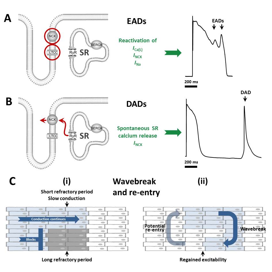
Notching in electrocardiography refers to the presence of distinct deflections or irregularities in the waveform of an electrocardiogram (ECG or EKG), particularly within the P wave (electrocardiography), P wave, QRS complex (fragmented QRS (fQRS)), or T wave. These notches appear as abrupt changes in the direction or slope of the waveform and can provide critical diagnostic information about Cardiac condition, cardiac conditions. Notching in different components of the ECG waveform is associated with various cardiac conditions, ranging from benign variants to serious pathologies, such as Cardiac conduction system, conduction delays, atrial fibrillation, myocardial ischemia, or structural heart disease ('crochetage sign' in atrial septal defect (ASD)). Definition, characteristics Notching is identified as an abrupt change in the direction of an ECG waveform, resulting in a "notch" or dip that creates a bimodal or M-shaped appearance. It is distinct from slurring, which involves a ... [...More Info...] [...Related Items...] OR: [Wikipedia] [Google] [Baidu] |
QRS Notch
The QRS complex is the combination of three of the graphical deflections seen on a typical electrocardiography, electrocardiogram (ECG or EKG). It is usually the central and most visually obvious part of the tracing. It corresponds to the depolarization of the right and left ventricle (heart), ventricles of the heart and contraction of the large ventricular muscles. In adults, the QRS complex normally lasts ; in children it may be shorter. The Q, R, and S waves occur in rapid succession, do not all appear in all leads, and reflect a single event and thus are usually considered together. A Q wave is any downward deflection immediately following the P wave (electrocardiography), P wave. An R wave follows as an upward deflection, and the S wave is any downward deflection after the R wave. The T wave follows the S wave, and in some cases, an additional U wave follows the T wave. To measure the QRS interval start at the end of the PR interval (or beginning of the Q wave) to the end ... [...More Info...] [...Related Items...] OR: [Wikipedia] [Google] [Baidu] |
Bundle Branch Block
A bundle branch block is a partial or complete interruption in the flow of electrical impulses in either of the bundle branches of the heart's electrical system. Anatomy and physiology The heart's electrical activity begins in the sinoatrial node (the heart's natural pacemaker), which is situated on the upper right atrium. The impulse travels next through the left and right atria and summates at the atrioventricular node. From the AV node the electrical impulse travels down the bundle of His and divides into the right and left bundle branches. The right bundle branch contains one fascicle. The left bundle branch subdivides into two fascicles: the left anterior fascicle, and the left posterior fascicle. Other sources divide the left bundle branch into three fascicles: the left anterior, the left posterior, and the left septal fascicle. The thicker left posterior fascicle bifurcates, with one fascicle being in the septal aspect. Ultimately, the fascicles divide into mill ... [...More Info...] [...Related Items...] OR: [Wikipedia] [Google] [Baidu] |
Myocardial Fibrosis
Cardiac fibrosis commonly refers to the excess deposition of extracellular matrix in the cardiac muscle, but the term may also refer to an abnormal thickening of the heart valves due to inappropriate proliferation of cardiac fibroblasts. Fibrotic cardiac muscle is stiffer and less compliant and is seen in the progression to heart failure. The description below focuses on a specific mechanism of valvular pathology but there are other causes of valve pathology and fibrosis of the cardiac muscle. Fibrocyte cells normally secrete collagen, and function to provide structural support for the heart. When over-activated this process causes thickening and fibrosis of the valve, with white tissue building up primarily on the tricuspid valve, but also occurring on the pulmonary valve. The thickening and loss of flexibility eventually may lead to valvular dysfunction and right-sided heart failure. Types Following are types of myocardial fibrosis: * Interstitial fibrosis, which is unspecific, ... [...More Info...] [...Related Items...] OR: [Wikipedia] [Google] [Baidu] |
Purkinje Fibers
The Purkinje fibers, named for Jan Evangelista Purkyně, ( ; ; Purkinje tissue or subendocardial branches) are located in the inner ventricular walls of the heart, just beneath the endocardium in a space called the subendocardium. The Purkinje fibers are specialized conducting fibers composed of electrically excitable cells. They are larger than cardiomyocytes with fewer myofibrils and many mitochondria. They conduct cardiac action potentials more quickly and efficiently than any of the other cells in the heart's electrical conduction system. Purkinje fibers allow the heart's conduction system to create synchronized contractions of its ventricles, and are essential for maintaining healthy and consistent heart rhythm. Histology Purkinje fibers are a unique cardiac end-organ. Further histologic examination reveals that these fibers are split in ventricles walls. The electrical origin of atrial Purkinje fibers arrives from the sinoatrial node. Given no aberrant channe ... [...More Info...] [...Related Items...] OR: [Wikipedia] [Google] [Baidu] |
Sampling Rate
In signal processing, sampling is the reduction of a continuous-time signal to a discrete-time signal. A common example is the conversion of a sound wave to a sequence of "samples". A sample is a value of the signal at a point in time and/or space; this definition differs from the term's usage in statistics, which refers to a set of such values. A sampler is a subsystem or operation that extracts samples from a continuous signal. A theoretical ideal sampler produces samples equivalent to the instantaneous value of the continuous signal at the desired points. The original signal can be reconstructed from a sequence of samples, up to the Nyquist limit, by passing the sequence of samples through a reconstruction filter. Theory Functions of space, time, or any other dimension can be sampled, and similarly in two or more dimensions. For functions that vary with time, let s(t) be a continuous function (or "signal") to be sampled, and let sampling be performed by measuring t ... [...More Info...] [...Related Items...] OR: [Wikipedia] [Google] [Baidu] |
Electrolyte Imbalance
Electrolyte imbalance, or water-electrolyte imbalance, is an abnormality in the concentration of electrolytes in the body. Electrolytes play a vital role in maintaining homeostasis in the body. They help to regulate heart and neurological function, fluid balance, oxygen delivery, acid–base balance and much more. Electrolyte imbalances can develop by consuming too little or too much electrolyte as well as excreting too little or too much electrolyte. Examples of electrolytes include calcium, chloride, magnesium, phosphate, potassium, and sodium. Electrolyte disturbances are involved in many disease processes and are an important part of patient management in medicine. The causes, severity, treatment, and outcomes of these disturbances can differ greatly depending on the implicated electrolyte. The most serious electrolyte disturbances involve abnormalities in the levels of sodium, potassium or calcium. Other electrolyte imbalances are less common and often occur in conjunction ... [...More Info...] [...Related Items...] OR: [Wikipedia] [Google] [Baidu] |
Long QT Syndrome Type 2
Long QT syndrome (LQTS) is a condition affecting repolarization (relaxing) of the heart after a heartbeat, giving rise to an abnormally lengthy QT interval. It results in an increased risk of an irregular heartbeat which can result in fainting, drowning, seizures, or sudden death. These episodes can be triggered by exercise or stress. Some rare forms of LQTS are associated with other symptoms and signs including deafness and periods of muscle weakness. Long QT syndrome may be present at birth or develop later in life. The inherited form may occur by itself or as part of larger genetic disorder. Onset later in life may result from certain medications, low blood potassium, low blood calcium, or heart failure. Medications that are implicated include certain antiarrhythmics, antibiotics, and antipsychotics. LQTS can be diagnosed using an electrocardiogram (EKG) if a corrected QT interval of greater than 450–500 milliseconds is found, but clinical findings, other EKG feature ... [...More Info...] [...Related Items...] OR: [Wikipedia] [Google] [Baidu] |
Myocardial Scarring
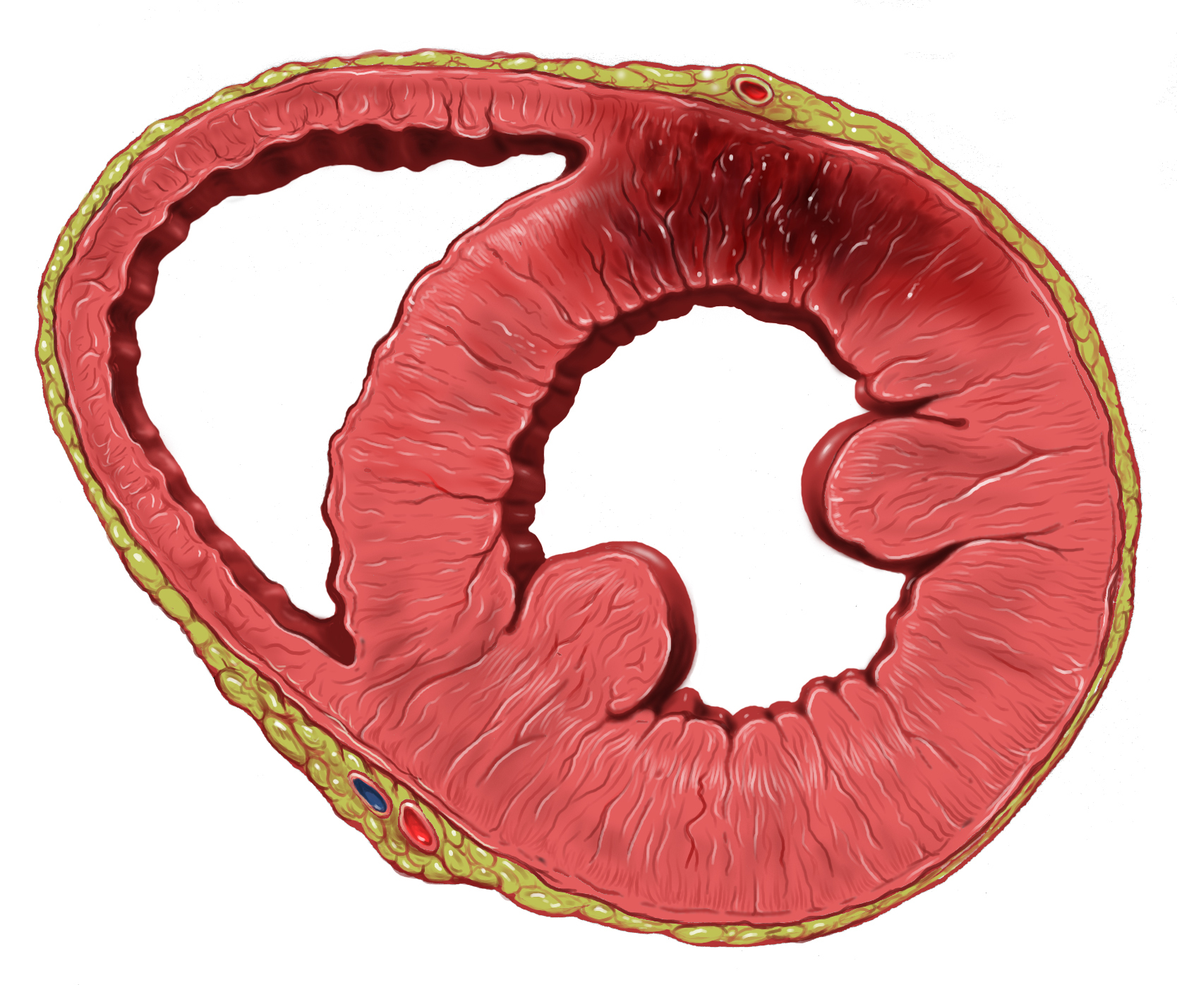
Myocardial scarring is the accumulation of fibrous tissue resulting after some form of trauma to the cardiac tissue. Fibrosis is the formation of excess tissue in replacement of necrotic or extensively damaged tissue. Fibrosis in the heart is often hard to detect because fibromas, scar tissue or small tumors formed in one cell line, are often formed. Because they are so small, they can be hard to detect by methods such as magnetic resonance imaging. A cell line is a path of fibrosis that follow only a line of cells. Causes Myocardial infarction A myocardial infarction, also known as a heart attack, often result in the formation of fibrosis. A myocardial infarction is an ischemic event, or a restriction of blood flow to body tissue, such as by atherothrombosis. Without blood flow to the myocardium, it is deprived of oxygen, causing tissue death and irreversible damage. The tissue destroyed by the infarction is replaced with non-functioning fibrosis, restoring some of the struc ... [...More Info...] [...Related Items...] OR: [Wikipedia] [Google] [Baidu] |
Myocardial Infarction
A myocardial infarction (MI), commonly known as a heart attack, occurs when Ischemia, blood flow decreases or stops in one of the coronary arteries of the heart, causing infarction (tissue death) to the heart muscle. The most common symptom is retrosternal Angina, chest pain or discomfort that classically radiates to the left shoulder, arm, or jaw. The pain may occasionally feel like heartburn. This is the dangerous type of acute coronary syndrome. Other symptoms may include shortness of breath, nausea, presyncope, feeling faint, a diaphoresis, cold sweat, Fatigue, feeling tired, and decreased level of consciousness. About 30% of people have atypical symptoms. Women more often present without chest pain and instead have neck pain, arm pain or feel tired. Among those over 75 years old, about 5% have had an MI with little or no history of symptoms. An MI may cause heart failure, an Cardiac arrhythmia, irregular heartbeat, cardiogenic shock or cardiac arrest. Most MIs occur d ... [...More Info...] [...Related Items...] OR: [Wikipedia] [Google] [Baidu] |
Cardiac Resynchronization Therapy
Cardiac resynchronisation therapy (CRT or CRT-P) is the insertion of electrodes in the left and right ventricles of the heart, as well as on occasion the right atrium, to treat heart failure by coordinating the function of the left and right ventricles via a pacemaker, a small device inserted into the anterior chest wall. CRT is indicated in patients with a low ejection fraction (typically 120 ms. The insertion of electrodes into the ventricles is done under local anesthetic, with access to the ventricles most commonly via the subclavian vein, although access may be conferred from the axillary or cephalic veins. Right ventricular access is direct, while left ventricular access is conferred via the coronary sinus (CS). CRT defibrillators (CRT-D) also incorporate the additional function of an implantable cardioverter-defibrillator (ICD), to quickly terminate an abnormally fast, life-threatening heart rhythm. CRT and CRT-D have become increasingly important therapeutic optio ... [...More Info...] [...Related Items...] OR: [Wikipedia] [Google] [Baidu] |
Early Repolarization
Benign early repolarization (BER) or early repolarization is found on an electrocardiogram (ECG) in about 1% of those with chest pain. It is diagnosed based on an elevated J-point / ST elevation with an end-QRS notch or end-QRS slur and where the ST segment concave up. It is believed to be a normal variant. Benign early repolarization that occurs as some patterns is associated with ventricular fibrillation. The association, revealed by research performed in the late 2000s, is very small. Types Benign early repolarization, very prevalent in younger people and healthy male athletes, can be divided into 3 subtypes: * Type 1 – BER pattern seen in lateral precordial leads. * Type 2 – BER pattern seen in inferior or inferolateral leads. * Type 3 – BER pattern seen globally (inferior, lateral, right precordial leads). Associations with serious conditions Research in the late 2000s has linked this finding to ventricular fibrillation, particularly in those who have fainted o ... [...More Info...] [...Related Items...] OR: [Wikipedia] [Google] [Baidu] |


